. 2012;7(2):e32087.
doi: 10.1371/journal.pone.0032087. Epub 2012 Feb 23.

Sprouty4 is an endogenous negative modulator of TrkA signaling and neuronal differentiation induced by NGF

Affiliations

Sprouty4 is an endogenous negative modulator of TrkA signaling and neuronal differentiation induced by NGF

Fernando C Alsina et al. PLoS One. 2012.

Abstract

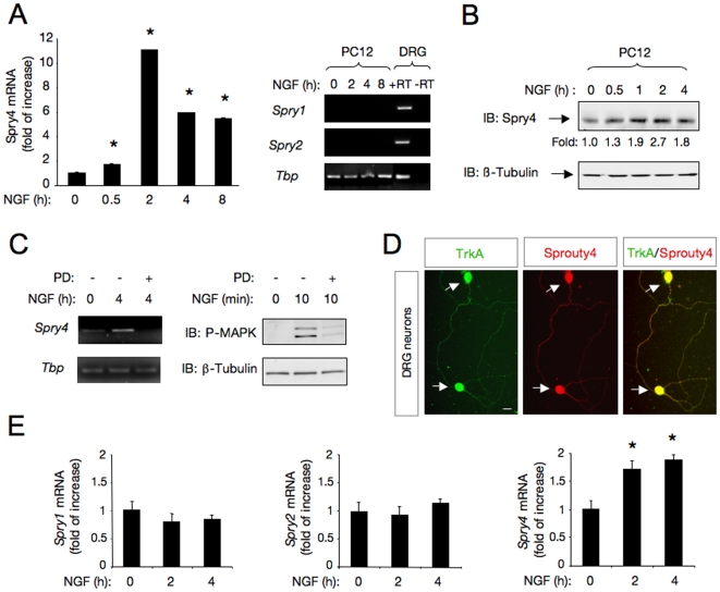
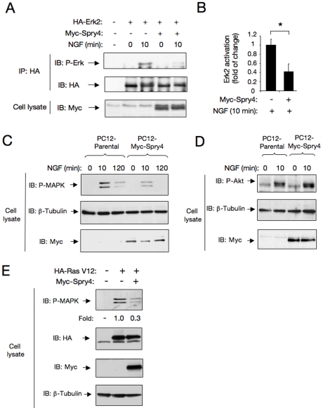
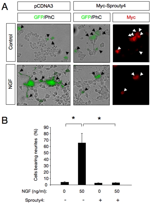
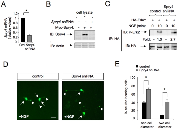
The Sprouty (Spry) family of proteins represents endogenous regulators of downstream signaling pathways induced by receptor tyrosine kinases (RTKs). Using real time PCR, we detect a significant increase in the expression of Spry4 mRNA in response to NGF, indicating that Spry4 could modulate intracellular signaling pathways and biological processes induced by NGF and its receptor TrkA. In this work, we demonstrate that overexpression of wild-type Spry4 causes a significant reduction in MAPK and Rac1 activation and neurite outgrowth induced by NGF. At molecular level, our findings indicate that ectopic expression of a mutated form of Spry4 (Y53A), in which a conserved tyrosine residue was replaced, fail to block both TrkA-mediated Erk/MAPK activation and neurite outgrowth induced by NGF, suggesting that an intact tyrosine 53 site is required for the inhibitory effect of Spry4 on NGF signaling. Downregulation of Spry4 using small interference RNA knockdown experiments potentiates PC12 cell differentiation and MAPK activation in response to NGF. Together, these findings establish a new physiological mechanism through which Spry4 regulates neurite outgrowth reducing not only the MAPK pathway but also restricting Rac1 activation in response to NGF.


Conflict of interest statement

Competing Interests: The authors have declared that no competing interests exist.

Figures

Figure 1
Figure 1. NGF signaling induces Sprouty4 in neuronal cells.
A) Left panel, quantitative analysis of Sprouty4 mRNA expression by real-time PCR in PC12 cells treated with NGF (50 ng/ml) during the indicated times. The levels of Sprouty4 (Spry4) mRNA were normalized using the expression of the housekeeping gene Tbp (TATA binding protein). Shown are averages ± SD of triplicate determinations. *p<0.001 versus control (Ctrl) group (one-way ANOVA followed by Dunnett's test). Right panel, expression of Spry1 and Spry2 mRNAs examined by RT-PCR (35 cycles) in DRG and PC12 cells treated with NGF (50 ng/ml) for different time-points. Expression of the housekeeping gene Tbp was used as loading control. The experiment was repeated two times with similar results. B) Western blot analysis of Sprouty4 expression in PC12 cells treated with NGF (50 ng/ml). Reprobing control was done with antibodies against -tubulin. Fold change relative to -tubulin is indicated. C) Left panel, expression of Spry4 mRNA examined by semiquantitative RT-PCR (27 cycles) in PC12 cells treated with the specific MEK inhibitor PD98059 (50 M) and stimulated with NGF (50 ng/ml) as indicated. Expression of the housekeeping gene Tbp was used as loading control. Right panel shows a control experiment performed in parallel to verify the inhibitory activity of PD98059 on MAPK pathway. PD98059 activity was controlled measuring MAPK activation by immunoblotting of PC12 cells stimulated for 10 min with NGF (50 ng/ml). Reprobing control was done with antibodies against -tubulin. D) Colocalization of Sprouty4 and TrkA in DRG dissociated neurons obtained from E14.5 rat embryos detected by immunofluorescence. Scale bars: 10 m.E) Quantitative analysis of Spry1, Spry2 and Spry4 mRNA expression by real-time PCR in DRG neurons, treated with NGF (50 ng/ml) during the indicated times. The Sproutys mRNA levels were normalized using the expression of the housekeeping gene Tbp. Shown are averages ± SD of triplicate determinations. *p<0.01 versus control (Ctrl) group (one-way ANOVA followed by Dunnett's test).
Figure 2
Figure 2. Sprouty4 restricts Erk/MAPK activation in response to NGF.
A) Erk2/MAPK activation (Phospho-Erk2/MAPK) was evaluated by transient transfection of HA-tagged Erk2 plasmid with a control or a Myc-tagged Sprouty4 vector into PC12 cells. After 36 h cells were serum-starved and stimulated with or without NGF for 10 min. The level of Erk2 activation (P-Erk2) was evaluated by HA-immunoprecipitation followed by immunoblotting with a specific antibody that recognizes the phosphorylated forms of ERK/MAPK. Reprobing of the same blot with anti-HA and anti-Myc antibodies is shown. B) Histogram shows quantification of Erk2/MAPK activation. Results are presented as averages ± SD from three independent experiments. *p<0.05 (Student's t test). C) Erk/MAPK activation (P-MAPK) in cell lysates of parental and PC12-Spry4 (Clon S2) cells treated with NGF (50 ng/ml) and detected by immunoblot (IB). Reprobing of the same blot with anti–tubulin and anti-Myc antibodies is shown. D) Akt activation (P-Akt) in cell lysates of parental and stable transfected PC12-Spry4 (Clon S2) cells treated with NGF (50 ng/ml) and detected by IB. Reprobing of the same blot with anti–tubulin and anti-Myc antibodies is shown. E) Ligand-independent Erk/MAPK activation (P-MAPK) was evaluated by transient transfection of HA-tagged Ras-V12 plasmid (constitutively active Ras) together with a control or a Myc-Sprouty4 vector into PC12 cells. After 36 h, cells were serum-starved and the levels of Erk/MAPK activation were evaluated by immunoblot with a specific antibody that recognizes the phosphorylated forms of ERK/MAPK. Reprobing of the same blot with anti-HA, anti-Myc and anti–tubulin antibodies is shown. Fold change relative to the level of constitutive active HA-Ras V12 is indicated.
Figure 3
Figure 3. Sprouty4 restricts Rac1 activation in response to NGF.
A) Rac1 activation (Rac1-GTP) in cell lysates of parental PC12 cells and clones overexpressing Spry4 (PC12-Spry4 cells) stimulated with NGF (50 ng/ml) for 10 min. Rac1 activation was assessed by GST-Pak-CRIB pull-down assay, followed by immunoblot (IB) with anti-Rac1 antibodies. The bottom panels show total Rac1 and Myc-tagged Sprouty4 levels present in cell lysates. B) Histogram shows quantification of Rac1 activation. Results are presented as averages ± SEM from three independent experiments. *p<0.05 (Student's t test). C) Co-immunoprecipitation between endogenous Rac1 and Myc-tagged Sprouty4 transiently transfected in Cos cells. Mock and Myc-tagged Sprouty4 transfected cell lysates were immunoprecipitated (IP) with anti-Myc antibodies. Immunoprecipitated proteins were resolved by SDS-PAGE and the filters immunoblotted with anti-Rac1 antibodies. The bottom panel shows equal level of -tubulin in both sample inputs.
Figure 4
Figure 4. Sprouty4 inhibits neuronal differentiation of PC12 cells in response to NGF.
A) Photomicrographs show PC12 cells transfected with control or Myc-tagged Sprouty4 plasmid together with a GFP expression vector. After 72 h of NGF treatment (50 ng/ml), cells were fixed and stained with anti-Myc antibodies. Arrowheads indicate neuronal cell bodies and arrows denote neurite tips. Scale bar: 10 m. B) Histogram shows quantification of the relative number of GFP positive PC12 cells bearing neurites longer than 1.5 cell body diameters after 72 h of treatment with NGF. The results are presented as averages SD of a representative experiment performed in triplicate. *p<0.001 (ANOVA followed by Student Newman Keuls). The experiment was repeated three times with similar results.
Figure 5
Figure 5. Sprouty4 inhibits neurite outgrowth of dorsal root ganglion neurons (DRG) in response to NGF.
A) Dissociated DRG neurons transfected with GFP in the absence (Control) or in the presence of an excess of Myc-tagged Sprouty4 (Spry4) construct were cultured with NGF (50 ng/ml). After 36 h in culture, neurons were fixed and stained with anti–tubulin antibodies. Scale bar represents 20 m. Arrows indicate neuronal cell bodies and arrowheads denote neurite tips. B) Left panel, histogram showing the inhibition of neurite outgrowth in DRG neurons by exogenous expression of Sprouty4. The results are averages SEM of a representative experiment measured in six wells per experimental group, *, p<0.05 (Student's t test). The experiment was repeated three times with similar results. Right panel, histogram showing the survival of DRG neurons by exogenous expression of Sprouty4. Neuronal survival was evaluated using the nuclear staining DAPI. GFP-positive neurons containing fragmented or condensed nuclear staining were scored as apoptotic cells. The results are averages SEM of a representative experiment performed in triplicate. C) Histogram shows the distribution of neurons carrying neurites in different length categories after transfection with GFP in the absence (Control) or in the presence of Myc-tagged Sprouty4. A total of 43 control- and 40 Sprouty4-transfected neurons from a representative assay were evaluated. Note the noticeable shift to the left of the distribution of neurons that received the Sprouty4 construct.
Figure 6
Figure 6. Sprouty4 Y53A mutant loses its ability to block both Erk/MAPK pathway and neurite outgrowth of PC12 cells in response to NGF.
A) Schematic representation of the domain structures of mammalian Sprouty family members. The figure shows an amino acid alignment of a conserved motif located at the N-terminal half of the molecule. There, a conserved tyrosine residue (Y) is indicated in red. The conserved C-terminal cysteine-rich domain is also shown. B) Erk/MAPK activation (P-MAPK) in cell lysates of parental and PC12-Myc-Spry4 Y53A cells treated with NGF (50 ng/ml) and detected by IB with a specific antibody that recognizes the phosphorylated forms of ERK/MAPK. Reprobing of the same blot with anti–tubulin and anti-Myc antibodies is shown. The experiment was repeated two times with similar results. C) Photomicrographs show PC12 cells transfected with control or Myc-tagged Sprouty4 Y53A mutant together with a GFP expression vector. After 72 h of NGF treatment, the cells were fixed and stained with anti-Myc. Arrows indicate neuronal cell bodies and arrowheads denote neurite tips. Scale bar: 10 m. D) The histogram shows the quantification of the relative number of GFP positive PC12 cells bearing neurites longer than 1.5 cell body diameters after 72 h of treatment with NGF. The results are presented as averages SD of a representative experiment performed in triplicate. *p<0.05 (ANOVA followed by Student Newman Keuls). The experiment was repeated three times with similar results.
Figure 7
Figure 7. Knockdown of Sprouty4 potentiates MAPK activation and PC12 cell differentiation in response to NGF.
A) Spry4 mRNA levels were analyzed by real-time PCR in PC12 cells transfected with scrambled (control, Ctrl) or Spry4 shRNA constructs. Transfected cells were enriched by puromycin treatment in order to increase the population of cells expressing scrambled or Spry4 shRNA constructs. Quantitative analysis is shown as averages SD of triplicate determinations. The levels of Spry4 mRNA were normalized using the expression of the housekeeping gene Tbp. *p<0.001 (Student's t test). B) Spry4 protein levels were analyzed by IB in Cos cells transfected with Spry4 shRNA or a control vector together with Myc-Spry4. Actin is shown as loading control. C) Spry4 knockdown on Erk2/MAPK activation was analyzed in PC12 cells treated with NGF (50 ng/ml) for 10 min. Erk2/MAPK activation was evaluated by transient transfection of HA-tagged Erk2 plasmid with a control or Spry4 shRNA construct into PC12 cells. Erk2 activation (P-Erk2) was evaluated by HA-immunoprecipitation (IP) followed by IB with a specific antibody that recognizes the phosphorylated forms of ERK/MAPK. Reprobing of the same blot with anti-HA antibody is shown. Numbers below the lanes are normalized to the levels of HA-Erk2. D) Morphological differentiation of PC12 cells transfected with scramble (Ctrl) or Spry4 shRNA cloned in the retroviral vector pGFP-V-RS and treated with NGF (50 ng/ml) for 24 h. E) The histogram shows the quantification of the relative number of GFP positive neurite-bearing cells longer than 1 or 2 cell diameters in the different experimental conditions. The results are shown as averages SD of a representative experiment performed in quadruplicates. *p<0.001 (Student's t test).
Figure 8
Figure 8. Model describing the role of Sprouty4 in NGF-induced TrkA receptor signaling and biology.
NGF-TrkA signaling induces Sprouty4 expression via MAPK pathway. Then, Sprouty4 acting through a negative feedback loop specifically inhibits MAPK pathway and Rac1 activation, thereby preventing neuronal differentiation of PC12 and DRG neurons.

Similar articles

Cited by

References

    1. Segal RA. Selectivity in neurotrophin signaling: theme and variations. Annu Rev Neurosci. 2003;26:299–330. - PubMed
    1. Huang EJ, Reichardt LF. Trk receptors: roles in neuronal signal transduction. Annu Rev Biochem. 2003;72:609–642. - PubMed
    1. Dikic I, Giordano S. Negative receptor signalling. Curr Opin Cell Biol. 2003;15:128–135. - PubMed
    1. Ledda F, Paratcha G. Negative Regulation of Receptor Tyrosine Kinase (RTK) Signaling: A Developing Field. Biomark Insights. 2007;2:45–58. - PMC - PubMed
    1. Mason JM, Morrison DJ, Basson MA, Licht JD. Sprouty proteins: multifaceted negative-feedback regulators of receptor tyrosine kinase signaling. Trends Cell Biol. 2006;16:45–54. - PubMed

Publication types