. 2012;7(2):e32305.
doi: 10.1371/journal.pone.0032305. Epub 2012 Feb 22.

The TAL effector PthA4 interacts with nuclear factors involved in RNA-dependent processes including a HMG protein that selectively binds poly(U) RNA

Affiliations

The TAL effector PthA4 interacts with nuclear factors involved in RNA-dependent processes including a HMG protein that selectively binds poly(U) RNA

Tiago Antonio de Souza et al. PLoS One. 2012.

Erratum in

Abstract

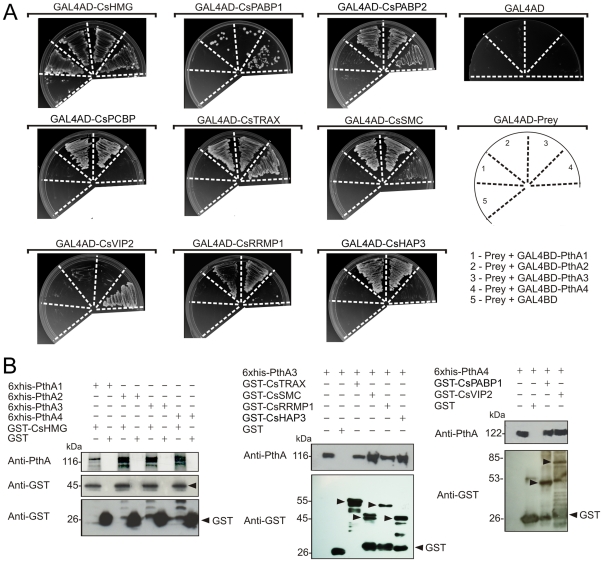
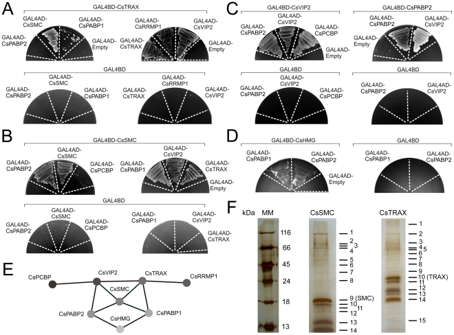
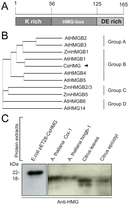
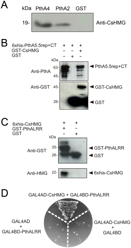
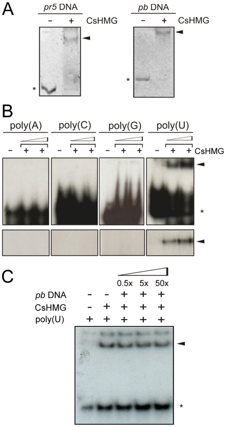
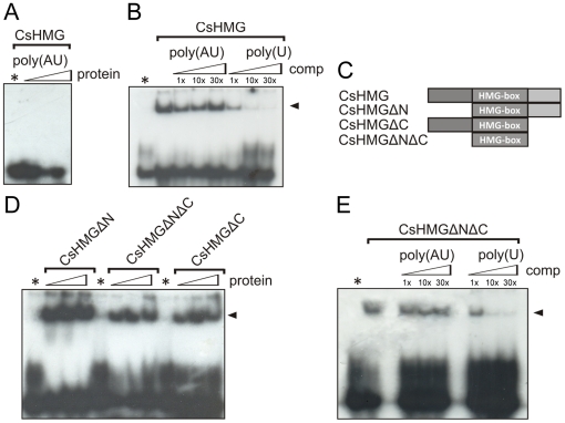
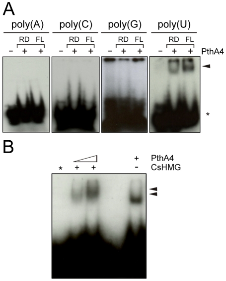
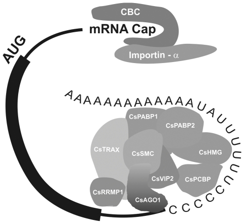
Plant pathogenic bacteria utilize an array of effector proteins to cause disease. Among them, transcriptional activator-like (TAL) effectors are unusual in the sense that they modulate transcription in the host. Although target genes and DNA specificity of TAL effectors have been elucidated, how TAL proteins control host transcription is poorly understood. Previously, we showed that the Xanthomonas citri TAL effectors, PthAs 2 and 3, preferentially targeted a citrus protein complex associated with transcription control and DNA repair. To extend our knowledge on the mode of action of PthAs, we have identified new protein targets of the PthA4 variant, required to elicit canker on citrus. Here we show that all the PthA4-interacting proteins are DNA and/or RNA-binding factors implicated in chromatin remodeling and repair, gene regulation and mRNA stabilization/modification. The majority of these proteins, including a structural maintenance of chromosomes protein (CsSMC), a translin-associated factor X (CsTRAX), a VirE2-interacting protein (CsVIP2), a high mobility group (CsHMG) and two poly(A)-binding proteins (CsPABP1 and 2), interacted with each other, suggesting that they assemble into a multiprotein complex. CsHMG was shown to bind DNA and to interact with the invariable leucine-rich repeat region of PthAs. Surprisingly, both CsHMG and PthA4 interacted with PABP1 and 2 and showed selective binding to poly(U) RNA, a property that is novel among HMGs and TAL effectors. Given that homologs of CsHMG, CsPABP1, CsPABP2, CsSMC and CsTRAX in other organisms assemble into protein complexes to regulate mRNA stability and translation, we suggest a novel role of TAL effectors in mRNA processing and translational control.


Conflict of interest statement

Competing Interests: The authors have declared that no competing interests exist.

Figures

Figure 1
Figure 1. Protein-protein interactions between PthAs and citrus nuclear proteins.
(A) Citrus preys fused to yeast GAL4-AD (GAL4AD-prey) or control plasmid (GAL4AD) were moved into yeast cells carrying one of the four PthA variants fused to GAL4-BD domain as shown in the diagram (1 to 4, respectively). Yeast double-transformants were grown on SC -Trp -Leu -His -Ade in the presence of 5 mM of 3AT. None of prey fusions transactivated the reporter genes when co-transformed with empty bait vector (5). The PthA baits also did not transactivate the reporter genes when co-transformed with the empty prey vector in the same growth conditions (GAL4AD). (B) Western blot detection of eluted fractions from GST pulldown assays using the purified 6xHis-PthAs 1–4 as prey and immobilized GST or GST-fusion proteins as baits. Arrows indicate bands corresponding to the expected size for the GST-fusion proteins CsHMG (∼45 kDa), CsTRAX (∼55 kDa), CsSMC (∼45 kDa), CsRRPMP1 (∼50 kDa), CsRRMP2 (∼46 kDa), CsPABP1 (∼53 kDa) and CsVIP2 (∼85 kDa) detected by the GST anti-serum. PthA proteins (∼116–122 kDa) were detected using the anti-PthA serum. Recombinant PthAs 3 and 4 were added as references in the first lanes of the gels shown in the middle and right panels, respectively.
Figure 2
Figure 2. Protein-protein interactions among the PthA4 interactors detected by yeast two-hybrid and mass spectrometry.
Yeast cells double-transformed with the indicated prey-bait constructs were grown in SC -Trp -Leu -His -Ade in the presence of 5 mM 3AT. (A) Positive interactions observed between CsTRAX and CsSMC, CsPABP1, CsTRAX, CsRRMP1 and CsVIP2. (B) Protein-protein interactions observed between CsSMC and CsPABP2, CsSMC, CsPABP1, CsVIP2 and CsTRAX, but not between CsSMC and CsKH. (C) Interactions of CsVIP2 with CsKH, reciprocal interactions between CsPABP2 and CsVIP2, and self interactions of CsVIP2 and CsPABP2. (D) Weak interactions between CsHMG and the poly(A)-binding proteins CsPABP2 and CsPABP1. (E) A diagram illustrating the network of interactions observed among the citrus PthA targets. (F) Silver-stained SDS polyacrylamide gels of citrus proteins trapped in cobalt beads carrying the recombinant 6xHis-tagged CsSMC or CsTRAX as baits (bands 9 and 10, respectively). Protein bands excised from the gels, indicated by the numbers, were identified by mass spectrometry (see Table 2 for details). The molecular markers (MM) are shown on the left.
Figure 3
Figure 3. CsHMG shows identity to plant HMGBs of group B.
(A) Schematic representation of the CsHMG primary structure showing its central HMG-box domain flanked by the basic K-rich N-terminal and the acidic DE-rich C-terminal. (B) Phylogenetic analysis of plant HMGB proteins showing that CsHMG belongs to group B HMGBs. (C) Western-blot detection of the recombinant 6xHis-CsHMG (∼22 kDa) made in bacteria compared to bands detected in citrus cell extracts with the expected molecular size for the endogenous CsHMG (∼16 kDa). The anti-CsHMG serum also cross-reacted with a band of similar size in the cell extracts of A. thaliana wild-type and heterozygous hmg-b1 mutant. This band, which has the expected molecular weight for AtHMGB1 (∼18 kDa), is less pronounced in the heterozygous hmgb-1 mutant, thus indicating that CsHMG is structurally related to AtHMGB1.
Figure 4
Figure 4. PthA binds to CsHMG in vivo through its invariable LRR region.
(A) PthA2-GST, PthA4-GST or GST alone bound to glutathione resins were incubated with a citrus cell lisate. Bound proteins were separated by gel electrophoresis and CsHMG was detected by the anti-CsHMG serum in the PthA samples only. (B) Western blot of GST-pulldown assay of immobilized GST or GST-CsHMG as baits and purified 6xHis-PthA5.5rep+CT2 as prey. The eluted 6xHis-PthA5.5rep+CT2 (∼63 kDa) was detected by the anti-PthA serum only when GST-CsHMG was used as bait. The purified 6xHis-PthA5.5rep+CT2 was added in the first lane of the gel as reference. (C) Western blot analysis of eluted fractions of GST-pulldown assay of immobilized GST or GST-PthALRR as baits and purified 6xHis-CsHMG as prey. The eluted 6xHis-CsHMG (∼22 kDa) was detected only when GST-PthALRR was used as bait. (D) Yeast two-hybrid assay showing the interaction between CsHMG and the PthA LRR domain. Yeast double-tranformants, including controls (GAL4AD+GAL4BD-PthALRR and GAL4BD+GAL4AD-CsHMG), were grown in SC -Trp -Leu -His -Ade in the presence of 5 mM 3AT.
Figure 5
Figure 5. CsHMG shows DNA and RNA-binding activities in vitro.
(A) EMSA using the recombinant 6xHis-CsHMG protein (5 µg) and DNA probes (200 ng) derived from the citrus pr5 promoter and the multiple-cloning site of the pBluescript vector. The DNA-protein complexes and free probes detected by ethidium bromide staining are indicated by the arrows and asterisks, respectively. (B) Upper panel, EMSA using 32P-labelled single strand RNA probes at a final concentration of 12.5 nM and increased amounts of purified CsHMG (0.1 and 0.5 µg). Shifted bands corresponding to CsHMG:RNA complexes observed with the poly(U) RNA and the free probes are indicated by the arrow and asterisk, respectively. Bottom panel, SDS-PAGE of UV-crosslink EMSA showing the selective binding of CsHMG to the poly(U) RNA probe (arrow). (C) EMSA using the 32P-labelled poly(U) RNA as probe at a final concentration of 12.5 nM, 100 ng of CsHMG and increasing amounts of the double strand DNA (multiple-cloning site of the pBluescript vector) as competitor. Shifted bands corresponding to CsHMG:RNA complexes and the free probe are indicated by the arrow and asterisk, respectively.
Figure 6
Figure 6. CsHMG selectively binds to poly(U) RNAs through its HMG-box domain.
(A) EMSA showing that CsHMG (0.5 and 1.0 µg) does not bind to the AU-rich RNA probe (5′- UUAUUAUUUAUUUAUUUAUU-3′). (B) Competition EMSA showing that the AU-rich RNA probe at 1, 10 and 30 times molar excess does not compete with the poly(U) probe for the binding to CsHMG, as opposed to the cold poly(U) probe. (C) Schematic representation of the truncated versions of CsHMG used in the experiments depicted in D and E. (D) All CsHMG truncations were capable of binding to the poly(U) RNA probe. (E) Competition EMSA showing that the AU-rich RNA probe at 1, 10 and 30 times molar excess does not compete with the poly(U) probe for the binding to the HMG-box (CsHMGΔNΔC), as opposed to the cold poly(U) probe, indicating that the HMG-box of CsHMG is sufficient to confer the poly(U) RNA-binding specificity.
Figure 7
Figure 7. PthA4 and its repetitive domain bind to poly(U) RNA.
(A) EMSA using 32P-labelled single strand RNA probes at a final concentration of 12.5 nM and 100 ng of the purified full-length (FL) PthA4 or its internal repetitive domain (RD). Shifted bands corresponding to the RNA:protein complexes and the free probes are indicated by the arrow and asterisks, respectively. (B) EMSA using the 32P-labelled single strand poly(U) probe and the recombinant full length PthA4 and CsHMG. PthA4 forms higher molecular weight bands in the presence of CsHMG (arrows). The CsHMG-poly(U) complex is not resolved from the free probe (*) in the 6% polyacrylamide gel.
Figure 8
Figure 8. Schematic model of the citrus multiprotein complex comprising the PthA4-interacting partners.
Protein-protein and protein-RNA contacts involving the PthA4 interactors based on the yeast two-hybrid, GST-pulldown and gel-shift assays described here, and literature data. The citrus multiprotein complex is reminiscent of that of mammalian miRISC involved in miRNA-mediated deadenylation . Importin-α, which interacts with all PthA variants is also a component of the cap-binding complex (CBC) which inhibits mRNA deadenylation when in the presence of a poly(A)-specific ribonucleases –. It is suggested that by interacting with such proteins and with poly(U) RNA (not necessarily simultaneously), PthA proteins may displace some of the components of this complex thought to promote deadenylation and mRNA decay and thus increase mRNA stabilization and translation initiation. U-rich sequences found in both 5′ and 3′ ends of mRNAs could represent binding sites of CsHMG and PthA4.

Similar articles

Cited by

References

    1. Mansfield JW. From bacterial avirulence genes to effector functions via the hrp delivery system: an overview of 25 years of progress in our understanding of plant innate immunity. Mol Plant Pathol. 2009;10:721–734. - PMC - PubMed
    1. Boch J, Bonas U. Xanthomonas AvrBs3 family-type III effectors: discovery and function. Annu Rev Phytopathol. 2010;48:419–436. - PubMed
    1. Kay S, Hahn S, Marois E, Hause G, Bonas U. A bacterial effector acts as a plant transcription factor and induces a cell size regulator. Science. 2007;318:648–651. - PubMed
    1. Römer P, Hahn S, Jordan T, Strauss T, Bonas U, et al. Plant pathogen recognition mediated by promoter activation of the pepper Bs3 resistance gene. Science. 2007;318:645–648. - PubMed
    1. Kay S, Hahn S, Marois E, Wieduwild R, Bonas U. Detailed analysis of the DNA recognition motifs of the Xanthomonas type III effectors AvrBs3 and AvrBs3Deltarep16. Plant J. 2009;59:859–871. - PubMed

Publication types

MeSH terms