. 2012;7(2):e32333.
doi: 10.1371/journal.pone.0032333. Epub 2012 Feb 22.

Macrophage sub-populations and the lipoxin A4 receptor implicate active inflammation during equine tendon repair

Affiliations

Macrophage sub-populations and the lipoxin A4 receptor implicate active inflammation during equine tendon repair

Stephanie Georgina Dakin et al. PLoS One. 2012.

Abstract

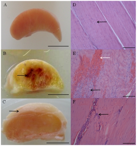
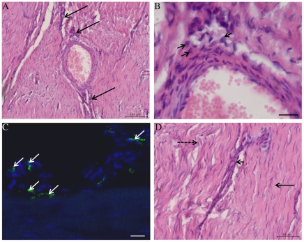
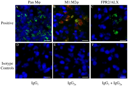
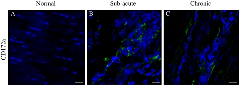
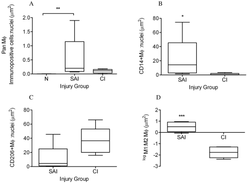
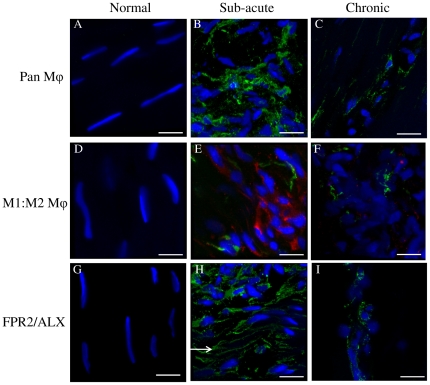
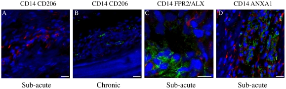
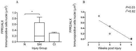
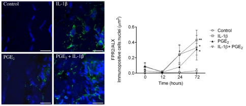
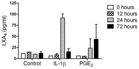
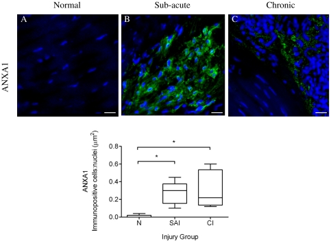
Macrophages (Mφ) orchestrate inflammatory and reparatory processes in injured connective tissues but their role during different phases of tendon healing is not known. We investigated the contribution of different Mφ subsets in an equine model of naturally occurring tendon injury. Post mortem tissues were harvested from normal (uninjured), sub-acute (3-6 weeks post injury) and chronically injured (>3 months post injury) superficial digital flexor tendons. To determine if inflammation was present in injured tendons, Mφ sub-populations were quantified based on surface antigen expression of CD172a (pan Mφ), CD14(high)CD206(low) (pro-inflammatory M1Mφ), and CD206(high) (anti-inflammatory M2Mφ) to assess potential polarised phenotypes. In addition, the Lipoxin A(4) receptor (FPR2/ALX) was used as marker for resolving inflammation. Normal tendons were negative for both Mφ and FPR2/ALX. In contrast, M1Mφ predominated in sub-acute injury, whereas a potential phenotype-switch to M2Mφ polarity was seen in chronic injury. Furthermore, FPR2/ALX expression by tenocytes was significantly upregulated in sub-acute but not chronic injury. Expression of the FPR2/ALX ligand Annexin A1 was also significantly increased in sub-acute and chronic injuries in contrast to low level expression in normal tendons. The combination of reduced FPR2/ALX expression and persistence of the M2Mφ phenotype in chronic injury suggests a potential mechanism for incomplete resolution of inflammation after tendon injury. To investigate the effect of pro-inflammatory mediators on lipoxin A(4) (LXA(4)) production and FPR2/ALX expression in vitro, normal tendon explants were stimulated with interleukin-1 beta and prostaglandin E(2). Stimulation with either mediator induced LXA(4) release and maximal upregulation of FPR2/ALX expression after 72 hours. Taken together, our data suggests that although tenocytes are capable of mounting a protective mechanism to counteract inflammatory stimuli, this appears to be of insufficient duration and magnitude in natural tendon injury, which may potentiate chronic inflammation and fibrotic repair, as indicated by the presence of M2Mφ.


Conflict of interest statement

Competing Interests: SGD's PhD is funded by Biotechnology and Biological Sciences Research Council (BBSRC) UK and Ceva (France). This does not alter the authors' adherence to all the PLoS ONE policies on sharing data and materials. The authors have declared that no other competing interests exist.

Figures

Figure 1
Figure 1. Typical macroscopic appearance of normal and injured equine flexor tendons.
(A) Normal superficial digital flexor tendon (SDFT) from a 12 year old horse; (B) sub-acutely injured SDFT 3 weeks post injury from a 4 year old horse, exhibiting a haemorrhagic granular central core (arrow) and (C) chronically injured SDFT >3 months post injury with a thickened fibrosed paratenon (arrow) from a 12 year old horse. Scale bar for macroscopic images = 1 cm. Corresponding longitudinal histology sections stained with Haematoxylin and Eosin: (D) normal SDFT showing regular arrangement of collagen fibrils (arrow); (E) sub-acutely injured SDFT with marked cellular infiltration (black arrow) and haemorrhage (white arrow); (F) chronically injured SDFT with increased cellularity in peri-vascular regions (arrow). Histology scale bar = 12.5 µm.
Figure 2
Figure 2. Haematoxylin and Eosin stained longitudinal histology sections of chronic injured SDFT (>3 months post injury) from a 7 year old horse (A, B and D).
(A) Reactive fibroplasia with increased cellularity in peri-vascular region (arrows). Scale bar = 100 µm. (B) Higher magnification of (A) showing presence of macrophages (arrowheads) in peri-vascular areas. Scale bar = 20 µm. (C) Corresponding 3-dimensional reconstructed Z stack image of dual antibody labelling for CD14 (red) and CD206 (green). Blue represents Hoechst nuclear counter stain. White arrows show CD14lowCD206high M2Mϕ located in peri-vascular endotenon regions. Scale bar = 20 µm. (D) Histological appearance of more normal SDFT to the right of the image (straight arrow) in contrast to irregular arrangement of collagen fibrils on the left (dashed arrow). The linear interface between more normal and injured zones of tendon is demarcated by an area of increased cellularity containing macrophages (arrowhead). Scalebar = 50 µm.
Figure 3
Figure 3. Representative Z stack images of antibody control cryosections of equine spleen.
Panel A–C represents positive controls for: (A)CD172a Mϕ (green); (B) Dual antibody labelling with CD14 (M1Mϕ red) and CD206 (M2Mϕ green) showing co-expression of both markers (CD14highCD206high) in splenic monocytes/macrophages; (C) Lipoxin A4 receptor (FPR2/ALX, green). Panel D–F represents negative controls, consisting of murine isotype matched primary control antibodies: (D) IgG1, (E) IgG2a, (F) IgG1 and IgG2a. Blue represents Hoechst nuclear counter stain. Scale bar = 12 µm.
Figure 4
Figure 4. Panel of representative 3-dimensional reconstructed Z stack immunofluorescent low magnification images of equine SDFT sections.
Pan Mϕ (CD172a) staining is shown for is for (A) normal, (B) sub-acute and (C) chronic injured tendons. Immunopositive cells are green; blue represents Hoechst nuclear counter stain. Scale bar = 20 µm.
Figure 5
Figure 5. Box plots illustrating ratios of areas (µm2) of immunopositive cells: counterstained nuclei.
(A) CD172a pan Mϕ in normal (uninjured), sub-acute and chronic injured equine tendons; (B) CD14high (M1Mϕ) and (C) CD206high (M2Mϕ) expression in sub-acute and chronic injured tendons respectively. (D) Log transformed ratio of areas of positive immuno-reactivity for M1∶M2 Mϕ from dual labelled CD14 and CD206 sections of sub-acute and chronic injured tendons. SAI = sub-acute injury (n = 5), CI = chronic injury (n = 5), N = normal tendon (n = 5). All values represent median with maximum and minimum range. *** P<0.001, ** P<0.01, * P<0.05.
Figure 6
Figure 6. Panel of representative 3-dimensional reconstructed Z stack immunofluorescent images of equine SDFT sections.
(A–C) CD172a Mϕ antibody, immunopositive cells are green; (D–F) Dual antibody labelling for CD14 (M1Mϕ red) and CD206 (M2Mϕ green) showing CD14highCD206low Mϕ in sub-acute injury (E) and CD14lowCD206high Mϕ in chronic injury (F); (G–I) FPR2/ALX, immunopositive cells are green. Arrow in panel H shows FPR2/ALX expression on tenocyte cytoplasmic extensions. Panels A, D, & G are from normal uninjured tendon; B, E & H are from sub-acutely injured tendon; C, F & I are from chronically injured tendon. Blue represents Hoechst nuclear counter stain. Scale bar = 12 µm.
Figure 7
Figure 7. Panel of representative 3-dimensional reconstructed Z stack immunofluorescent images of injured SDFT sections.
(A) Dual labelled CD14 (red) and CD206 (green) showing predominance of CD14high Mϕ in sub-acute tendon injury. Scale bar = 20 µm. (B) Dual labelled CD14 (red) and CD206 (green) showing predominance of CD206high Mϕ in chronic tendon injury. Scale bar = 20 µm. (C) Dual labelled CD14 (red) and FPR2/ALX (green), showing FPR2/ALX expression by tenocytes but not M1Mϕ in sub-acute tendon injury. Scale bar = 12 µm. (D) Dual labelled CD14 (red) and Annexin A1 (green), showing Annexin A1 expression by tenocytes but not M1Mϕ in sub-acute tendon injury. Scale bar = 20 µm. Blue represents Hoechst nuclear counter stain.
Figure 8
Figure 8. FPR2/ALX expression in normal and injured equine tendons.
(A) Box plot illustrating ratio of areas (µm2) of immunopositive cells: counterstained nuclei of expression of the Lipoxin A4 receptor (FPR2/ALX) in normal (N, n = 5), sub-acute (SAI, n = 5) and chronic injured (CI n = 5) equine tendons. Values represent median with maximum and minimum range. * P<0.05. (B) FPR2/ALX expression in sub-acutely injured tendons (n = 5) showing significant decline in FPR2/ALX protein expression with time after injury (P = 0.03, r2 = 0.82).
Figure 9
Figure 9. Panel of representative 2-dimensional images illustrating FPR2/ALX expression in tendon explants showing non-stimulated (vehicle only) control compared to explants stimulated with 5 ng ml−1 IL-1β and 1.0 µM PGE2 either alone or in combination.
Immunopositive staining is green, with Hoechst nuclear counter stain in blue. Scale bar = 12 µm. Graph showing the effect of pro-inflammatory mediators on tendon FPR2/ALX expression in vitro. Data represent average FPR2/ALX expression in tendon explants derived from 3 normal (uninjured) horses, whereby 2 replicates were analysed for each experimental condition and time point per horse. FPR2/ALX expression was determined at time points 0, 12, 24 and 72 hours after stimulation and compared to non-stimulated controls. Significant up-regulation of FPR2/ALX occurs 24 hours after stimulation with IL-1β or a combination of both mediators, and maximally 72 hours after stimulation with IL-1β, PGE2 or combination of both mediators compared to controls. ** P<0.01, * P<0.05. Error bars denote SEM.
Figure 10
Figure 10. The effect of pro-inflammatory mediators on release of lipoxin A4 (LXA4) in media from tendon explants.
Data represent average LXA4 levels from 3 normal (uninjured) horses. LXA4 levels were determined at time points 0, 12, 24 and 72 hours after stimulation and compared to non-stimulated controls. There was a difference in the temporal response showing increased LXA4 release 24 hours after stimulation with IL-1β (P<0.001) and 72 hours after stimulation with PGE2 (P = 0.048) compared to non-stimulated controls. Values for significance were based on a linear model.
Figure 11
Figure 11. Panel of representative 3-dimensional reconstructed Z stack immunofluorescent images of equine SDFT sections.
Annexin A1 (ANXA1) staining is shown for is for (A) normal, (B) sub-acute and (C) chronic injured tendons. Immunopositive cells are green; blue represents Hoechst nuclear counter stain. Scale bar = 20 µm. Box plot shows significantly increased Annexin A1 expression in sub-acute and chronic injured tendons compared to low level expression in normal tendons. SAI = sub-acute injury (n = 5), CI = chronic injury (n = 5), N = normal tendon (n = 5). Values represent median with maximum and minimum range. * P<0.05.

Similar articles

Cited by

References

    1. Kujala UM, Sarna S, Kaprio J. Cumulative incidence of achilles tendon rupture and tendinopathy in male former elite athletes. Clin J Sport Med. 2005;15:133–135. - PubMed
    1. Welsh RP, Clodman J. Clinical survey of Achilles tendinitis in athletes. Can Med Assoc J. 1980;122:193–195. - PMC - PubMed
    1. Avella CS, Ely ER, Verheyen KL, Price JS, Wood JL, et al. Ultrasonographic assessment of the superficial digital flexor tendons of National Hunt racehorses in training over two racing seasons. Equine Vet J. 2009;41:449–454. - PubMed
    1. Jarvinen M, Jozsa L, Kannus P, Jarvinen TL, Kvist M, et al. Histopathological findings in chronic tendon disorders. Scand J Med Sci Sports. 1997;7:86–95. - PubMed
    1. Jozsa L, Reffy A, Kannus P, Demel S, Elek E. Pathological alterations in human tendons. Arch Orthop Trauma Surg. 1990;110:15–21. - PubMed

Publication types

MeSH terms