. 2012;7(2):e32378.
doi: 10.1371/journal.pone.0032378. Epub 2012 Feb 22.

Spleen tyrosine kinase regulates AP-1 dependent transcriptional response to minimally oxidized LDL

Affiliations

Spleen tyrosine kinase regulates AP-1 dependent transcriptional response to minimally oxidized LDL

Soo-Ho Choi et al. PLoS One. 2012.

Abstract

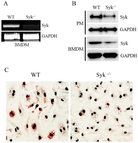
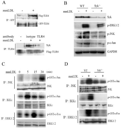
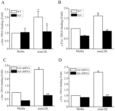
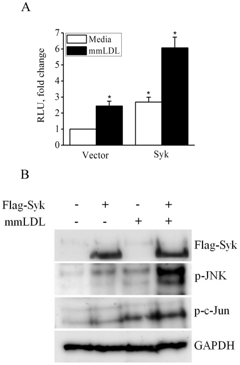
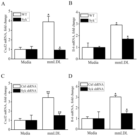
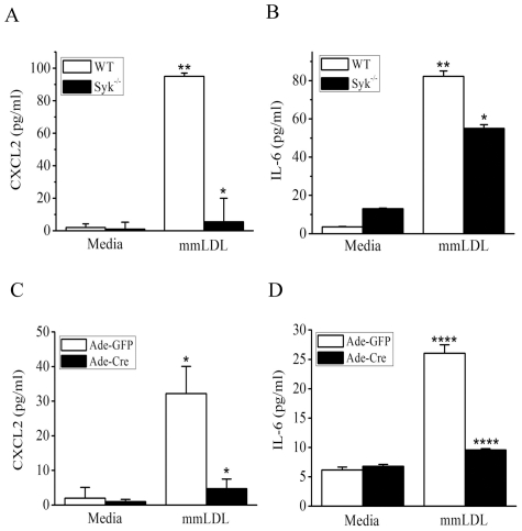
Oxidative modification of low-density lipoprotein (LDL) turns it into an endogenous ligand recognized by pattern-recognition receptors. We have demonstrated that minimally oxidized LDL (mmLDL) binds to CD14 and mediates TLR4/MD-2-dependent responses in macrophages, many of which are MyD88-independent. We have also demonstrated that the mmLDL activation leads to recruitment of spleen tyrosine kinase (Syk) to TLR4 and TLR4 and Syk phosphorylation. In this study, we produced a macrophage-specific Syk knockout mouse and used primary Syk(-/-) macrophages in our studies. We demonstrated that Syk mediated phosphorylation of ERK1/2 and JNK, which in turn phosphorylated c-Fos and c-Jun, respectively, as assessed by an in vitro kinase assay. c-Jun phosphorylation was also mediated by IKKε. c-Jun and c-Fos bound to consensus DNA sites and thereby completed an AP-1 transcriptional complex and induced expression of CXCL2 and IL-6. These results suggest that Syk plays a key role in TLR4-mediated macrophage responses to host-generated ligands, like mmLDL, with subsequent activation of an AP-1 transcription program.


Conflict of interest statement

Competing Interests: The authors have declared that no competing interests exist.

Figures

Figure 1
Figure 1. Reduced Syk expression in macrophages from Sykflox/flox/LysM-Cre mice.
(A) Total RNA was isolated from BMDM of Sykflox/flox/LysM-Cre(−) (WT) and Sykflox/flox/LysM-Cre(+) (Syk−/−) mice, and RT-PCR with Syk and GAPDH primers was performed. (B) Lysates of resident peritoneal macrophages (PM) and BMDM were run on SDS-PAGE, transferred to a membrane and immunoblotted with anti-Syk and anti-GAPDH antibodies. (C) WT and Syk−/− BMDM were incubated with 50 µg/ml mmLDL (to induce macropinocytosis) and 200 µg/ml of native LDL (lipid carrier) for 40 hours. The cells were stained for neutral lipid with Oil Red O.
Figure 2
Figure 2. MAP kinases and transcription factors phosphorylation in WT and Syk−/− macrophages stimulated with mmLDL and KLA.
(A) Upper blots: Ba/F3 cells stably expressing GFP-TLR4 and Flag-TLR4 were incubated with media or mmLDL (50 µg/ml) for 15 min. Cell lysates were immunoprecipitated with an anti-GFP antibody and immunoblotted with anti-Flag and anti-GFP antibodies. Lower blots: Ba/F3 cells were preincubated with 5 µg/ml of an anti-mouse TLR4/MD2 antibody (UT12) or an isotype IgG3κ control for 1 hour and then stimulated with mmLDL (50 µg/ml) for 15 min. Cell lysates were immunoprecipitated with a different anti-mouse TLR4 antibody (UT18) and immunoblotted with anti-Syk and anti-Flag antibodies. (B) WT and Syk−/− BMDM were incubated with media or mmLDL (50 µg/ml) for 15 min. Cell lysates were separated on SDS-PAGE and immunoblotted with antibodies against Syk, phospho-ERK1/2, phospho-c-Jun, phospho-JNK and GAPDH. (C) In vitro kinase assay in J774 macrophages. Cells were incubated with mmLDL (50 µg/ml) for indicated periods of time and then precipitated with anti-JNK, anti-IKKε and anti-ERK1/2 antibodies. Endogenous JNK and IKKε kinase activities were determined using GST-c-Jun (1–79 aa) as a substrate, and endogenous ERK1/2 kinase activity was determined using GST-c-Fos (300–380 aa) as a substrate. Protein levels of endogenous JNK, IKKε and ERK1/2 in the same cell lysates were determined by immunoblot. This experiment informed the optimal stimulation time periods for the experiment in panel D. (D) In vitro kinase assay in WT and Syk−/− BMDM. Cells were treated with media or mmLDL (50 µg/ml) for 30 min (JNK), 5 min (IKKε) or 15 min (ERK1/2). Kinase activities were determined using the same method as in C.
Figure 3
Figure 3. c-Jun and c-Fos DNA binding in WT and Syk−/− macrophages stimulated with mmLDL.
BMDM from WT or Syk−/− mice (A and B) and J774 expressing control of Syk-specific shRNA (C and D) were incubated for 1 hour with media or 50 µg/ml mmLDL. Nuclear extracts were isolated and used in a transcription factor DNA binding plate-based assay. Mean ± SEM (n = 4). *, p<0.05; **, p<0.005 WT vs. Syk−/−.
Figure 4
Figure 4. AP-1 activation in Syk-expressing CHO cells.
(A) CHO cells were transfected with 1 µg Flag-Syk (or empty vector), 200 ng AP-1-Luc reporter, and 200 ng β-galactosidase plasmid. After 36 hours, cells were incubated with media or mmLDL for 6 hours and luciferase activity was measured and normalized to β-galactosidase activity. Mean±SEM (n = 3). *, p<0.05 vector vs. Syk, and media vs. mmLDL. (B) CHO Cells were transfected with 300 ng empty vector or Flag-Syk. After 24 hours, cells were incubated with mmLDL for 6 hours and cell lysates were tested for expression of Syk (Flag) and for p-JNK, p-c-Jun and GAPDH.
Figure 5
Figure 5. mRNA expression of Cxcl2 and Il-6 in WT and Syk−/− macrophages stimulated with mmLDL.
BMDM from WT or Syk−/− mice (A and B) and J774 cells expressing control or Syk-specific shRNA (C and D) were incubated for 1 hour with media or 50 µg/ml mmLDL. Total RNA was isolated and expression of Cxcl2 and Il-6 mRNA was measured by qPCR and normalized to the GAPDH mRNA levels. Mean ± SEM (n = 3). *, p<0.05; **, p<0.005 WT vs. Syk−/−.
Figure 6
Figure 6. Secretion of CXCL2 (MIP-2) and IL-6 by WT and Syk−/− macrophages stimulated with mmLDL.
BMDM from WT or Syk−/− mice (A and B) and resident peritoneal macrophages from Sykflox/flox mice infected with adenovirus expressing GFP or Cre (for 48 hours at 500 MOI) (C and D) were incubated for 6 hours with media or 50 µg/ml mmLDL. Cell culture media were collected and CXCL2 and IL-6 protein levels were measured by ELISA. Mean ± SEM (n = 3). *, p<0.05; ****, p<0.00005 WT vs. Syk−/−.

Similar articles

Cited by

References

    1. Mocsai A, Ruland J, Tybulewicz VLJ. The SYK tyrosine kinase: a crucial player in diverse biological functions. Nat Rev Immunol. 2010;10:387–402. - PMC - PubMed
    1. Jakus Z, Fodor S, Abram CL, Lowell CA, Mocsai A. Immunoreceptor-like signaling by [beta]2 and [beta]3 integrins. Trends in Cell Biology. 2007;17:493–501. - PubMed
    1. Woodside DG, Obergfell A, Talapatra A, Calderwood DA, Shattil SJ, et al. The N-terminal SH2 Domains of Syk and ZAP-70 Mediate Phosphotyrosine-independent Binding to Integrin Cytoplasmic Domains. J Biol Chem. 2002;277:39401–39408. - PubMed
    1. Bae YS, Lee JH, Choi SH, Kim S, Almazan F, et al. Macrophages Generate Reactive Oxygen Species in Response to Minimally Oxidized Low-Density Lipoprotein: Toll-Like Receptor 4- and Spleen Tyrosine Kinase-Dependent Activation of NADPH Oxidase 2. Circ Res. 2009;104:210–218. - PMC - PubMed
    1. Choi S-H, Harkewicz R, Lee JH, Boullier A, Almazan F, et al. Lipoprotein accumulation in macrophages via toll-like receptor-4-dependent fluid phase uptake. Circ Res. 2009;104:1355–1363. - PMC - PubMed

Publication types

MeSH terms