. 2012;7(2):e32386.
doi: 10.1371/journal.pone.0032386. Epub 2012 Feb 22.

IP3 3-kinase opposes NGF driven neurite outgrowth

Affiliations

IP3 3-kinase opposes NGF driven neurite outgrowth

Richard Eva et al. PLoS One. 2012.

Abstract

The inositol (1,4,5) trisphosphate 3-kinases comprise a family of enzymes (A, B, and C) that phosphorylate the calcium mobilising molecule inositol (1,4,5) trisphosphate (IP(3)) to generate inositol (1,3,4,5) tetrakisphosphate. This molecule can function as a second messenger, but its roles are not completely understood. The A isoform of inositol (1,4,5) trisphosphate 3-kinase localises to filamentous actin within dendritic spines in the hippocampus and is implicated in the regulation of spine morphology and long term potentiation, however the mechanisms through which it signals in neuronal cells are not completely understood. We have used NGF driven neurite outgrowth from PC12 cells as a platform to examine the impact of signaling via inositol (1,4,5) trisphosphate 3-kinase activity in a neuronal cell. We have found that the catalytic activity of the enzyme opposes neurite outgrowth, whilst pharmacological inhibition of inositol (1,4,5) trisphosphate 3-kinase leads to a significant increase in neurite outgrowth, and we show that the reduction in neurite outgrowth in response to inositol (1,4,5) trisphosphate 3-kinase activity correlates with reduced ERK activity as determined by western blotting using phosphorylation-specific antibodies. Our findings suggest a novel neuronal signaling pathway linking metabolism of IP(3) to signaling via ERK.


Conflict of interest statement

Competing Interests: The authors have read the journal's policy and have the following conflicts: Funded in part by a commercial funder (GlaxoSmithKline), who provided part funding for a Ph.D. studentship, which has now been completed. At present, no authors are employed by GlaxoSmithKline, and are not involved with consultancy, patents or products in development or marketed products related to GlaxoSmithKline. The authors are free to adhere to the PLoS ONE policies on sharing data and materials.

Figures

Figure 1
Figure 1. IP3 3-KA co-localises with F-actin at the PC12 cell growth cone, and inhibits neurite outgrowth.
(A) Growth cone of PC12 cell stably expressing high levels of IP3 3-KA-GFP, fixed ten days after culture in the presence of 100 ng/ml NGF, and labeled with phalloidin-594. IP3 3-KA-GFP co-localises with F-actin. Scale bar is 10 µm. (B) Quantitative analysis of the effects of IP3 3-KA-GFP expression on neurite outgrowth. Neurite outgrowth was quantified using the Cellomics Arrayscan Neurite Outgrowth algorithm to measure the percentage of cells with neurites, and average neurite length (described in full in materials and methods). IP3 3-KA expression results in an inhibition of neurite outgrowth. Data are presented as mean +/− SEM. ***p<0.001, by ANOVA with post-hoc analysis. (C) Arrayscan images of PC12 cells expressing varying levels of IP3 3-KA-GFP, fixed four days after culture in the presence of 100 ng/ml NGF, and labeled using Hoescht 33342 and the Cellomics Neurite Outgrowth HitKit to identify neuritis. Scale bar is 100 µm.
Figure 2
Figure 2. The catalytic activity of IP3 3-KA opposes NGF-driven neurite outgrowth from PC12 cells.
(A) Localisation of mutant IP3 3-KA constructs in PC12 cell neurites. IP3 3-KA D260A/K262A-GFP is enriched at the growth cone, whilst IP3 3-KA 66-459-GFP is not. Scale bar is 10 µm. (B) Quantitative analysis of the effects of mutated IP3 3-KA expression on neurite outgrowth. D260A/K262A-GFP overexpression results in an increase in the number of cells with neurites and decreases neurite length. IP3 3-KA 66-459-GFP expression results in a decrease in both indices of neurite outgrowth. Data are presented as mean +/− SEM. *p<0.05, ***p<0.001, by ANOVA with post-hoc analysis. (C) Arrayscan images of PC12 cells expressing mutant IP3 3-KA-GFP, fixed four days after culture in the presence of 100 ng/ml NGF, and labeled using Hoescht 33342 and the Cellomics Neurite Outgrowth HitKit to identify neuritis. Scale bar is 100 µm.
Figure 3
Figure 3. Inhibition of IP3 3-kinase increases neurite outgrowth from PC12 cells, and IP3 3-KA-PC12 cells.
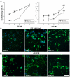
(A) Analysis of the effects of C5 on wild type PC12 cells and cells expressing high levels of IP3 3-KA. Differentiating PC12 cells in the presence of C5 results in an increase in two indices of neurite outgrowth. Data are presented as mean +/− SEM. ***p<0.001, by ANOVA with post-hoc analysis. (B) Arrayscan images of PC12 cells expressing high levels of IP3 3-KA, fixed four days after culture in the presence of 100 ng/ml NGF, and labeled using Hoescht 33342 and the Cellomics Neurite Outgrowth HitKit to identify neurites (using a 10× objective). (C) Arrayscan images of PC12 cells, fixed four days after culture in the presence of 100 ng/ml NGF, and labeled as for (B). Scale bar is 100 µm.
Figure 4
Figure 4. IP3 3-KA activity results in attenuation of ERK phosphorylation.
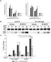
IP3 does not appear to be required for NGF driven neurite outgrowth. (A) Analysis of the effects of XeC blockade of the IP3 receptor, on PC12 cells expressing high levels of IP3 3-KA and on wild type PC12 cells. XeC has no effect on control PC12 cells, but partially overcomes the inhibition of neurite outgrowth caused by IP3 3-KA expression. Data are presented as mean +/− SEM. **p<0.01, by ANOVA with post-hoc analysis. (B) Determination of ERK activation in control PC12 cells and IP3 3-KA expressing cells using a suboptimal dose of NGF, in the presence of either 40 µM C5 or vehicle. Lower blot represents ERK activation as detected by phospho-specific antibodies against ERK1 and ERK2. Upper blot represents total ERK, as detected using an antibody that detects both ERK1 and ERK2. All antibodies were visualised with an HRP conjugated secondary antibody. The blot shown is representative of three independent experiments. (C) Densitometric quantification of ERK activation, as depicted in (B). **p<0.01, Data are presented as mean +/− SEM. ***p<0.001, by ANOVA with post-hoc analysis. IP3 3-KA expression attenuates ERK activation, whilst C5 inhibition of IP3 3-kinase results in increased ERK activity.

Similar articles

Cited by

References

    1. Berridge MJ. Unlocking the secrets of cell signaling. Annu Rev Physiol. 2005;67:1–21. - PubMed
    1. Irvine RF, Lloyd-Burton SM, Yu JC, Letcher AJ, Schell MJ. The regulation and function of inositol 1,4,5-trisphosphate 3-kinases. Adv Enzyme Regul. 2006;46:314–323. - PMC - PubMed
    1. Schell MJ. Inositol trisphosphate 3-kinases: focus on immune and neuronal signaling. Cell Mol Life Sci. 2010;67:1755–1778. - PMC - PubMed
    1. Miller AT, Chamberlain PP, Cooke MP. Beyond IP3: roles for higher order inositol phosphates in immune cell signaling. Cell Cycle. 2008;7:463–467. - PubMed
    1. Pattni K, Banting G. Ins(1,4,5)P3 metabolism and the family of IP3-3Kinases. Cell Signal. 2004;16:643–654. - PubMed

Publication types

MeSH terms

Substances