. 2012;7(2):e32479.
doi: 10.1371/journal.pone.0032479. Epub 2012 Feb 27.

Mechanisms of loss of functions of human angiogenin variants implicated in amyotrophic lateral sclerosis

Affiliations

Mechanisms of loss of functions of human angiogenin variants implicated in amyotrophic lateral sclerosis

Aditya K Padhi et al. PLoS One. 2012.

Abstract

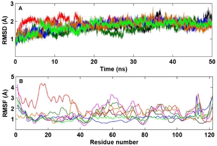
Background: Mutations in the coding region of angiogenin (ANG) gene have been found in patients suffering from Amyotrophic Lateral Sclerosis (ALS). Neurodegeneration results from the loss of angiogenic ability of ANG (protein coded by ANG). In this work, we performed extensive molecular dynamics (MD) simulations of wild-type ANG and disease associated ANG variants to elucidate the mechanism behind the loss of ribonucleolytic activity and nuclear translocation activity, functions needed for angiogenesis.

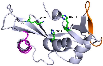
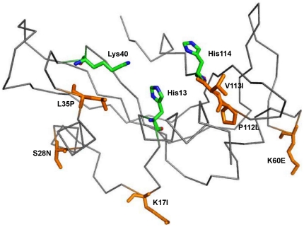
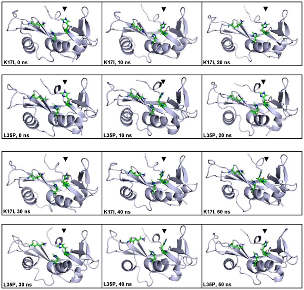
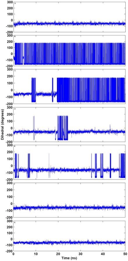
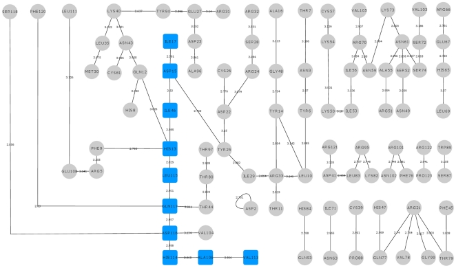
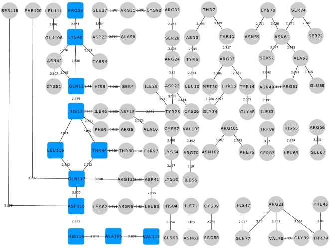
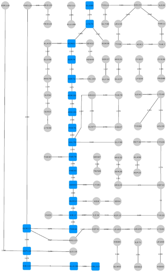
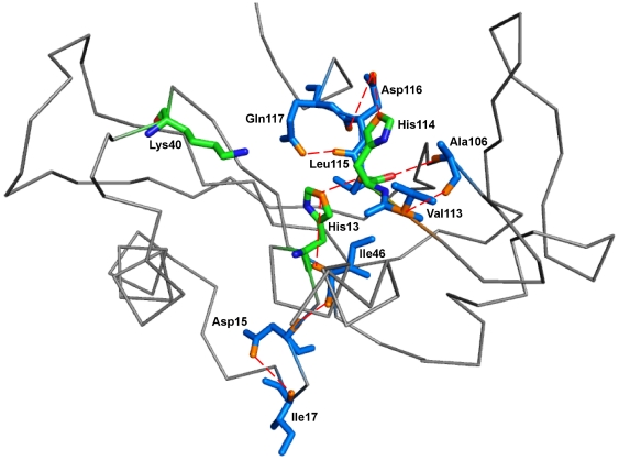
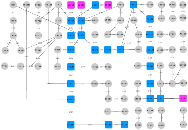
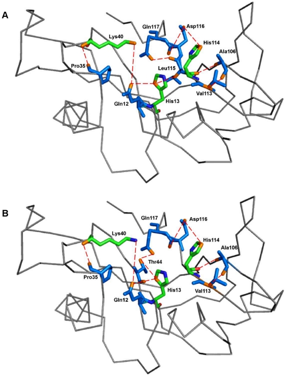
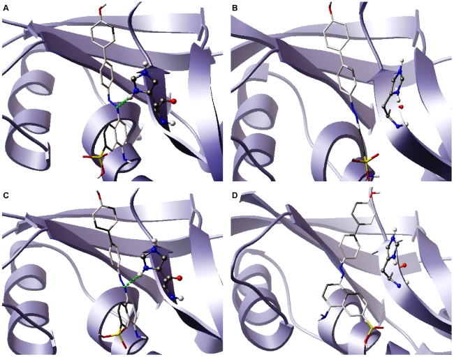
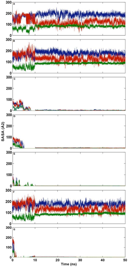
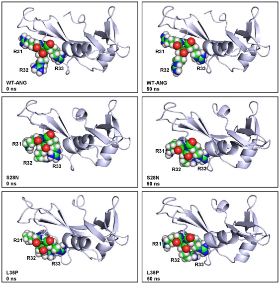
Methodology/principal findings: MD simulations were carried out to study the structural and dynamic differences in the catalytic site and nuclear localization signal residues between WT-ANG (Wild-type ANG) and six mutants. Variants K17I, S28N, P112L and V113I have confirmed association with ALS, while T195C and A238G single nucleotide polymorphisms (SNPs) encoding L35P and K60E mutants respectively, have not been associated with ALS. Our results show that loss of ribonucleolytic activity in K17I is caused by conformational switching of the catalytic residue His114 by 99°. The loss of nuclear translocation activity of S28N and P112L is caused by changes in the folding of the residues (31)RRR(33) that result in the reduction in solvent accessible surface area (SASA). Consequently, we predict that V113I will exhibit loss of angiogenic properties by loss of nuclear translocation activity and L35P by loss of both ribonucleolytic activity and nuclear translocation activity. No functional loss was inferred for K60E. The MD simulation results were supported by hydrogen bond interaction analyses and molecular docking studies.

Conclusions/significance: Conformational switching of catalytic residue His114 seems to be the mechanism causing loss of ribonucleolytic activity and reduction in SASA of nuclear localization signal residues (31)RRR(33) results in loss of nuclear translocation activity in ANG mutants. Therefore, we predict that L35P mutant, would exhibit loss of angiogenic functions, and hence would correlate with ALS while K60E would not show any loss.


Conflict of interest statement

Competing Interests: The authors have declared that no competing interests exist.

Figures

Figure 1
Figure 1. Cartoon representation of X-ray structure for Human Angiogenin (PDB code: 1B1I).
Cartoon representation of the structure of Human Angiogenin (PDB entry 1B1I) showing its functional sites; catalytic triad residues are represented as stick models, nuclear localization signal is represented in magenta color and receptor binding site is represented in orange color. Figure produced using PyMOL .
Figure 2
Figure 2. Ribbon representation of mutational sites in Human Angiogenin.
Ribbon representation of structure of Human Angiogenin (PDB entry 1B1I) with mutational sites; mutations are labeled and represented as stick models in orange color. Figure produced using PyMOL .
Figure 3
Figure 3. RMSD and RMSF profile for WT-ANG and mutants.
(A) Control plots representing the stability of the models during the molecular dynamics run. The root mean square deviation (RMSD) of the backbone atoms from the equilibrated conformation (0 ns) is presented as a function of time. The RMSD time profiles for WT-ANG, K17I, S28N, P112L, L35P, K60E and V113I are shown in black, red, dark green, blue, orange, pink, and light green, respectively. (B) Root mean square fluctuation (RMSF) values of atomic positions computed for the backbone atoms are shown as a function of residue number. The RMSF values for WT-ANG, K17I, S28N, P112L, L35P, K60E and V113I are shown in black, red, dark green, blue, orange, pink, and light green, respectively.
Figure 4
Figure 4. Conformational switching of catalytic residue His114 in K17I and L35P mutants.
Orientation of the catalytic triad residue His114 at a regular interval of 10 ns during the MD simulations of K17I and L35P mutants. In these figures, T = 0 ns is the start of production phase post-equilibration. Figure produced using PyMOL .
Figure 5
Figure 5. Computed change in HA-CA-CB-CG dihedral angle of His114 for WT-ANG and mutants.
The HA-CA-CB-CG dihedral angle change of catalytic residue His114 computed as a function of time (A) WT-ANG (B) K17I (C) S28N (D) P112L (E) L35P (F) K60E and (G) V113I.
Figure 6
Figure 6. Hydrogen bond interaction network for K17I variant.
The hydrogen bond interactions between contiguous amino acid residues based on a 3.2 Å cut-off has been presented here. The path leading from the site of mutation Ile17 to catalytic residue His114 has been shown in blue square boxes. Other hydrogen bond interactions are shown in grey circles. The bond length is given on the edge between the nodes of amino acid residues. The path is mediating through Leu115 which plays a role in His114 conformational switching.
Figure 7
Figure 7. Hydrogen bond interaction network for S28N variant.
The hydrogen bond interactions between contiguous amino acid residues based on a 3.2 Å cut-off has been presented here. The path leading from the site of mutation Asn28 to catalytic residue His114 has been shown in blue square boxes. Other hydrogen bond interactions are shown in grey circles. The bond length is given on the edge between the nodes of amino acid residues. The path is mediating through Leu115 which plays a role in His114 conformational switching.
Figure 8
Figure 8. Hydrogen bond interaction network for L35P mutant.
The hydrogen bond interactions between contiguous amino acid residues based on a 3.2 Å cut-off has been presented here. The paths leading from the site of mutation Pro35 to catalytic residue His114 have been shown in blue square boxes. Other hydrogen bond interactions are shown in grey circles. The bond length is given on the edge between the nodes of amino acid residues. One of the path is mediating through Leu115 which plays a role in His114 conformational switching and the other path is mediating through Thr44 similar to that of WT-ANG.
Figure 9
Figure 9. Hydrogen bond interaction network for K60E mutant.
The hydrogen bond interactions between contiguous amino acid residues based on a 3.2 Å cut-off has been presented here. The path leading from the site of mutation Glu60 to catalytic residue His114 has been shown in blue square boxes. Other hydrogen bond interactions are shown in grey circles. The bond length is given on the edge between the nodes of amino acid residues. The path is mediating through Thr44 similar to that of WT-ANG and there is no conformational switching of His114.
Figure 10
Figure 10. Residues interacting through hydrogen bonds from the site of mutation to His114 in K17I variant.
Amino acid residues of K17I variant through which hydrogen bonds exert influence from the site of mutation on the catalytic site His114. The ribbon representation shows the shortest path traced by the contiguous amino acid sequence Ile17-Asp15-Ile46-His13-Leu115-Gln117-Asp116-His114-Ala106-Val113 and the hydrogen bond interactions. Residues have been shown in stick model in marine blue color. Catalytic triad residues have been shown as stick model and represented in green color. Hydrogen bonds between residues have been shown in red dashed-lines. Figure produced using PyMOL .
Figure 11
Figure 11. Hydrogen bond interaction network for WT-ANG.
The hydrogen bond interactions between contiguous amino acid residues based on a 3.2 Å cut-off has been presented here. All hydrogen bond interaction paths up to catalytic residue His114 have been compared with the hydrogen bond interaction paths of other mutants. These hydrogen bond interactions are shown in blue square boxes. Other hydrogen bond interactions are shown in grey circles. The starting residues through which these paths led to His114 are shown in pink square boxes. The bond length is given on the edge between the nodes of amino acid residues. All the hydrogen bond interaction paths are mediating through Thr44 and there is no conformational switching of His114.
Figure 12
Figure 12. Residues interacting through hydrogen bonds from the site of mutation to His114 in L35P mutant.
Amino acid residues of L35P mutant through which hydrogen bonds exert influence from the site of mutation on the catalytic site His114. There were two shortest paths identified. The first mediated through Leu115 and the second mediated through Thr44. Residues have been shown as stick models in marine blue color. Catalytic triad residues have been shown as stick model and represented in green color. Hydrogen bonds between residues have been shown in red dashed-lines. (A) The ribbon representation shows the path traced by the contiguous amino acid sequence Pro35-Lys40-Gln12-His13-Leu115-Gln117-Asp116-His114-Ala106-Val113 (B) The ribbon representation shows the path traced by the contiguous amino acid sequence Pro35-Lys40-Gln12-His13-Thr44-Gln117-Asp116-His114-Ala106-Val113. Figure produced using PyMOL .
Figure 13
Figure 13. Lowest-energy AutoDock poses of NCI-65828 with His114 in K17I and L35P mutants.
Stereo views of lowest-energy AutoDock poses of K17I and L35P mutants using NCI-65828. The backbone trace of ANG is shown along with the His114 residue as stick model depicting the hydrogen bond between the azo-group of the inhibitor and His114 (in green dashed line). In K17I, (A) presence hydrogen bond in native conformation with His114 (B) and its absence in the altered conformation of His114 were visualized. Similarly, in L35P, (C) presence hydrogen bond in native conformation with His114 (D) and its absence in the altered conformation of His114 are shown. Also shown is how the conformational switching of His114 affects substrate binding at the catalytic site.
Figure 14
Figure 14. Computed change in solvent accessible surface area for WT-ANG and mutants.
Variation of solvent accessible surface area (SASA) of nuclear localization signal residues R31, R32 and R33 over the period of simulation (A) WT-ANG (B) K17I (C) S28N (D) P112L (E) L35P (F) K60E and (G) V113I. (R31: blue, R32: red and R33: green).
Figure 15
Figure 15. Changes in folding of residues 31RRR33 for WT-ANG, S28N and L35P mutants.
Comparison of the changes in the folding of the residues R31, R32 and R33 at initial condition and at 50 ns of simulation. The residues are in open conformation for WT-ANG throughout the duration of simulation. For S28N and L35P mutants, a closed conformation is dominant. Figure produced using PyMOL .

Similar articles

Cited by

References

    1. Wijesekera LC, Leigh PN. Amyotrophic lateral sclerosis. Orphanet J Rare Dis. 2009;4:3. - PMC - PubMed
    1. Bento-Abreu A, Van Damme P, Van Den Bosch L, Robberecht W. The neurobiology of amyotrophic lateral sclerosis. Eur J Neurosci. 2010;31:2247–2265. - PubMed
    1. DeJesus-Hernandez M, Mackenzie IR, Boeve BF, Boxer AL, Baker M, et al. Expanded GGGGCC hexanucleotide repeat in noncoding region of C9ORF72 causes chromosome 9p-linked FTD and ALS. Neuron. 2011;72:245–256. - PMC - PubMed
    1. Renton AE, Majounie E, Waite A, Simón-Sánchez J, Rollinson S, et al. A hexanucleotide repeat expansion in C9ORF72 is the cause of chromosome 9p21-linked ALS-FTD. Neuron. 2011;72:257–268. - PMC - PubMed
    1. Oosthuyse B, Moons L, Storkebaum E, Beck H, Nuyens D, et al. Deletion of the hypoxia-response element in the vascular endothelial growth factor promoter causes motor neuron degeneration. Nat Genet. 2001;28:131–138. - PubMed

Publication types

MeSH terms