. 2011 Dec;1(7):607-13.
doi: 10.1534/g3.111.001438. Epub 2011 Dec 1.

A Genetic Screen for Dominant Enhancers of the Cell-Cycle Regulator α-Endosulfine Identifies Matrimony as a Strong Functional Interactor in Drosophila

A Genetic Screen for Dominant Enhancers of the Cell-Cycle Regulator α-Endosulfine Identifies Matrimony as a Strong Functional Interactor in Drosophila

Jessica R Von Stetina et al. G3 (Bethesda). 2011 Dec.

Abstract

The coordination of cell-cycle events with developmental processes is essential for the reproductive success of organisms. In Drosophila melanogaster, meiosis is tightly coupled to oocyte development, and early embryos undergo specialized S-M mitoses that are supported by maternal products. We previously showed that the small phosphoprotein α-endosulfine (Endos) is required for normal oocyte meiotic maturation and early embryonic mitoses in Drosophila. In this study, we performed a genetic screen for dominant enhancers of endos(00003) and identified several genomic regions that, when deleted, lead to impaired fertility of endos(00003)/+ heterozygous females. We uncovered matrimony (mtrm), which encodes a Polo kinase inhibitor, as a strong dominant enhancer of endos. mtrm(126) +/+ endos(00003) females are sterile because of defects in early embryonic mitoses, and this phenotype is reverted by removal of one copy of polo. These results provide compelling genetic evidence that excessive Polo activity underlies the strong functional interaction between endos(00003) and mtrm(126). Moreover, we show that endos is required for the increased expression of Mtrm in mature oocytes, which is presumably loaded into early embryos. These data are consistent with the model that maternal endos antagonizes Polo function in the early embryo to ensure normal mitoses through its effects on Mtrm expression during late oogenesis. Finally, we also identified genomic deletions that lead to loss of viability of endos(00003)/+ heterozygotes, consistent with recently published studies showing that endos is required zygotically to regulate the cell cycle during development.

Keywords: Drosophila; early embryonic cell cycle; matrimony; polo; α-endosulfine.


Figures

Figure 1
Figure 1
Coordination between the cell cycle and development during meiotic maturation and early embryonic mitoses. (A) Stage 10 egg chamber diagram, exemplifying a stage in which the oocyte is arrested in prophase I. (B) Diagram of a stage 14 oocyte, which has progressed to metaphase I as a result of meiotic maturation. (C) Early embryo showing nuclei that undergo mitotic divisions in a shared cytoplasm, relying on maternal stockpiles of RNA and proteins. Stage 10 follicle cells are shown in gray, germline and early embryo cytoplasm in pink, and nuclei in red. Double arrows between (A) and (B) represent egg chamber development and oocyte meiotic maturation at stage 13. Double arrows between (B) and (C) represent completion of meiosis (upon egg activation) and fertilization. DAPI-stained DNA from a stage 10 oocyte arrested in prophase I (A′), a stage 14 oocyte in early (B′) or late (B′′) metaphase I, and from early embryonic nuclei in interphase (C′) or mitosis (C′′) are shown below corresponding diagrams. Scale bar, 5 μm.
Figure 2
Figure 2
Chromosomal distribution of deficiencies that dominantly enhance endos00003/+. Gray bars depict chromosome arms (X, 2L, 2R, 3L, and 3R), and black numbers indicate polytene chromosome divisions. Deficiencies shown in red dominantly cause sterility or semisterility of endos00003 heterozygous females, whereas those shown in blue dominantly cause lethality of endos00003 heterozygous animals. Red and blue bars above deficiency indicate their deleted genomic regions according to FlyBase (http://flybase.org). Asterisk indicates endos location.
Figure 4
Figure 4
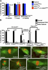
endos and mtrm show dominant genetic interactions during early embryonic mitoses. (A) Hatch rates for embryos derived from y w control, endos00003/+, mtrm126/+, or mtrm126 +/+ endos00003 females in wild-type (2 copies of polo), polo16-1/+ (one copy of polo) or GFP::polo (three copies of polo) background. Three hundred embryos were analyzed per genotype. a vs. a, P = 2.8 × 10⁻⁷. b vs. b, P = 6.8 × 10−76. c vs. c, P = 2.1 × 10⁻¹². d vs. d, P = 2.5 × 10−78. e vs. e, P = 4.3 × 10−106. (B) Quantification of percentage of nuclei from embryos derived from y w control, endos00003/+, mtrm126/+, or mtrm126 +/+ endos00003, or mtrm126 + polo16-1/+ endos00003 + females displaying normal or defective DNA and spindle morphology. In addition to misaligned DNA, other defects include attached spindles, highly condensed DNA, or no mitotic nuclei with two rosettes present. Numbers of nuclei (n) analyzed are shown above bars. Ten to fifteen embryos were analyzed for each genotype. (C–H) Examples of y w control embryo nucleus with normal DNA morphology (C), or nuclei from embryos produced by mtrm126 +/+ endos00003 female showing misaligned DNA and abnormal spindle (D, E) or highly condensed and disorganized DNA (F,G), and normal nucleus from embryo produced by mtrm126 + polo16-1/+ endos00003 + female (H) are shown. Scale bar, 5 μm.
Figure 3
Figure 3
endos and mtrm do not show genetic interactions during oocyte meiosis. (A) Quantification of percentage of oocytes remaining in prophase I at stages 12, early 13, mid 13, and late 13 and 14 of oogenesis. Numbers of oocytes analyzed for y w control, endos00003/+, mtrm126/+, and mtrm126 +/+ endos00003 are as follows, respectively. Stage 12: 69, 56, 68, 41; early stage 13: 101, 163, 171, 109; mid stage 13: 27, 92, 65, 61; late stage 13: 128, 247, 189, 265; stage 14: 106, 239, 312, 773. (B) Quantification of percentage of stage 14 oocytes showing abnormal DNA morphology. Numbers of oocytes analyzed for y w control, endos00003/+, mtrm126/+, and mtrm126 +/+ endos00003 are 106, 239, 312, and 773, respectively. *P = 0.002. **P = 0.0005. ***P = 1 × 10⁻¹⁴. ns, no significant difference. (C–E) Examples of stage 14 oocyte DNA morphology for control (C, normal), mtrm126 (D, abnormal) and mtrm126 +/+ endos00003 (E, abnormal). Scale bar, 5 μm.
Figure 5
Figure 5
endos is required for maternal expression of Mtrm. (A) Western blot showing Mtrm protein expression at different stages of oogenesis in y w control, endos00003, twe1, or elgi1 homozygous females. g-St. 11, germarium through stage 11; St. 12, stage 12; St. 13, stage 13; St. 14, stage 14. Actin was used as a loading control. One hundred egg chambers (or g-St.11 sets) were loaded per lane. (B) Mtrm Western blot of y w control, mtrm126, endos00003, and elgi1 stage 14 oocytes. Tubulin was used as a loading control. We were unable to examine Mtrm levels in elgi1 endos00003 double homozygous females due to the poor health of this genotype. (C) Model for genetic interaction between endos and mtrm. In wild-type females (Control), Endos promotes expression of Mtrm, a known negative regulator of Polo, during oogenesis resulting in normal levels of Polo function in the early embryo. In mtrm126/+ heterozygous females, reduced levels of maternal Mtrm loaded into the embryo lead to increased Polo activity and abnormal embryonic mitoses. Removal of one copy of endos in mtrm126 +/+ endos00003 double heterozygous females results in early embryos with further reduced levels of Mtrm, leading to even higher levels of Polo activity and more severe defects during early embryonic mitoses.

Similar articles

Cited by

References

    1. Archambault V., Zhao X., White-Cooper H., Carpenter A. T., Glover D. M., 2007. Mutations in Drosphila Greatwall/Scant reveal its roles in mitosis and meiosis and interdependence with Polo kinase. PLoS Genet. 3: e200. - PMC - PubMed
    1. Donaldson M. M., Tavares A. A., Ohkura H., Deak P., Glover D. M., 2001. Metaphase arrest with centromere separation in polo mutants of Drosphila. J. Cell Biol. 153: 663–676 - PMC - PubMed
    1. Drummond-Barbosa D., Spradling A. C., 2004. Alpha-endosulfine, a potential regulator of insulin secretion, is required for adult tissue growth control in Drosphila. Dev. Biol. 266: 310–321 - PubMed
    1. Gharbi-Ayachi A., Labbe J. C., Burgess A., Vigneron S., Strub J. M., et al. , 2010. The substrate of Greatwall kinase, Arpp19, controls mitosis by inhibiting protein phosphatase 2A. Science 330: 1673–1677 - PubMed
    1. Goshima G., Wollman R., Goodwin S. S., Zhang N., Scholey J. M., et al. , 2007. Genes required for mitotic spindle assembly in Drosphila S2 cells. Science 316: 417–421 - PMC - PubMed

LinkOut - more resources