. 2012 Feb;2(2):235-48.
doi: 10.1534/g3.111.001537. Epub 2012 Feb 1.

Extent With Modification: Leg Patterning in the Beetle Tribolium castaneum and the Evolution of Serial Homologs

Extent With Modification: Leg Patterning in the Beetle Tribolium castaneum and the Evolution of Serial Homologs

David R Angelini et al. G3 (Bethesda). 2012 Feb.

Abstract

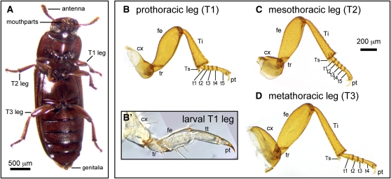
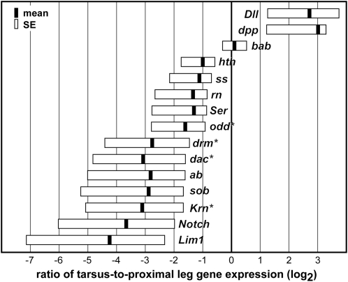
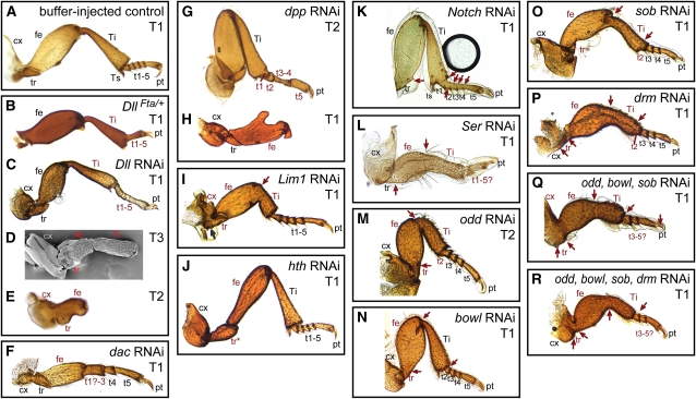
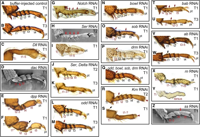
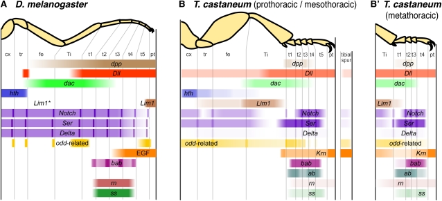
Serial homologs are similar structures that develop at different positions within a body plan. These structures share some, but not all, aspects of developmental patterning, and their evolution is thought to be constrained by shared, pleiotropic gene functions. Here we describe the functions of 17 developmental genes during metamorphic development of the legs in the red flour beetle, Tribolium castaneum. This study provides informative comparisons between appendage development in Drosophila melanogaster and T. castaneum, between embryonic and adult development in T. castaneum, and between the development of serially homologous appendages. The leg gap genes Distal-less and dachshund are conserved in function. Notch signaling, the zinc-finger transcription factors related to odd-skipped, and bric-à-brac have conserved functions in promoting joint development. homothorax knockdown alters the identity of proximal leg segments but does not reduce growth. Lim1 is required for intermediate leg development but not distal tarsus and pretarsus development as in D. melanogaster. Development of the tarsus requires decapentaplegic, rotund, spineless, abrupt, and bric-à-brac and the EGF ligand encoded by Keren. Metathoracic legs of T. castaneum have four tarsomeres, whereas other legs have five. Patterns of gene activity in the tarsus suggest that patterning in the middle of the tarsal region, not the proximal- or distal-most areas, is responsible for this difference in segment number. Through comparisons with other recent studies of T. castaneum appendage development, we test hypotheses for the modularity or interdependence of development during evolution of serial homologs.

Keywords: adult development; appendage patterning; developmental constraint; metamorphosis; serial homology.


Figures

Figure 1
Figure 1
The ventral appendages of Tribolium castaneum include the antennae, mouthparts (mandibles, maxillae, and labium), thoracic legs, and genitalia. (A) Ventral view of an adult female. The thoracic legs of T. castaneum are roughly similar, with six true adult podomeres: coxa, trochanter, femur, tibia, tarsus, and pretarsus. The tarsus is subdivided by joints into tarsomeres, which lack independent muscles. The prothoracic leg (B) and mesothoracic leg (C) have five tarsomeres, whereas the metathoracic leg has four tarsomeres (D). The coxae of each type of leg have distinct shapes and sizes. (B′) Larval legs consist of five main segments: the coxa, trochanter, femur, tibiotarsus, and pretarsus. Abbreviations: cx, coxa; fe, femur; pt, pretarsus; t1-5, tarsomeres 1-5; T1–T3, thoracic segments 1-3; Ti, tibia; Ts, tibial spur; tt, tibiotarsus; tr, trochanter.
Figure 2
Figure 2
Relative expression of candidate genes is compared in the tarsus and proximal leg of pupae. Expression ratios are given in log2 scale. Zero represents equal expression in each region; genes appearing to the right are enriched in the tarsus, relative to the rest of the leg. Black bars indicate the mean expression ratio, whereas boxes indicate standard error. Genes with expression ratios significantly different from 1 (equal expression in both regions) are denoted with an asterisk (Welch’s t-test, P < 0.05).
Figure 3
Figure 3
RNA interference effects on adult legs. (A) Control treatments were indistinguishable from unmanipulated beetles. (B) DllFta heterozygotes develop with reduced tarsi, lacking joints. (C) This phenotype is similar to mild Dll RNAi specimens. (D) Dll RNAi also resulted in stronger phenotypes, in which structures distal to the tibia are deleted. (E) In the most severe Dll-depleted individuals, the legs are truncated within the femur, which is reduced. Joints are also absent from the remaining segments. (F) Severe dac RNAi specimens had deletions of the distal femur, tibia, and proximal tarsomeres. (G) RNAi targeting dpp caused alterations of the proximal tarsomeres in mildly affected specimens. (H) In more severe dpp RNAi specimens, the legs are truncated in the mid-femur. (I) Lim1 RNAi caused the loss of proximal leg joints (red arrow) and a reduction of the distal femur and proximal tibia. Although the femur−tibia joint did not form normally, an anatomical boundary was present at this position. (J) Knockdown of hth caused a homeotic transformation of the coxa, trochanter and proximal femur toward more distal morphologies. (K) Notch RNAi eliminated most joints from the leg (red arrows). (L) Ser RNAi eliminated joints (red arrows) and reduced the overall length of the leg. (M-R) Depletion of odd-related genes caused reduction of the leg and loss of proximal joints (red arrows), as well as deletion of proximal tarsomeres. Abbreviations: cx, coxa; fe, femur; pt, pretarsus; t1-5, tarsomeres 1-5; T1, prothoracic leg; T2, mesothoracic leg; T3, metathoracic leg; Ti, tibia; tr, trochanter; Ts, tibial spur. Structures with a defect are labeled in red.
Figure 4
Figure 4
Phenotypes in the tarsus produced through RNAi. (A) Normal tarsus anatomy in the pro- and mesothoracic legs includes five tarsomeres. The distal tibia is also visible, showing the distal tibial spurs. (B) The metathoracic legs normally have four tarsomeres. (C) Dll RNAi produces mild specimens in which the tarsomeres lack joints and the region is reduced. (D) Depletion of dac produced fusions in the proximal tarsus. (E) Knockdown of dpp reduced t1-t4. (F) Rarely dpp RNAi caused a transformation of the first tarsomere (t1*) toward a distal tibia identity, as indicated by an ectopic tibial spur (black arrow). (G) Notch RNAi caused the loss of joints in the tarsus (red arrows). (H) Mild Ser-depleted specimens also lacked joints between tarsomeres (red arrows). (I) Moderate Ser knockdown phenotypes included fusion and reduction of the tarsus. (J–K) Simultaneous depletion of Ser and Delta produced phenotypes similar to Ser RNAi alone. (L—Q) RNAi targeting the odd-related genes caused fusion of proximal tarsomeres with the tibia (L, M, O), or adjacent tarsomeres (O). The first tarsomere (L, N, P) and sometimes also the second (Q) were deleted in moderate and severe specimens. (R) Krn RNAi eliminated the pretarsus (red arrowhead), and caused occasional loss of joints (red arrow) in mild specimens. (S) More severely affected Krn RNAi specimens had a reduction of the tarsus with complete loss of joints. Tibial spurs were also deleted, although ectopic tibial spurs could sometimes be found (black arrow). (T–U) Knockdown of bab caused loss of joints in the tarsus (red arrows) and fusion of tarsomeres. (V) Depletion of ab caused fusions of tarsomeres. (W) Rarely partial ectopic joint formation was observed in ab RNAi (black arrow). (X) RNAi targeting rn caused a reduction of the tarsus and failure of joint formation. (Y) In severely affected rn RNAi specimens the pretarsus also failed to form. (Z) Knockdown of ss also caused fusions of tarsomeres (red arrow). Abbreviations are as in Figure 3.
Figure 5
Figure 5
Summary of RNAi phenotypes in the legs. (A) Functional domains of genes in the leg of D. melanogaster. For each gene, colored bars represent the region on the corresponding adult leg in which the gene is expressed and/or functions, focusing on the late third instar larval imaginal leg disc. Dll, EGF, and dpp are also required in the embryo for initiation of imaginal leg disc primordia. odd-related genes are expressed more broadly in the tarsus earlier in development, and are required for proper formation of tarsomere joints. In D. melanogaster, ab has been observed in the leg discs, but its expression pattern has not been described in detail (Hu et al. 1995). *Lim1 expression has also been detected in multiple domains in the proximal imaginal leg disc, including the femur (Tsuji et al. 2000). (B) Patterning of the pro- and mesothoracic legs of T. castaneum. Intensity of expression indicates phenotypic penetrance of RNAi effects. The tibial spur is represented by the column at right. (B′) The metathoracic leg has only four tarsomeres, and its patterning of the intermediate tarsomeres (t2-t3) differs from the more anterior legs.

Similar articles

Cited by

References

    1. Abu-Shaar M., Mann R. S., 1998. Generation of multiple antagonistic domains along the proximodistal axis during Drosophila leg development. Development 125: 3821–3830 - PubMed
    1. Abzhanov A., Kaufman T. C., 2000. Homologs of Drosophila appendage genes in the patterning of arthropod limbs. Dev. Biol. 227: 673–689 - PubMed
    1. Amin A., Li Y., Finkelstein R., 1999. Hedgehog activates the EGF receptor pathway during Drosophila head development. Development 126: 2623–2630 - PubMed
    1. Angelini D. R., Kaufman T. C., 2004. Functional analyses in the hemipteran Oncopeltus fasciatus reveal conserved and derived aspects of appendage patterning in insects. Dev. Biol. 271: 306–321 - PubMed
    1. Angelini D. R., Kaufman T. C., 2005a Functional analyses in the milkweed bug Oncopeltus fasciatus (Hemiptera) support a role for Wnt signaling in body segmentation but not appendage development. Dev. Biol. 283: 409–423 - PubMed

LinkOut - more resources