. 2012 Feb;2(2):261-70.
doi: 10.1534/g3.111.001479. Epub 2012 Feb 1.

Whole-Genome Sequencing of Sordaria macrospora Mutants Identifies Developmental Genes

Affiliations

Whole-Genome Sequencing of Sordaria macrospora Mutants Identifies Developmental Genes

Minou Nowrousian et al. G3 (Bethesda). 2012 Feb.

Abstract

The study of mutants to elucidate gene functions has a long and successful history; however, to discover causative mutations in mutants that were generated by random mutagenesis often takes years of laboratory work and requires previously generated genetic and/or physical markers, or resources like DNA libraries for complementation. Here, we present an alternative method to identify defective genes in developmental mutants of the filamentous fungus Sordaria macrospora through Illumina/Solexa whole-genome sequencing. We sequenced pooled DNA from progeny of crosses of three mutants and the wild type and were able to pinpoint the causative mutations in the mutant strains through bioinformatics analysis. One mutant is a spore color mutant, and the mutated gene encodes a melanin biosynthesis enzyme. The causative mutation is a G to A change in the first base of an intron, leading to a splice defect. The second mutant carries an allelic mutation in the pro41 gene encoding a protein essential for sexual development. In the mutant, we detected a complex pattern of deletion/rearrangements at the pro41 locus. In the third mutant, a point mutation in the stop codon of a transcription factor-encoding gene leads to the production of immature fruiting bodies. For all mutants, transformation with a wild type-copy of the affected gene restored the wild-type phenotype. Our data demonstrate that whole-genome sequencing of mutant strains is a rapid method to identify developmental genes in an organism that can be genetically crossed and where a reference genome sequence is available, even without prior mapping information.

Keywords: Sordaria macrospora; developmental mutants; next-generation sequencing.


Figures

Figure 1
Figure 1
Strategy for whole-genome sequencing of pooled DNA from mutants and wild type. Mutants pro23 and pro44 were crossed against the spore color mutant fus. Single spore isolates derived from black and light-brown ascospores were screened for fertility and color; for both crosses, 40 spore isolates with a sterile/light-brown phenotype were chosen to represent double mutants pro23/fus and pro44/fus, respectively. For the resequencing of the wild type, 20 isolates with a fertile/black phenotype were chosen from each cross. The pooled DNA from 40 spore isolates for each genotype was used for sequencing. As it turned out during the analysis, mutant pro44 had acquired a second, unrelated mutation that also led to sterility. Therefore, the mutant name is given in brackets to indicate that this sample was no longer used for the analysis of pro44 but only for the analysis of the fus mutation. For the pro44 analysis, the mutant was outcrossed again to obtain a clean pro44 strain; this strain was used for crossing to obtain single spore isolates for sequencing according to the same principle (Figure S2).
Figure 2
Figure 2
Genomic structure of the pro41 gene locus in the wild-type and mutant pro23. Numbers in the wild type correspond to nucleotides in contig 2.1 (S. macrospora genome version 02). An approximately 1.1-kb region (green in the wild type) is deleted in mutant pro23. Several other regions (orange, red, and light and dark blue) adjacent to the deleted region are duplicated in the mutant strain and occupy the position of the deleted region. In most cases, the orientation of the duplicated regions is the same as in the wild type, with the exception of one case in which a small region is inverted in pro23 (indicated by an arrowhead within the duplicated region). The deleted region comprises the C-terminal part of pro41 (black arrow).
Figure 3
Figure 3
Complementation of mutant pro23 with the pro41 ORF. Mutant pro23 was transformed with plasmid pE3-5Mr that carries the pro41 ORF under control of the respective gpd and trpC promoter and terminator sequences of A. nidulans. The figure shows the wild-type (forms perithecia; black dots in the photographs of the petri dishes), mutant pro23 (sterile, does not form perithecia), and two complemented transformants (produce perithecia). Strains were grown on corn meal agar, and photographs were taken after 6 d (A) or 8 d (B). Scale bar in (B) indicates 2 mm.
Figure 4
Figure 4
A point mutation in the tih gene causes a splice defect in the fus mutant strain. (A) PCR and reverse-transcription PCR analysis of the tih gene in the wild type and fus mutant. Small arrows in (B) indicate the oligonucleotide primers used to amplify a tih gene fragment. “+” and “−” indicate experiments with or without reverse transcriptase, respectively. Each sample was analyzed in duplicate with two independent biological replications. As expected, a 1.1-kb fragment was amplified from genomic DNA of both strains. From wild-type cDNA, the expected 0.6-kb fragment (after splicing of two introns) was amplified. PCR from cDNA of the fus strain yielded two fragments of 0.7 and 0.3 kb. (B) Two misspliced tih transcripts were identified in the fus mutant strain. The wild-type tih gene is shown as a white arrow with introns indicated in gray. Below are the derived coding regions, and above predicted peptides from the wild type and mutant fus. Sequence analysis of the PCR fragments from (A) confirmed the point mutation in the first base of the second intron in the fus mutant (nt 1040, G to A transition). The mutation leads to a splice defect of the second intron resulting in two transcripts in the fus strain (fus-T1 and fus-T2). In the first transcript (fus-T1), the second intron is not spliced, leading to a longer transcript represented by the 0.7 kb RT-PCR fragment in (A), and a premature stop codon, and therefore a shorter coding sequence, and a frame-shifted, shorter peptide (indicated in light blue in peptide fus-P1). In the fus-T2 transcript, a second intron is spliced using an incorrect 5′ splice site at nt 718, which results in a shorter transcript represented by the 0.3 kb RT-PCR fragment in (A), and a shorter, frame-shifted peptide (indicated in dark blue in peptide fus-P2). The lengths of the coding sequences of the wild-type and the two fus transcripts are given on the right; adh_short indicates the alcohol dehydrogenase domain within the predicted TIH peptide which is not fully present in the derived peptides fus-P1 and fus-P2 of the mutant strain.
Figure 5
Figure 5
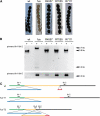
Complementation of the fus mutant with a wild-type copy of the tih gene. Mutant fus was transformed with plasmid pEHN5-tih that carries the tih ORF under control of respecitve gpd and trpC promoter and terminator sequences of A. nidulans. Single spore isolates from three independent complemented transformants (R4557, R7055, and R7111) were analyzed for phenotype and expression of the tih gene. (A) Strains were grown on corn meal agar, and photographs of ascospores were taken after 9 d. The fus mutant has light-brown ascospores whereas the wild type and the complemented transformants have black ascospores. Scale bars indicate 20 μm. (B) RT-PCR analysis of tih expression. The three complemented strains express the spliced wild-type transcript of the tih gene (0.6-kb fragment with primers tih-1/2, position of primers indicated in panel C), whereas in the fus recipient strain, only the misspliced/non-spliced (0.3 kb and 0.7 kb, respectively) fragments were detected. To demonstrate that the complemented transformants still produce the misspliced transcripts (in addition to the complementing wild-type copy), RT-PCR analysis was performed with oligonucleotides tih-1/5 that specifically amplify the misspliced transcript. The corresponding fragment (0.3 kb) can be amplified from the fus strain and the transformants, but not from the wild type. (C) Position of the oligonucleotide primers and the spliced introns in the wild type transcript, and the two transcripts from the mutant strain.
Figure 6
Figure 6
A point mutation in the pro44 gene (SMAC_03223) shifts the stop codon in the mutant strain. Schematic representation of the 2.4-kb pro44 mRNA and the derived wild-type and mutant proteins (above). The A to G mutation in the wild-type stop codon leads to the formation of a longer polypeptide in the mutant strain. The additional 107 amino acids are shown in blue; the predicted GATA zinc finger domain is shown in black.
Figure 7
Figure 7
Complementation of mutant pro44 with constructs containing SMAC_03223. Mutant pro44 was transformed with plasmid pIG3146-37 that carries the SMAC_03223 ORF under control of its own promoter and terminator sequences (transformant pro44::SMAC_03223_native) or plasmid pIG3147-1 that carries the SMAC_03223 ORF under control of the respective gpd and trpC promoter and terminator sequences of Aspergillus nidulans (transformant pro44::SMAC_03223_constitutive). The figure shows a side view (longitudinal section) of the region comprising the agar/air interface from cultures of the wild type, the sterile mutant pro44 and two complemented transformants. The wild type forms mature perithecia at the agar/air interface, whereas mutant pro44 only forms protoperithecia that are submerged in the agar (greater magnification shown in small inserted photograph, arrowheads indicate protoperithecia). Complemented transformants produce mature perithecia at the agar/air interface like the wild type; however, some protoperithecia and even mature perithecia are still formed submerged in the growth medium. Strains were grown on corn meal agar; photographs were taken after 8 d; scale bar indicates 1 mm, and 200 µm in the inserted pro44 photograph.

References

    1. Balzer S., Malde K., Jonassen I., 2011. Systematic exploration of error sources in pyrosequencing flowgram data. Bioinformatics 27: i304–i309 - PMC - PubMed
    1. Bautz E., Freese E., 1960. On the mutagenic effect of alkylating agents. Proc. Natl. Acad. Sci. U S A. 46: 1585–1594 - PMC - PubMed
    1. Bell A. A., Wheeler M. H., 1986. Biosynthesis and functions of fungal melanins. Annu. Rev. Phytopathol. 24: 411–451
    1. Bloemendal S., Lord K. M., Rech C., Hoff B., Engh I., et al. , 2010. A mutant defective in sexual development produces aseptate ascogonia. Eukaryot. Cell 9: 1856–1866 - PMC - PubMed
    1. Borghouts C., Kimpel E., Osiewacz H. D., 1997. Mitochondrial DNA rearrangements of Podospora anserina are under control of the nuclear gene grisea. Proc. Natl. Acad. Sci. U S A. 94: 10768–10773 - PMC - PubMed

LinkOut - more resources