Role of endometrial immune cells in implantation
- PMID: 22384430
- PMCID: PMC3283071
- DOI: 10.5653/cerm.2011.38.3.119
Role of endometrial immune cells in implantation
Abstract
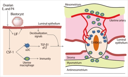
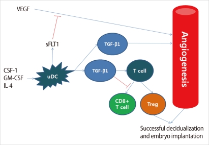
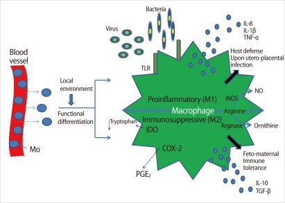
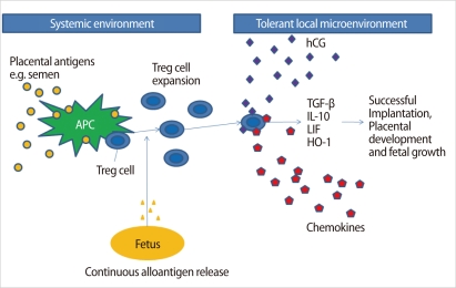
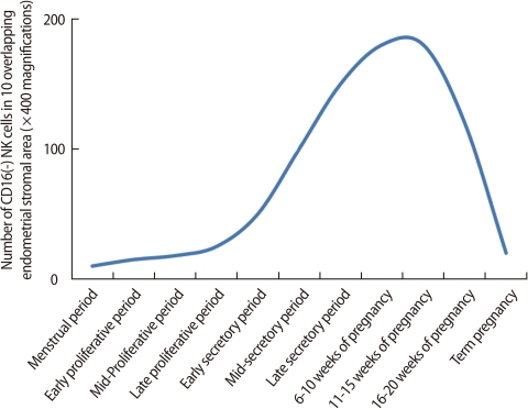
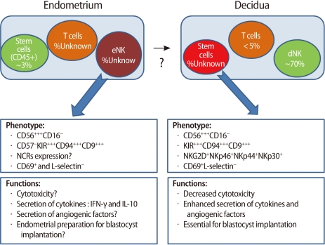
Implantation of an embryo occurs during the mid-secretory phase of the menstrual cycle, known as the "implantation window." During this implantation period, there are significant morphologic and functional changes in the endometrium, which is followed by decidualization. Many immune cells, such as dendritic and natural killer (NK) cells, increase in number in this period and early pregnancy. Recent works have revealed that antigen-presenting cells (APCs) and NK cells are involved in vascular remodeling of spiral arteries in the decidua and lack of APCs leads to failure of pregnancy. Paternal and fetal antigens may play a role in the induction of immune tolerance during pregnancy. A balance between effectors (i.e., innate immunity and helper T [Th] 1 and Th17 immunity) and regulators (Th2 cells, regulatory T cells, etc.) is essential for establishment and maintenance of pregnancy. The highly complicated endocrine-immune network works in decidualization of the endometrium and at the fetomaternal interface. We will discuss the role of immune cells in the implantation period and during early pregnancy.
Keywords: Decidua; Dendritic Cells; Endometrium; Human; Implantation; Lymphocytes; Macrophages; Natural Killer Cells; Regulatory T Cells; Th17 Cells.
Conflict of interest statement
No potential conflict of interest relevant to this article was reported.
Figures
Similar articles
-
Corin, an enzyme with a putative role in spiral artery remodeling, is up-regulated in late secretory endometrium and first trimester decidua.Hum Reprod. 2013 May;28(5):1172-80. doi: 10.1093/humrep/det028. Epub 2013 Feb 21. Hum Reprod. 2013. PMID: 23434834
-
Endometrial Immunity for Embryo Implantation and Pregnancy Establishment.Tohoku J Exp Med. 2020 Jan;250(1):49-60. doi: 10.1620/tjem.250.49. Tohoku J Exp Med. 2020. PMID: 31996497 Review.
-
Ovarian and endometrial immunity during the ovarian cycle.J Reprod Immunol. 2019 Jun;133:7-14. doi: 10.1016/j.jri.2019.04.001. Epub 2019 Apr 25. J Reprod Immunol. 2019. PMID: 31055226 Review.
-
Immune Tolerance of the Human Decidua.J Clin Med. 2021 Jan 18;10(2):351. doi: 10.3390/jcm10020351. J Clin Med. 2021. PMID: 33477602 Free PMC article. Review.
-
Endometrial TGFβ signaling fosters early pregnancy development by remodeling the fetomaternal interface.Am J Reprod Immunol. 2023 Dec;90(6):e13789. doi: 10.1111/aji.13789. Am J Reprod Immunol. 2023. PMID: 38009061 Free PMC article. Review.
Cited by
-
Immune Tolerance of Embryo Implantation and Pregnancy: The Role of Human Decidual Stromal Cell- and Embryonic-Derived Extracellular Vesicles.Int J Mol Sci. 2022 Nov 2;23(21):13382. doi: 10.3390/ijms232113382. Int J Mol Sci. 2022. PMID: 36362169 Free PMC article. Review.
-
Relationship between maternal immunological response during pregnancy and onset of preeclampsia.J Immunol Res. 2014;2014:210241. doi: 10.1155/2014/210241. Epub 2014 Jun 2. J Immunol Res. 2014. PMID: 24987708 Free PMC article. Review.
-
Uterine natural killer cell biology and role in early pregnancy establishment and outcomes.F S Rev. 2021 Oct;2(4):265-286. doi: 10.1016/j.xfnr.2021.06.002. Epub 2021 Jun 23. F S Rev. 2021. PMID: 35756138 Free PMC article.
-
Recurrent Implantation Failure-Is It the Egg or the Chicken?Life (Basel). 2021 Dec 27;12(1):39. doi: 10.3390/life12010039. Life (Basel). 2021. PMID: 35054432 Free PMC article. Review.
-
Role of the innate immunity in female reproductive tract.Adv Biomed Res. 2014 Jan 9;3:1. doi: 10.4103/2277-9175.124626. eCollection 2014. Adv Biomed Res. 2014. PMID: 24592358 Free PMC article. Review.
References
-
- Dey SK, Lim H, Das SK, Reese J, Paria BC, Daikoku T, et al. Molecular cues to implantation. Endocr Rev. 2004;25:341–373. - PubMed
-
- Laird SM, Tuckerman EM, Cork BA, Linjawi S, Blakemore AI, Li TC. A review of immune cells and molecules in women with recurrent miscarriage. Hum Reprod Update. 2003;9:163–174. - PubMed
-
- Flynn L, Byrne B, Carton J, Kelehan P, O'Herlihy C, O'Farrelly C. Menstrual cycle dependent fluctuations in NK and T-lymphocyte subsets from non-pregnant human endometrium. Am J Reprod Immunol. 2000;43:209–217. - PubMed
LinkOut - more resources
Full Text Sources
Miscellaneous