. 2012 Jul;136(3):334-43.
doi: 10.1111/j.1365-2567.2012.03585.x.

Enhanced humoral and HLA-A2-restricted dengue virus-specific T-cell responses in humanized BLT NSG mice

Affiliations

Enhanced humoral and HLA-A2-restricted dengue virus-specific T-cell responses in humanized BLT NSG mice

Smita Jaiswal et al. Immunology. 2012 Jul.

Abstract

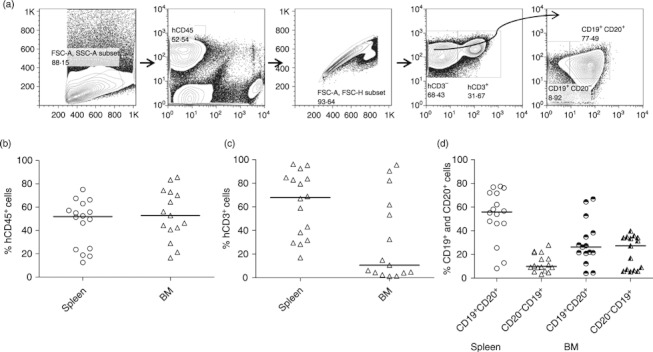
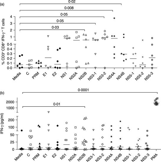
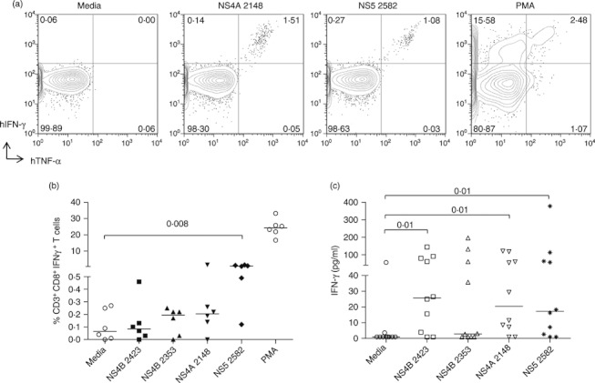
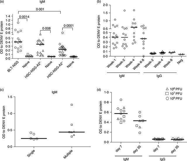
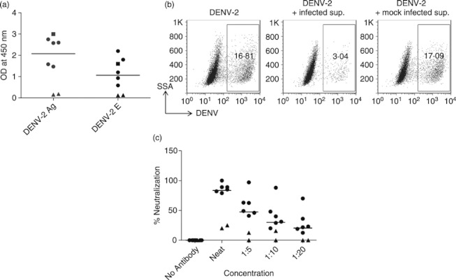
Dengue is a mosquito-borne viral disease of humans, and animal models that recapitulate human immune responses or dengue pathogenesis are needed to understand the pathogenesis of the disease. We recently described an animal model for dengue virus (DENV) infection using humanized NOD-scid IL2rγ(null) mice (NSG) engrafted with cord blood haematopoietic stem cells. We sought to further improve this model by co-transplantation of human fetal thymus and liver tissues into NSG (BLT-NSG) mice. Enhanced DENV-specific antibody titres were found in the sera of BLT-NSG mice compared with human cord blood haematopoietic stem cell-engrafted NSG mice. Furthermore, B cells generated during the acute phase and in memory from splenocytes of immunized BLT-NSG mice secreted DENV-specific IgM antibodies with neutralizing activity. Human T cells in engrafted BLT-NSG mice secreted interferon-γ in response to overlapping DENV peptide pools and HLA-A2 restricted peptides. The BLT-NSG mice will allow assessment of human immune responses to DENV vaccines and the effects of previous immunity on subsequent DENV infections.


Figures

Figure 1
Figure 1
Human haematolymphoid cells in organs of engrafted BLT-NSG mice. (a) Approximately 3 months after engraftment of BLT-NSG mice with human fetal liver and thymus tissue, engraftment was assessed in target organs using flow cytometry. Initial gating strategy to identify cells in the lymphocyte gate was based on forward and side scatter profiles. Human CD45+ cells were next selected using antibodies directed against mouse and human CD45. Viable hCD45+ cells were gated by exclusion of the viability marker LIVE DEAD AQUA. T cells were next selected for by identifying CD3+ cells within the lymphocyte gate. B cells were identified using the phenotypic markers CD19 and CD20 on the CD3-negative population within the lymphocyte gate. Values on the y-axis represent frequencies of (b) hCD45+, (c) hCD3+ and (d) hCD19+ CD20+ and CD19+ CD20 cells within the lymphocyte gate in the spleen (n = 16) and all cells in the bone marrow (n = 16).
Figure 2
Figure 2
Human T-cell responses in BLT-NSG mice. (a) Interferon-γ (IFN-γ) response after primary subcutaneous infection with 1 × 10⁶ plaque-forming units (PFU) of dengue virus serotype 2 (DENV-2) NGC. Splenocytes from BLT-NSG mice (day 7 post-infection) were stimulated with overlapping peptide pools spanning the entire DENV-2 genome (BEI Resources). Cytokine production was assessed by intracellular cytokine staining. A representative graph indicates the frequency of hCD45+ CD3+ CD8+ IFN-γ-producing T cells. (b) IFN-γ levels in supernatants of splenocytes obtained after second immunization with 1 × 10⁶ PFU of DENV-2 NGC. Splenocytes from DENV-2-infected mice were stimulated with DENV-2 peptide pools for 5 days. Culture supernatants were assessed for human IFN-γ by ELISA (Quantikine; R&D Systems).
Figure 3
Figure 3
HLA-A2-restricted human T-cell responses in HLA-A2+ BLT-NSG mice. (a) Representative dot plots indicating the frequencies of interferon-γ-positive, tumour necrosis factor-α-positive (IFN-γ+ TNF-α+) T cells by intracellular cytokine staining of splenocytes from BLT-NSG mice infected by the subcutaneous route with 1 × 10⁶ plaque-forming units (PFU) dengue virus serotype 2 (DENV-2) NGC. Splenocytes were stimulated with 10 μg/ml of NS4A2148, NS52582 peptides, PMA or media and cytokine production was assessed in an intracellular cytokine staining assay. (b) Frequencies of hCD45+ CD3+ CD8+ IFN-γ+ secreting T cells at day 7 post-infection after ex vivo stimulation with HLA-A2-restricted peptides NS4A2148, NS4B2353, NS4B2423, NS52582, PMA and media. (c) IFN-γ levels in supernatants of splenocytes stimulated with HLA-A2-restricted DENV-2 peptides and peptide NS52582 after a second immunization with 1 × 10⁶ PFU DENV-2 NGC.
Figure 4
Figure 4
Detection of dengue virus (DENV) -specific IgM antibodies in the sera of infected engrafted BLT-NSG mice. Sera were obtained from DENV-2-infected and mock-infected mice. IgM or IgG antibodies against DENV-2 E protein (Hawaii Biotech) were detected by ELISA. (a) Comparison of IgM responses in the sera of infected BLT-NSG, NSG HLA-A2+ and NSG HLA-A2 mice 7 days after infection with 1 × 10⁶ plaque-forming units (PFU) of DENV-2 NGC. (b) Kinetics of IgM and IgG responses in the sera of BLT-NSG mice infected with DENV-2 NGC. (c) DENV-specific IgM responses in the sera of mice immunized once (open circles), twice (open triangles) or three times (open squares) with 1 × 10⁶ PFU of DENV-2 NGC. (d) IgM and IgG response in BLT-NSG mice infected by the subcutaneous route with 108 (open circles), 107 (open squares) or 106 (open triangles) PFU DENV-2 S16803.
Figure 5
Figure 5
Neutralization activity of dengue virus serotype 2 (DENV-2) -specific antibodies. Splenocytes from DENV-2 S16803-infected or naive mice were stimulated with CpG + interleukin-2 and Epstein–Barr virus. Culture supernatants obtained 14 days later were used as a source of antibodies to perform ELISA and neutralization assays. (a) IgM responses to DENV-2 antigen and DENV-2 envelope protein in culture supernatants from splenocytes of naive mice (closed triangles) and infected mice [108 plaque-forming units (PFU) closed circles; 107 PFU closed squares]. (b) Representative dot plots show the frequencies of DENV-2-infected cells in the presence or absence of DENV-2-specific antibodies. (c) Percent neutralization activity of serially diluted supernatants from stimulated splenocytes obtained from BLT-NSG mice infected with 108 PFU of DENV-2 S16803 (closed circles) and naive mice (closed triangles).

Similar articles

Cited by

References

    1. Gubler DJ. Dengue and dengue hemorrhagic fever. Clin Microbiol Rev. 1998;11:480–96. - PMC - PubMed
    1. Gubler DJ, Clark GG. Dengue/dengue hemorrhagic fever: the emergence of a global health problem. Emerg Infect Dis. 1995;1:55. - PMC - PubMed
    1. Endy TP, Nisalak A, Chunsuttitwat S, Vaughn DW, Green S, Ennis FA, Rothman AL, Libraty DH. Relationship of pre-existing dengue virus (DV) neutralizing antibody levels to viremia and disease severity in a prospective cohort study of DV infection in Thailand. J Infect Dis. 2004;189:990–1000. - PubMed
    1. Laoprasopwattana K, Libraty DH, Endy TP, et al. Antibody dependent cellular cytotoxicity in pre-secondary dengue virus serotype 3 (DV3) but not in DV2 infection plasma samples inversely correlated with viremia levels. J Infect Dis. 2007;195:1108–16. - PubMed
    1. Libraty DH, Acosta LP, Tallo V, et al. A prospective nested case–control study of dengue in infants: rethinking and refining the antibody-dependent enhancement dengue hemorrhagic fever model. PLoS Med. 2009;6:e1000171. - PMC - PubMed

Publication types

MeSH terms