. 2012 Sep 1;21(13):2508-19.
doi: 10.1089/scd.2011.0695. Epub 2012 Apr 20.

MicroRNA-193 pro-proliferation effects for bone mesenchymal stem cells after low-level laser irradiation treatment through inhibitor of growth family, member 5

Affiliations

MicroRNA-193 pro-proliferation effects for bone mesenchymal stem cells after low-level laser irradiation treatment through inhibitor of growth family, member 5

Jue Wang et al. Stem Cells Dev. .

Erratum in

Abstract

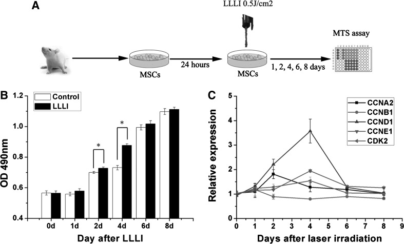
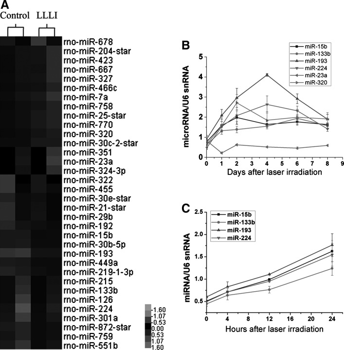
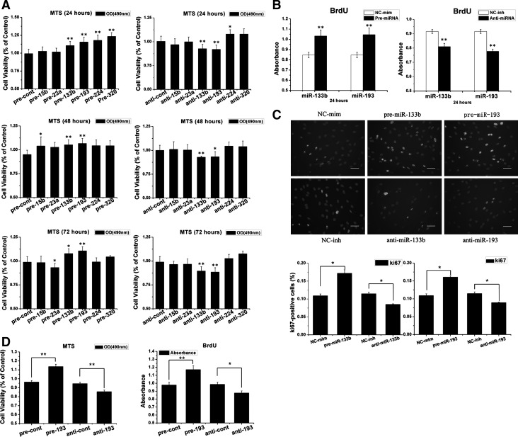
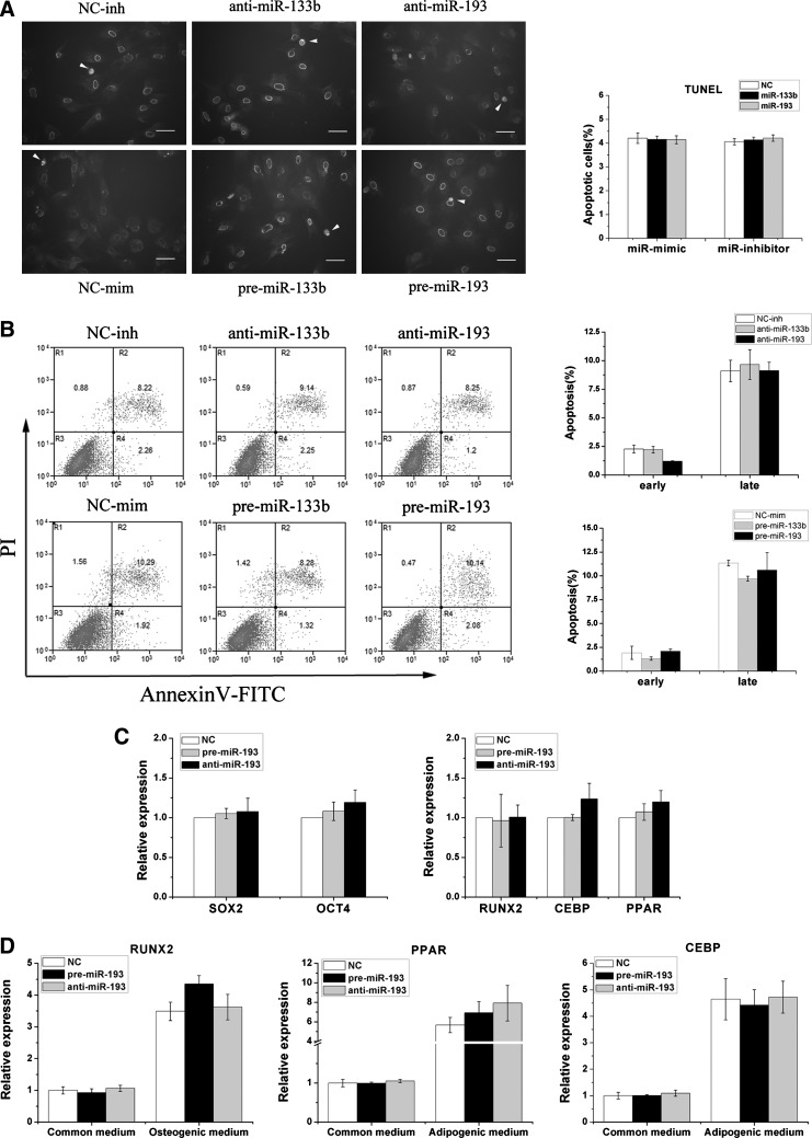
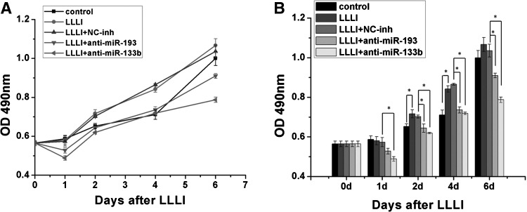
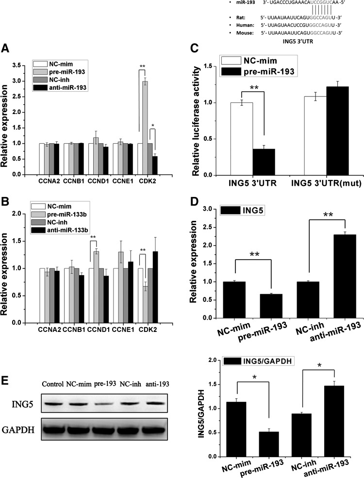
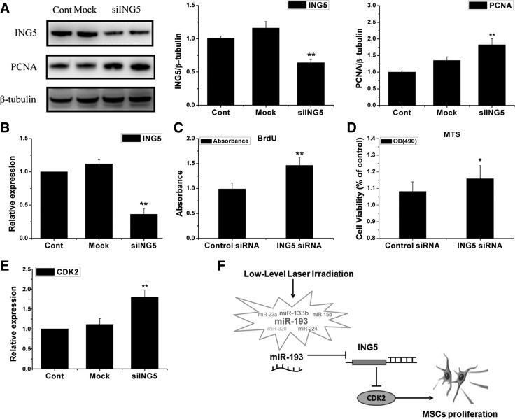
The enhanced proliferation of mesenchymal stem cells (MSCs) can be helpful for the clinical translation of cell therapy. Low-level laser irradiation (LLLI) has been demonstrated as regulating MSC proliferation. MicroRNAs (miRNAs) are involved in various pathophysiologic processes in stem cells, but the role of miRNAs in the LLLI-based promotion of MSC proliferation remains unclear. We found that the proliferation level and cell cycle-associated genes in MSCs were increased after LLLI treatment in a time-dependent manner. Microarray assays revealed subsets of miRNAs to be differentially regulated, and these dynamic changes were confirmed by quantitative real-time polymerase chain reaction (qRT-PCR) after LLLI. miR-193 was the most highly up-regulated miRNA, and the change in it was related with the proliferation level. Gain-loss function experiments demonstrated that miR-193 could regulate the proliferation of MSCs, including human's and rat's, but could not affect the apoptosis and differentiation level. Blockade of miR-193 repressed the MSC proliferation induced by LLLI. By qRT-PCR, we found that miR-193, in particular, regulated cyclin-dependent kinase 2 (CDK2) expression. Bioinformatic analyses and luciferase reporter assays revealed that inhibitor of growth family, member 5 (ING5) could be the best target of miR-193 to functionally regulate proliferation and CDK2 activity, and the mRNA and protein level of ING5 was regulated by miR-193. Furthermore, the ING5 inhibited by small interfering RNA (siRNA) could up-regulate the proliferation of MSCs and the expression of CDK2. Taken together, these results strongly suggest that miR-193 plays a critical part in MSC proliferation in response to LLLI stimulation, which is potentially amenable to therapeutic manipulation for clinical application.


Figures

FIG. 1.
FIG. 1.
Low-level laser irradiation (LLLI) promotes mesenchymal stem cell (MSC) proliferation and increases cell cycle-associated gene expression. (A) MSCs isolated from rats were passaged and placed in wells for 24 h. The MSCs were individually irradiated with a low-level laser. Proliferation of the MSCs was measured by 3-(4,5-dimethylthiazol-2-yl)-5-(3-carboxymethoxyphenyl)-2-(4-sulfophenyl)-2H-tetrazolium (MTS) analyses. (B) MSCs were irradiated by LLLI at an energy density of 0.5 J/cm2 (test group) and 0 (control group). After LLLI, the MTS assay was carried out, and OD490 was compared at day 0, 1, 2, 4, 6, and 8. Mean±standard error of the mean (SEM). *P<0.05 compared with control groups (n=3). (C) The expression of cyclin A2 (CCNA2), cyclin B1 (CCNB1), cyclin D1 (CCND1), cyclin E1 (CCNE1), and cyclin-dependent kinase 2 (CDK2) was detected by quantitative real-time-polymerase chain reaction (qRT-PCR) in the MSCs treated by LLLI in the same process as (B). CCNA2 was up-regulated on day 2, and CCND1/CCNE1 and CDK2 were up-regulated on day 4, compared with the control groups (n=3).
FIG. 2.
FIG. 2.
Profiling of microRNA (miRNA) expression after LLLI treatment in MSCs. (A) Expression patterns of differentially regulated miRNAs in MSCs 4 days after the irradiated LLLI were identified by global microarray analyses. A heat map showing miRNAs regulated between LLLI treatment and control groups (LLLI and control) in MSCs with a cutoff point of 1.2 (P<0.05) (gray represents lower-than-median expression levels; black represents equal-to-median expression levels; and black represents higher expression levels). (B) Day-wise temporal regulation of miRNAs after LLLI treatment in MSCs was analyzed by qRT-PCR. The expression levels of each miRNA are normalized to U6 and represented as a fold change relative to the control group (n=3). (C) Hour-wise temporal regulation of miRNAs in MSCs after LLLI, in the same process as that in (B) (n=3).
FIG. 3.
FIG. 3.
miR-133b/193 regulates MSC proliferation. (A) Cells were transfected with different miRNAs, and their viability was assessed by the MTS assay at different times (24, 48, and 72 h) after transfection. Results are the percentage of control (taken as 100%). Overall, there were significant differences among miR-193 and miR-133b compared with the control at different times (n=3). (B) 5-bromo-2′-deoxyuridine (BrdU) incorporation was used to detect the proliferation of MSCs transfected by miR-193 and miR-133b (n=3). (C) Representative images of Ki-67 immunostaining in MSCs transfected with miR-193 and miR-133b as well as empty xenografts that analyze cell proliferation. Bar=50 μm white indicates ki-67-positive cells; gray indicates staining by 4′,6-diamidino-2-phenylindole (DAPI). The percentage of Ki-67-positive cell number was divided by the DAPI number in the same zone. Mean±SEM. *P<0.05, **P<0.01 compared with control groups. (n=3). All experiments were repeated thrice (technical triplicates) with biological triplicates. (D) Proliferation level of human MSCs (hMSCs) that were transfected with miR-193 or its inhibitor was detected by BrdU incorporation and MTS assay (n=6). Results show that miR-193 could promote hMSCs proliferation.
FIG. 4.
FIG. 4.
miR-193 cannot regulate MSC apoptosis and differentiation. (A) Analyses of in-situ terminal deoxynucleotidyl transferase dUTP nick end labeling (TUNEL) detection of apoptotic cells in MSCs transfected with miR-193 and miR-133b mimics or inhibitors. Representative images of the TUNEL assay (left panel), and TUNEL-positive cells (right panel) of transfected miRNAs and control MSCs. White arrows indicate TUNEL-positive cells. Bars: 50 μm. Mean±SEM, n=3. (B) Apoptosis in MSC cells was detected by flow cytometry. Cells were transfected with miR-193 and miR-133b mimics or inhibitors, and cell suspensions were stained with annexin V/propidium iodide (PI) and subjected to flow cytometric analyses. The apoptosis rate was quantified (right panel). There were no significant differences between transfected MSCs and those in the control groups. Mean±SEM, n=3. (C) The pluripotency of undifferentiated MSCs was detected by the expression of OCT4 and SOX2. The differentiation of MSCs was detected by RUNX2 (marker of osteogenic), CEBP, and PPAR (marker of adipose) by qRT-PCR. Mean±SEM, n=3. (D) After transfection, the MSCs were transferred to either osteoblast- or adipocyte-inducing media for 10 days. Cells were collected, and the differentiation of MSCs was analyzed by qRT-PCR as mentioned in (C).
FIG. 5.
FIG. 5.
Effects of miR-193, miR-133b inhibitors on LLLI-induced cell proliferation. (A) Cell viability was assessed by the MTS assay 1, 2, 4, and 6 days after 0.5 J/cm2 LLLI in the presence or absence of miR-193/133b inhibitors. Changes in cell viability over time are shown in the left panel. (B) Differences between each group are shown in the right panel. miR-133b and miR-193 could significantly inhibit the MSC proliferation induced by LLLI. From 3 independent experiments. Mean±SEM. *P<0.05, n=3.
FIG. 6.
FIG. 6.
Functional assessment of miR-193/133b. (A, B) Expressions of cell-cycle genes were detected by qRT-PCR in MSCs treated with miR-133b (A) or miR-193 (B) mimics or inhibitors (n=3). (C) Luciferase reporter analyses of an inhibitor of growth family, member 5 (ING5) wild-type 3′ untranslated region (3′UTR), as well as 1 TargetScan miR-193 site mutated (ING5 3′UTR mute) together with expression plasmids for miR-193 or a scrambled miRNA (control miRNA). miR-193 overexpression decreased luciferase activity of the wild-type but not the mutant 3′UTR where the miR-193 binding site was deleted. (D) ING5 mRNA expression levels were measured by qRT-PCR in the treatment MSCs. (E) Expression of the ING5 protein was detected by western blotting with normalization to glyceraldehyde-3 phosphate dehydrogenase (GAPDH) in transfected MSCs. The overexpression of miR-193 led to decreased ING5 expression, whereas miR-193 inhibition led to increased ING5 expression. Mean±SEM. *P<0.05. **P<0.01, n=3.
FIG. 7.
FIG. 7.
Knockdown of ING5 by small interfering RNA promotes MSCs proliferation. (A) Expression of ING5 and proliferative cell nuclear antigen (PCNA) were determined by western blot and quantitative determination (n=3). (B) qRT-PCR analysis for ING5 mRNA expression (n=3). (C, D) MSCs proliferation was determined by BrdU (C) and MTS (D) (n=3). (E) The expression of CDK2 in MSCs transfected with si-ING5 was detected by qRT-PCR. Mean±SEM. *P<0.05. **P<0.01 compared with control siRNA groups (n=3). (F) Proposed model of miR-193 in regulating the process of LLLI promoting MSCs proliferation.

Similar articles

Cited by

References

    1. Pittenger MF. Mackay AM. Beck SC. Jaiswal RK. Douglas R. Mosca JD. Moorman MA. Simonetti DW. Craig S. Marshak DR. Multilineage potential of adult human mesenchymal stem cells. Science. 1999;284:143–147. - PubMed
    1. Baxter MA. Wynn RF. Jowitt SN. Wraith JE. Fairbairn LJ. Bellantuono I. Study of telomere length reveals rapid aging of human marrow stromal cells following in vitro expansion. Stem Cells. 2004;22:675–682. - PubMed
    1. Xin Y. Wang YM. Zhang H. Li J. Wang W. Wei YJ. Hu SS. Aging adversely impacts biological properties of human bone marrow-derived mesenchymal stem cells: implications for tissue engineering heart valve construction. Artif Organs. 2010;34:215–222. - PubMed
    1. Peplow PV. Chung TY. Ryan B. Baxter GD. Laser photobiomodulation of gene expression and release of growth factors and cytokines from cells in culture: a review of human and animal studies. Photomed Laser Surg. 2011;29:285–304. - PubMed
    1. Hou JF. Zhang H. Yuan X. Li J. Wei YJ. Hu SS. In vitro effects of low-level laser irradiation for bone marrow mesenchymal stem cells: proliferation, growth factors secretion and myogenic differentiation. Lasers Surg Med. 2008;40:726–733. - PubMed

Publication types

MeSH terms

LinkOut - more resources