Clinical Trial
. 2012 Jul;68(1):8-27.
doi: 10.1111/j.1600-0897.2012.01108.x. Epub 2012 Mar 2.

Methylome of fetal and maternal monocytes and macrophages at the feto-maternal interface

Affiliations
Clinical Trial

Methylome of fetal and maternal monocytes and macrophages at the feto-maternal interface

Sun Young Kim et al. Am J Reprod Immunol. 2012 Jul.

Abstract

Problem: Decidual macrophages (dMφ) of the mother and placental macrophages (Hofbauer cells, HC) of the fetus are deployed at a critical location: the feto-maternal interface. This study was conducted to compare the DNA methylome of maternal and fetal monocytes, dMφ, and HC and thereby to determine the immunobiological importance of DNA methylation in pregnancy.

Method of study: Paired samples were obtained from normal pregnant women at term not in labor and their neonates. Maternal monocytes (MMo) and fetal monocytes (FMo) were isolated from the peripheral blood of mothers and fetal cord blood, respectively. dMφ and HC were obtained from the decidua of fetal membranes and placentas, respectively. DNA methylation profiling was performed using the Illumina Infinium Human Methylation27 BeadChip. Quantitative real-time PCR and Western Blot were performed for validation experiments.

Results: (i) Significant differences in DNA methylation were found in each comparison (MMo versus FMo, 65 loci; dMφ versus HC, 266 loci; MMo versus dMφ, 199 loci; FMo versus HC, 1030 loci). (ii) Many of the immune response-related genes were hypermethylated in fetal cells (FMo and HC) compared to maternal cells (MMo and dMφ). (iii) Genes encoding markers of classical macrophage activation were hypermethylated, and genes encoding alternative macrophage activation were hypomethylated in dMφ and HC compared to MMo and FMo, respectively. (iv) mRNA expressions of DNMT1, DNMT3A, and DNMT3B were significantly lower in dMφ than in HC. (v) 5-azacytidine treatment increased expression of INCA1 in dMφ.

Conclusions: The findings herein indicate that DNA methylation patterns change during monocyte-macrophage differentiation at the feto-maternal interface. It is also suggested that DNA methylation is an important component of the biological machinery conferring an anti-inflammatory phenotype to macrophages at the feto-maternal interface.


Conflict of interest statement

Conflict of Interest

The authors have no financial conflicts of interest.

Figures
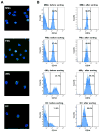
Figure 1
Figure 1
Identification of isolated monocytes and macrophages. (A) Immunoflourescent staining of CD14 (green) positive sorted maternal monocytes (MMo), Fetal monocytes (FMo), decidual macrophages (dMϕ) and Hofbauer cells (HC). The nuclei are stained with DAPI (blue). Sorted cells show mononuclear morphology. (B) The purity of isolated cells for methylation microarray by flow sorting was more than 80 % in each of MMo, FMo, dMϕ, and HC.
Figure 2
Figure 2
Methylation microarray analysis. (A) The distribution of loci methylation β values is shown for each of the 24 samples. The peaks near β=0 illustrates that most of loci are un-methylated in each sample, while the peaks near β=1 show that there is a sizable portion of loci with high methylation β values in each sample. A principal component analysis (PCA) plot (B) and a heat map (C) generated with data from 6 pairs of maternal monocytes (MMo, gray), fetal monocytes (FMo, blue), decidual macrophages (dMϕ, black) and Hofbauer cells (HC, green) show a clear segregation among the cell groups. The PCA plot (B) uses the data from all loci, while the loci chosen for the heat map (C) are the top 200 ones varying the most across all 24 samples (unbiased filtering) as described elsewhere.
Figure 3
Figure 3
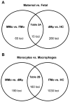
Venn diagram summary of differentially methylated loci between the groups. Differentially methylated loci were using a FDR cut-off of 5% and average DNA methylation difference (Δβ)>0.2. (A) Division of maternal vs. fetal groups. 65 loci were differentially methylated between maternal monocytes (MMo) and fetal monocytes (FMo), and 266 loci were differentially methylated between decidual macrophages (dMϕ) and Hofbauer cells (HC). Fifteen loci were shared between each comparison. (B) Division of blood monocytes vs. tissue macrophages. One hundred ninety-nine loci were differentially methylated between MMo and dMϕ, and 1,030 loci were listed for differentially methylated genes between FMo and HC. One hundred sixty loci were overlapped between each comparison.
Figure 4
Figure 4
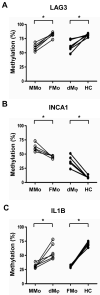
Bisulfite pyrosequencing results of (A) LAG3, (B) INCA1 and (C) IL-1β in 6 samples of genomic DNAs from maternal monocytes (MMo), fetal monocytes (FMo), decidual macrophages (dMϕ) and Hofbauer cells (HC). Bisulfite sequencing was performed across 3 CpG sites of each gene chosen from the region interrogated by methylation microarray, and the average percentages of methylation are well-correlated with microarray data. *p<0.05.
Figure 5
Figure 5
mRNA expressions of DNA methyltransferases by qRT-PCR show distinguished patterns in maternal monocytes (MMo), fetal monocytes (FMo), decidual macrophages (dMϕ), and Hofbauer cells (HC). The DNMTs mRNA expressions were normalized on the content of RPLPO for each. *p<0.05, n=5.
Figure 6
Figure 6
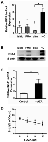
Differential INCA1 expression in monocytes or macrophages by function of DNA methylation. (A) qRT-PCR of INCA1 transcripts of maternal monocytes (MMo), fetal monocytes (FMo), macrophages from decidua (dMϕ), and Hofbauer cells (HC). mRNA expression of INCA1 is significantly down-regulated in dMϕ than in HC. The INCA1 mRNA expressions were normalized on the content of RPLPO. *p<0.05, n=5. (B) Western blot of INCA1 of total proteins from columned MMo, FMo, dMϕ, and HC shows that protein expression of INCA1 is higher in HC than in dMϕ, n=4. (C) qRT-PCR of INCA1 mRNA expression following treatment of 10 μM of 5-Azacytidine for 3 days to dMϕ. mRNA expression of INCA1 was increased in dMϕ exposed to 5-Azacytidine. The INCA1 mRNA expressions were normalized on the content of RPLPO. *p<0.05, n=5. (D) BrdU pulse labeling detection in 5-Azacytidine treated dMϕ. After treatment of 5-Azacytidine (0, 5, 10, or 50 μM), macrophages were pulsed with BrdU (10 μM) for 1 h, and stained with anti BrdU antibody. The percentages of BrdU positive cells were measured by FACS. BrdU labeling decreased with 5-azacytidine treatment in a dose-dependent manner. *p< 0.05 vs. control (0 μM), n=4.

Similar articles

Cited by

References

    1. Satosar A, Ramirez NC, Bartholomew D, Davis J, Nuovo GJ. Histologic correlates of viral and bacterial infection of the placenta associated with severe morbidity and mortality in the newborn. Hum Pathol. 2004;35:536–545. - PubMed
    1. Gomez R, Romero R, Ghezzi F, Yoon BH, Mazor M, Berry SM. The fetal inflammatory response syndrome. Am J Obstet Gynecol. 1998;179:194–202. - PubMed
    1. Qiu X, Zhu L, Pollard JW. Colony-stimulating factor-1-dependent macrophage functions regulate the maternal decidua immune responses against Listeria monocytogenes infections during early gestation in mice. Infect Immun. 2009;77:85–97. - PMC - PubMed
    1. Langrehr JM, White DA, Hoffman RA, Simmons RL. Macrophages produce nitric oxide at allograft sites. Ann Surg. 1993;218:159–166. - PMC - PubMed
    1. Kitchens WH, Chase CM, Uehara S, Cornell LD, Colvin RB, Russell PS, Madsen JC. Macrophage depletion suppresses cardiac allograft vasculopathy in mice. Am J Transplant. 2007;7:2675–2682. - PubMed

MeSH terms