. 2012 Apr;168(1):153-63.
doi: 10.1111/j.1365-2249.2012.04560.x.

Tolerance to lipopolysaccharide promotes an enhanced neutrophil extracellular traps formation leading to a more efficient bacterial clearance in mice

Affiliations

Tolerance to lipopolysaccharide promotes an enhanced neutrophil extracellular traps formation leading to a more efficient bacterial clearance in mice

V I Landoni et al. Clin Exp Immunol. 2012 Apr.

Abstract

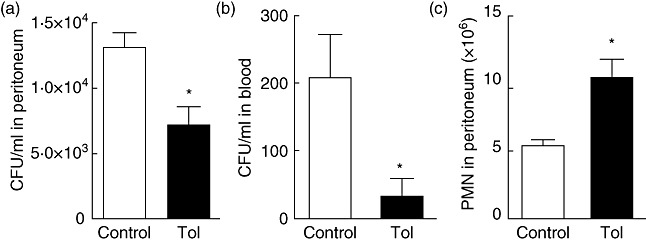
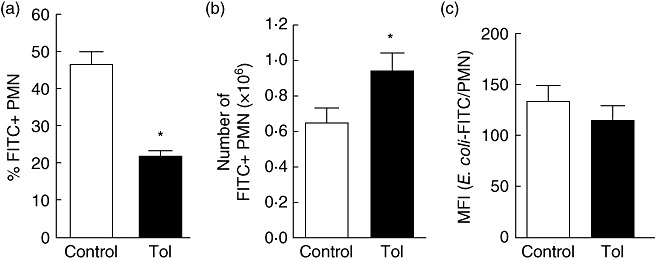
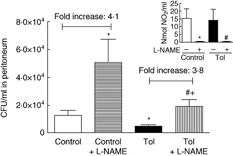
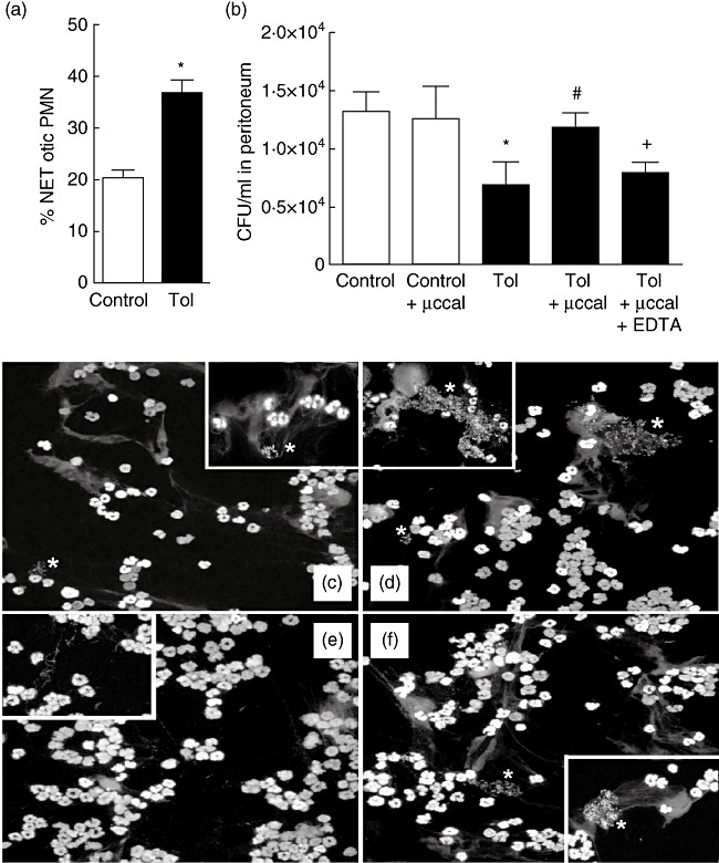
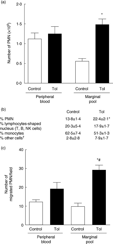
Tolerance to lipopolysaccharide (LPS) constitutes a stress adaptation, in which a primary contact with LPS results in a minimal response when a second exposure with the same stimulus occurs. However, active important defence mechanisms are mounted during the tolerant state. Our aim was to assess the contribution of polymorphonuclear neutrophils (PMN) in the clearance of bacterial infection in a mouse model of tolerance to LPS. After tolerance was developed, we investigated in vivo different mechanisms of bacterial clearance. The elimination of a locally induced polymicrobial challenge was more efficient in tolerant mice both in the presence or absence of local macrophages. This was related to a higher number of PMN migrating to the infectious site as a result of an increased number of PMN from the marginal pool with higher chemotactic capacity, not because of differences in their phagocytic activity or reactive species production. In vivo, neutrophils extracellular trap (NET) destruction by nuclease treatment abolished the observed increased clearance in tolerant but not in control mice. In line with this finding, in vitro NETs formation was higher in PMN from tolerant animals. These results indicate that the higher chemotactic response from an increased PMN marginal pool and the NETs enhanced forming capacity are the main mechanisms mediating bacterial clearance in tolerant mice. To sum up, far from being a lack of response, tolerance to LPS causes PMN priming effects which favour distant and local anti-infectious responses.


Figures

Fig. 1
Fig. 1
Increased clearance of a polymicrobial challenge in the absence of macrophages in mice tolerant to lipopolysaccharide (LPS). Colony-forming units (CFU) (5 × 10⁶) of live intestinal bacteria were injected intraperitoneally (i.p.) after depletion of macrophages (by lip-clod treatment) in control mice and mice rendered tolerant to LPS by intravenous (i.v.) inoculation of LPS (n = 12). After 4 h, CFU were evaluated in peritoneal lavage (a) and blood (b). (c) The absolute number of total polymorphonuclear neutrophils (PMN) in the peritoneal lavage was determined by optical microscopy. *P < 0·05 versus control.
Fig. 2
Fig. 2
Evaluation of in vivo phagocytosis of bacteria. Tolerance was developed by intravenous (i.v.) inoculation of lipopolysaccharide (LPS). Dead fluorescein isothiocyanate (FITC)–Escherichia coli was injected intraperitoneally (i.p.) in control and tolerant animals and 4 h later peritoneal lavage was performed (n = 12). The % (a) and mean fluorescence intensity (MFI, c) of FITC + polymorphonuclear neutrophils (PMN) (identified as Ly-6G+ cells) were determined by flow cytometry. The absolute number of FITC + PMN (b) was calculated using the absolute number of PMN in the peritoneal lavage and the % of FITC + PMN shown in (a); *P < 0·05 versus control.
Fig. 3
Fig. 3
The increased bacterial clearance in tolerant mice is not affected by inhibition of reactive nitrogen species. In vivo contribution of reactive nitrogen species production was determined by inoculating the inhibitor of reactive nitric oxide synthase, Nω-nitro-L-arginine methyl ester (L-NAME) (1·5 mg per mice) 10 min before bacterial challenge in control and tolerant mice. After 4 h, colony-forming units (CFU) were evaluated using agar MacConkey in peritoneal lavage. The numbers at the top of the bars indicate the fold increase of CFU/ml + L-NAME versus untreated mice. Inset: nmol of NO2/ml were determined in the experimental groups by the Griess method; n = 9, *P < 0·05 versus control, #P < 0·05 versus tolerant, +P < 0·05 versus control + L-NAME.
Fig. 4
Fig. 4
Enhanced neutrophil extracellular traps (NETs) formation is involved in the increased bacterial clearance in tolerant mice. (a) In vitro NETs formation capacity was evaluated on polymorphonuclear neutrophils (PMN) recovered from the peritoneal cavity 4 h after polymicrobial challenge. PMN were seeded and treated with phorbol myristate acetate (PMA) for 6 h. The percentage of PMN-forming NETs (NETotic PMN) was determined using epifluorescence microscopy by staining DNA with propidium iodide; n = 6, *P < 0·05 versus control. (b) In vivo NET contribution was determined by inoculating micrococcal nuclease (µccal) or µccal + ethylenediamine tetraacetic acid (EDTA) 10 min before bacterial challenge in control and tolerant mice. Four hours later colony-forming units (CFU) were evaluated using agar MacConkey in peritoneal lavage; n = 13, *P < 0·05 versus control, #P < 0·05 versus tolerant, +P < 0·05 versus tolerant + µccal. (c–f) Four hours after the baterial challenge peritoneal, PMN were carefully collected and seeded onto coverslides. DNA was stained with propidium iodide and NETs were visualized by confocal microscopy (×200 and insets ×600). Asterisks indicate propidium iodide-stained bacteria. Representative microphotographies of an independent experiment are shown from one control (c), tolerant (d), tolerant with µccal treatment (e) and tolerant with µccal inactivated with EDTA (f).
Fig. 5
Fig. 5
The marginal pool of polymorphonuclear neutrophils (PMN) is increased in tolerant mice. (a) The absolute number of PMN present in peripheral blood and marginal pool were determined as described in Materials and methods; n = 18, *P < 0·05 versus control marginal pool. (b) Cellular composition of the marginal pool as determined by light microscopy using May–Grünwald–Giemsa staining; n = 6, *P < 0·05 versus control. †Very large cells that could not be assigned to any category. (c) In vitro chemotactic migration of PMN against N-Formyl-Met-Leu-Phe (fMLP). Isolated PMN from peripheral blood and marginal pool (1 × 10⁵) were allowed to migrate through a polycarbonate filter for 90 min at 37°C with CO2 in response to fMLP (10−7M). Then, the filter was fixed and stained and the number of chemotactic cells was counted in five random high-power fields (×400) for each of triplicate samples. The results were expressed as the number of migrated PMN per field; n = 6 *P < 0·05 versus control marginal pool and peripheral blood, #P < 0·05 versus tolerant peripheral blood.

Similar articles

Cited by

References

    1. Natanson C, Hoffman WD, Suffredini AF, Eichacker PQ, Danner RL. Selected treatment strategies for septic shock based on proposed mechanisms of pathogenesis. Ann Intern Med. 1994;120:771–83. - PubMed
    1. Vincent JL, Sakr Y, Sprung CL, et al. Sepsis in European intensive care units: results of the SOAP study. Crit Care Med. 2006;34:344–53. - PubMed
    1. Fan H, Cook JA. Molecular mechanisms of endotoxin tolerance. J Endotoxin Res. 2004;10:71–84. - PubMed
    1. Van Epps HL. Ignoring endotoxin. J Exp Med. 2006;203:1137. - PMC - PubMed
    1. Cavaillon JM, Adib-Conquy M. Bench-to-bedside review: endotoxin tolerance as a model of leukocyte reprogramming in sepsis. Crit Care. 2006;10:233. - PMC - PubMed

Publication types

MeSH terms