. 2012 Mar 20;51(11):2258-64.
doi: 10.1021/bi201573k. Epub 2012 Mar 8.

Proteolytic regulation of the mitochondrial cAMP-dependent protein kinase

Affiliations

Proteolytic regulation of the mitochondrial cAMP-dependent protein kinase

Jennifer R Shell et al. Biochemistry. .

Abstract

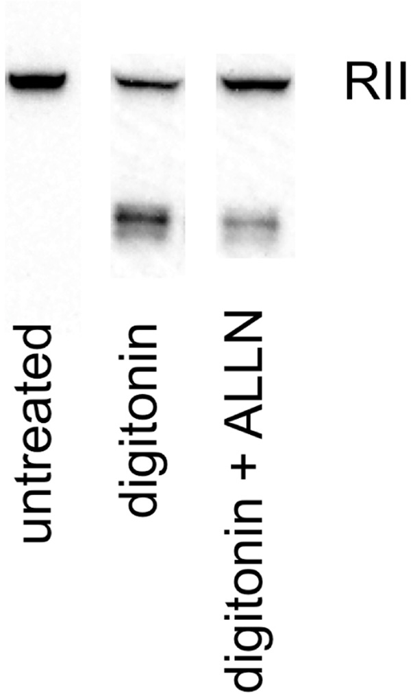
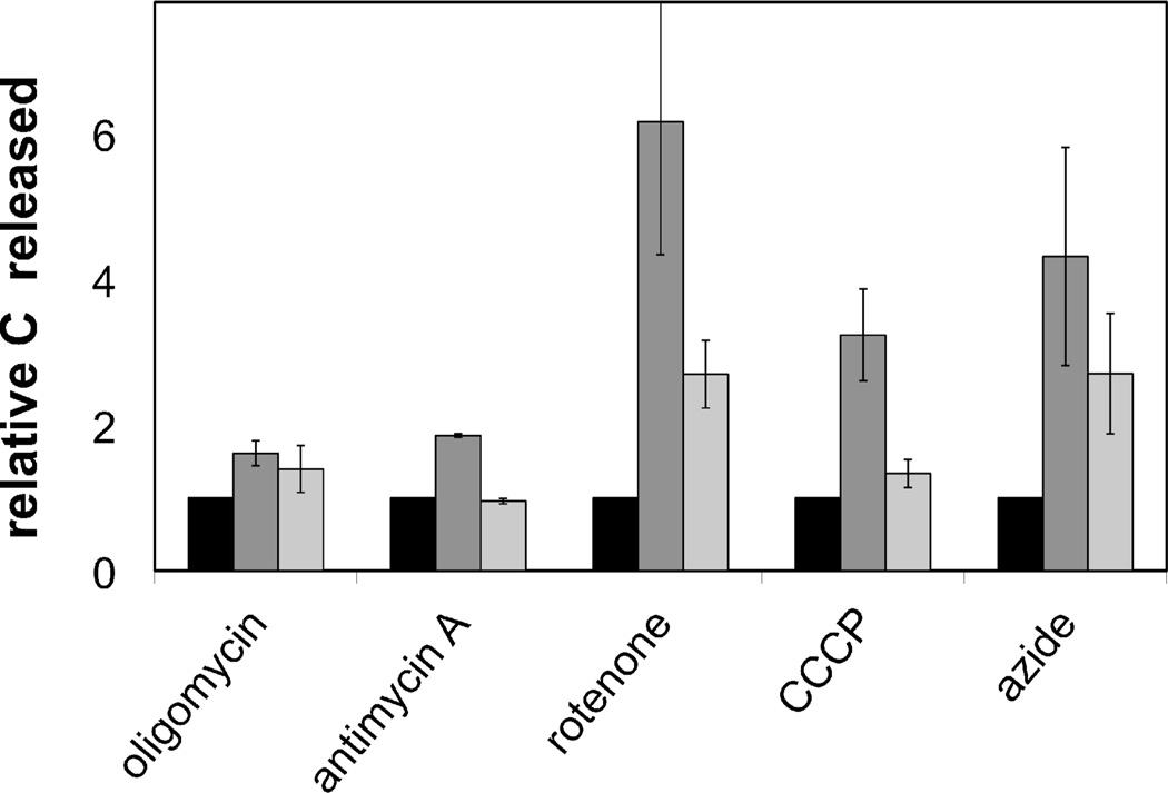
The mitochondrial cAMP-dependent protein kinase (PKA) is activatable in a cAMP-independent fashion. The regulatory (R) subunits of the PKA holoenzyme (R(2)C(2)), but not the catalytic (C) subunits, suffer proteolysis upon exposure of bovine heart mitochondria to digitonin, Ca(2+), and a myriad of electron transport inhibitors. Selective loss of both the RI- and RII-type subunits was demonstrated via Western blot analysis, and activation of the C subunit was revealed by phosphorylation of a validated PKA peptide substrate. Selective proteolysis transpires in a calpain-dependent fashion as demonstrated by exposure of the R and C subunits of PKA to calpain and by attenuation of R and C subunit proteolysis in the presence of calpain inhibitor I. By contrast, exposure of mitochondria to cAMP fails to promote R subunit degradation, although it does result in enhanced C subunit catalytic activity. Treatment of mitochondria with electron transport chain inhibitors rotenone, antimycin A, sodium azide, and oligomycin, as well as an uncoupler of oxidative phosphorylation, also elicits enhanced C subunit activity. These results are consistent with the notion that signals, originating from cAMP-independent sources, elicit enhanced mitochondrial PKA activity.


Figures

Figure 1
Figure 1. Calpain Digestion of Bovine PKA R and C Subunits
Calpain 1 was added to C or R (I and II) subunit in the presence or absence of ALLN. The reaction was initiated by the addition of variable concentrations of CaCl2, and incubated at 37 °C for 20 min: R subunit (♦), C subunit (▲), R subunit and ALLN (●), and C subunit and ALLN (X).
Figure 2
Figure 2. Calpain Digestion of Intact Bovine R2C2 Holoenzyme
Calpain 1 was added to the R2C2 holoenzyme in the presence of 100 µM CaCl2 for 0.5, 1.5, and 2.0 h and the C (○) and R (●) subunits subsequently quantified via western blot analysis (inset).
Figure 3
Figure 3. Digitonin and Ca2+-induced digestion of mitochondrial PKA
A. Bovine heart mitochondria incubated with digitonin in the presence or absence of ALLN where black bar = untreated mitochondria, dark gray bar = digitonin-exposed mitochondria, and the light gray bar = digitonin and ALLN (calpain inhibitor I)-exposed mitochondria. B. Bovine heart mitochondria incubated with digitonin and CaCl2 in the presence or absence of ALLN where black bar = untreated mitochondria, dark gray bar = digitonin/CaCl2-exposed mitochondria, and the light gray bar = digitonin/CaCl2 and ALLN (calpain inhibitor I)-exposed mitochondria. C. Calpain activity (assessed utilizing the fluorescent substrate N-succinyl-Leu-Tyr-7-amido-4-methylcoumarin) from bovine heart mitochondria incubated with digitonin (black trace), digitonin and CaCl2 (gray trace), or buffer alone (inset). D. Bovine heart mitochondria incubated with CaCl2 in the presence or absence of ALLN as a function of time where the black trace is the rate of proteolysis of RI and the gray trace the rate of proteolysis of RII. Immunoblots were imaged using an AlphaInnotech FC2, and quantified using Fluorchem FC2 software. The data represents the mean of 3 experiments ± SEM.
Figure 4
Figure 4. Mitochondrial R and C subunit integrity in response to cAMP and calpain
Mitochondria were treated with (A) cAMP or (B) calpain I and CaCl2. Calpain-exposed mitochondria were examined in the presence or absence of ALLN. Immunoblots were imaged using an AlphaInnotech FC2, and quantified using Fluorchem FC2 software. Quantification of untreated (black bar), calpain I-exposed (dark gray bar), and calpain I/ALLN-exposed (light gray bar) mitochondria are shown in C. The data represents the mean of three experiments ± SEM.
Figure 5
Figure 5. Mitoplast-associated R subunit content from mitochondria treated with digitonin in the absence and presence of ALLN
Mitochondria were treated with digitonin (0.5 mg/mg protein) and phospholipase A in the presence or absence of ALLN (200 µM) and incubated at 37°C for 2 h. The resultant mitoplasts were then pelleted at 11,000 × g for 15 min at 4 °C.
Figure 6
Figure 6. Ca2+-induced PKA activation: release of active C subunit
A. Mitochondria were treated with cAMP or CaCl2, centrifuged, and the pellet and supernatant processed for western blot analysis. B. Mitochondria were incubated with the soluble adenylate cyclase inhibitor KH7 for 30 min, then treated with CaCl2 in the presence or absence of ALLN, centrifuged, and the supernatant analyzed for C subunit by western blot: untreated (black bar), Ca2+-exposed (dark gray bar), and Ca2+/ALLN-exposed (light gray bar) mitochondria. C. PKA activity, using a previously described enzyme-coupled assay(21), in untreated (light gray trace) and cAMP-exposed (black trace) mitochondria. D. PKA activity in Ca2+-exposed (black trace), Ca2+/ALLN-exposed (light gray trace), and Ca2+/PKI-exposed mitochondria (dark gray trace).
Figure 7
Figure 7. Metabolic inhibitors induce activation of PKA in a calpain-dependent fashion
Mitochondria were exposed to oligomycin, antimycin A, rotenone, CCCP, or sodium azide in the presence or absence of ALLN for 30 min at 37 °C and subsequently centrifuged. The supernatants were analyzed by western blot for the presence of the C subunit. The relative amount of C subunit released from untreated (black bar), metabolic inhibitor-exposed (gray bar), and metabolic inhibitor/ALLN-exposed mitochondria. The data represents the mean of three experiments ± SEM.

Similar articles

Cited by

References

    1. Manning G, Whyte DB, Martinez R, Hunter T, Sudarsanam S. The protein kinase complement of the human genome. Science. 2002;298:1912–1934. - PubMed
    1. Gullingsrud J, Kim C, Taylor SS, McCammon JA. Dynamic binding of PKA regulatory subunit RI alpha. Structure. 2006;14:141–149. - PubMed
    1. Kim C, Xuong NH, Taylor SS. Crystal structure of a complex between the catalytic and regulatory (RIalpha) subunits of PKA. Science. 2005;307:690–696. - PubMed
    1. Zhong H, SuYang H, Erdjument-Bromage H, Tempst P, Ghosh S. The transcriptional activity of NF-kappaB is regulated by the IkappaB-associated PKAc subunit through a cyclic AMP-independent mechanism. Cell. 1997;89:413–424. - PubMed
    1. Dulin NO, Niu J, Browning DD, Ye RD, Voyno-Yasenetskaya T. Cyclic AMP-independent activation of protein kinase A by vasoactive peptides. J Biol Chem. 2001;276:20827–20830. - PubMed

Publication types

Substances