Review
. 2012 Aug;42(8):808-20.
doi: 10.3109/00498254.2012.663515. Epub 2012 Mar 2.

Understanding substrate selectivity of human UDP-glucuronosyltransferases through QSAR modeling and analysis of homologous enzymes

Affiliations
Review

Understanding substrate selectivity of human UDP-glucuronosyltransferases through QSAR modeling and analysis of homologous enzymes

Dong Dong et al. Xenobiotica. 2012 Aug.

Abstract

The UDP-glucuronosyltransferase (UGT) enzyme catalyzes the glucuronidation reaction which is a major metabolic and detoxification pathway in humans. Understanding the mechanisms for substrate recognition by UGT assumes great importance in an attempt to predict its contribution to xenobiotic/drug disposition in vivo. Spurred on by this interest, 2D/3D-quantitative structure activity relationships and pharmacophore models have been established in the absence of a complete mammalian UGT crystal structure. This review discusses the recent progress in modeling human UGT substrates including those with multiple sites of glucuronidation. A better understanding of UGT active site contributing to substrate selectivity (and regioselectivity) from the homologous enzymes (i.e. plant and bacterial UGTs, all belong to family 1 of glycosyltransferase (GT1)) is also highlighted, as these enzymes share a common catalytic mechanism and/or overlapping substrate selectivity.


Figures
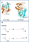
Figure 1
Figure 1. Panel A: Cartoon representation of glycosyltransferase GT-A and GT-B folds
N-terminal domain is colored cyan and C-terminal domain orange. The GT-A fold consists of two dissimilar β/α/β domains, with the N-terminal domain binding the nucleotide sugar and the highly variable C-terminal domain binding the acceptor substrate. The GT-B fold consists of two similar Rossman fold domains, with the highly variable N-terminal domain binding the acceptor substrate and the C-terminal domain binding the nucleotide sugar. The enzyme for GT-A fold demonstration is the nucleotide-diphospho-sugar transferase SpsA from Bacillus subtilis (PDB code: 1H7L). The enzyme for GT-B fold demonstration is the triterpene UDP-glucosyltransferase UGT71G1 from Medicago truncatula (PDB code: 2ACW). Panel B: Two main catalytic mechanisms of glycosyltransferase-mediated reaction: retention and inversion of the anomeric configuration. Human UGTs are inverting glycosyltransferases, like all other members of the GT1 family. X = -OH, -COOH, -NH, -NH2, or -SH.
Figure 2
Figure 2. The different catalytic mechanisms for O- and N-glucuronidation in human UGTs
Panel A: Proposed catalytic mechanism for O-glucuronidation (so-called “serine hydrolase like mechanism”) involving two catalytic resides histidine and aspartate. Panel B: N-glucuronidation does not required proton abstraction because N-necleophiles can readily develop positive charged amine as the transition state. The question mark denotes that the role of the “catalytic” dyad histidine-aspartate in N-glucuronidation remains unknown.
Figure 3
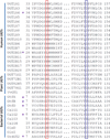
Figure 3. A sequence alignment between human UGTs and other GT1 enzymes with solved crystal structures
The putative catalytic base is highlighted in red box and the corresponding acid in blue box. Stars denote the enzymes which do not use histidine as the catalytic base.
Figure 4
Figure 4. The common features pharmacophore for human UGT1A1, 1A4, and 1A9 (Miners et al., 2004)
Panel A: The common features pharmacophore include two hydrophobic regions (H) in addition to the glucuronidation site (G). Panel B: the pharmacophore was mapped to selective substrates for UGT1A1, 1A4 and 1A9.
Figure 5
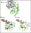
Figure 5. A homology model for human UGT1A9 and the superimposition of the CoMFA/CoMISA steric maps with the modeled active site (Wu et al., 2012)
Panel A shows the constructed model for UGT1A9. The model consists of N- (in gray) and C-terminal (in light green) domains. The N- and C-terminal domains contain central stranded parallel sheets flanked by α-helices on both sides. The substrate binding pocket is almost entirely formed by the N-terminal residues, although some C-terminal residues also contributed to the formation of the pocket. The pocket is primarily formed by LoopN1, Nα1, Nα3-2, LoopN4, Nα5-1, Nα5-2, Loop C1 and Loop C5. The cofactor is present at the left side of pocket. The catalytic residue histidine (in green stick model) was located at the start of helix Nα1. The substrate kaempferol (in a 3-OH catalysis mode) is shown in stick-and-ball model. Panel B/C: Superposition of the CoMFA (B) and CoMSIA (C) steric maps over the active site of the homology-modeled UGT1A9 structure based on a simulated binding model of kaempferol (3-OH). The UGT1A9 protein is shown in a stick model. Kaempferol is indicated in a ball-and-stick model and the cofactor is shown in a ball-and-stick model with a molecular surface. Green: Areas in which bulky groups are sterically favorable for glucuronidation; Yellow: Areas in which bulky groups are unfavorable for glucuronidation.
Figure 6
Figure 6. Comparisons of the active sites (in a green solid surface) of crystal structures for 5 plant UGTs: (A) UGT71G1 (PDB code, 2ACW), (B) UGT72B1 (PDB code, 2VCE), (C) UGT78G1 (PDB code, 3HBF), (D) UGT85H2 (PDB code, 2PQ6), and (E) Vv GT1 (PDB code, 2C1Z)
The volume of UGT72B1 active site is estimated at 41 Å3, much smaller than those of UGT71G1, UGT78G1, UGT85H2, or VvGT1. Please note that estimates of the active site volumes (indicated by question marks) for the latters are inaccurate (greatly less than the actual values) as the active sites are open to the solvent. The bars denote a rough distance between the active site and the helix Nα5, suggesting that residues from Nα5 creates a more significant steric hindrance for UGT71G1 than for UGT78G1, UGT85H2, or VvGT1.
Figure 7
Figure 7. Comparisons of the active sites of GT1 enzymes reveals significant conformational differences are required to accommodate distinct substrates
Panel A: An active site comparison of UGT72B1-TCP (in cyan) and VvGT1-kaempferol (in white) complexes. Panel B: An active site comparison of OleI-oleandomycin (in blue) and VvGT1-kaempferol (in white) complexes. The PDB codes for the crystal structures can be found in Table 1. Star denotes the unique loop C2 in UGT72B1. Dashed line denotes the hydrogen bond. TCP: 2,4,5-trichlorophenol.
Figure 8
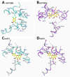
Figure 8. Accommodation of substrates in GT1 enzymes requires conformational changes in the protein in relation to the substrate free enzymes
The changes mainly occur at the side chains of residues forming the active site. Panel A: a comparison of UGT72B1 binding site for TCP binding (in cyan) with the substrate-free binding site (in white). Panel B: a comparison of UGT78G1 structures for myricetin binding (in purple) with the substrate-free binding site (in white). Panel C: A comparison of VvGT1 structures for kaempferol binding (in cyan) with the substrate-free binding site (in white). Panel C: A comparison of VvGT1 structures for quercetin binding (in purple) with the substrate-free binding site (in white). The PDB codes for the crystal structures can be found in Table 1. Star denotes the site of glycosylation. Dashed line denotes the hydrogen bond. TCP: 2,4,5-trichlorophenol.
Figure 9
Figure 9. A hypothetical binding model of flavonols (quercetin is shown as an example) to human UGT1A1
Panels A-B show the molecular basis for regioselectivity of 4 plant UGTs towards quercetin deduced from crystal structures and mutagenesis studies (Osmani et al., 2009). In the presence of steric hindrance from Nα5 (i.e., F148), positioning of 3-OH of quercetin in UGT71B1 is disfavored. In the absence of significant steric hindrance from Nα5, positioning of 3-OH of quercetin in UGT78G1, UGT85H2, and VvGT1 is favored. Please also refer to Figure 6 for the comparisons of substrate-binding pockets among plant UGTs. Panel B (VvGT crystal structure is used) is shown for a better understanding of the geometry of the substrate-binding pocket (i.e., the green meshed surface) within the plant UGT protein, as well as its relative coordinates to the helix Nα5 (situates at the end of the pocket), catalytic residue histidine, cofactor, and solvent. Panel C shows the hypothetical binding modes of quercetin to human UGT1A1. The inner part (or the end) of substrate-binding pocket provides steric bulks (possible aromatic rings), which disfavor the positioning of flavonols for 3-OH catalysis. Comparing to 7-OH, 3’-OH is more preferred by the potential H-bond formation (dashed green line) between 4’-OH and the active site residues.

References

    1. Battaglia E, Senay C, Fournel-Gigleux S, Herber R, Siest G, Magdalou J. The chemical modification of human liver UDP-glucuronosyltransferase UGT1*6 reveals the involvement of a carboxyl group in catalysis. FEBS Lett. 1994;346(2–3):146–150. - PubMed
    1. Berger I, Guttman C, Amar D, Zarivach R, Aharoni A. The molecular basis for the broad substrate specificity of human sulfotransferase 1A1. PLoS One. 2011;6(11):e26794. - PMC - PubMed
    1. Bolam DN, Roberts S, Proctor MR, Turkenburg JP, Dodson EJ, Martinez-Fleites C, Yang M, Davis BG, Davies GJ, Gilbert HJ. The crystal structure of two macrolide glycosyltransferases provides a blueprint for host cell antibiotic immunity. Proc Natl Acad Sci U S A. 2007;104(13):5336–5341. - PMC - PubMed
    1. Bosma PJ. Inherited disorders of bilirubin metabolism. J Hepatol. 2003;38:107–117. - PubMed
    1. Strassburg CP. Pharmacogenetics of Gilbert’s syndrome. Pharmacogenomics. 2008;9:703–715. - PubMed

Substances

LinkOut - more resources