

Langerhans cells are critical in epicutaneous sensitization with protein antigen via thymic stromal lymphopoietin receptor signaling
- PMID: 22385635
- PMCID: PMC4600611
- DOI: 10.1016/j.jaci.2012.01.063
Langerhans cells are critical in epicutaneous sensitization with protein antigen via thymic stromal lymphopoietin receptor signaling
Abstract
Background: The clarification of cutaneous dendritic cell subset and the role of thymic stromal lymphopoietin (TSLP) signaling in epicutaneous sensitization with protein antigens, as in the development of atopic dermatitis, is a crucial issue.
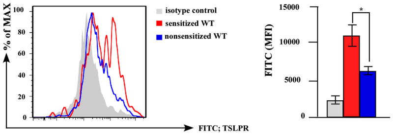
Objectives: Because TSLP is highly expressed in the vicinity of Langerhans cells (LCs), we sought to clarify our hypothesis that LCs play an essential role in epicutaneous sensitization with protein antigens through TSLP signaling.
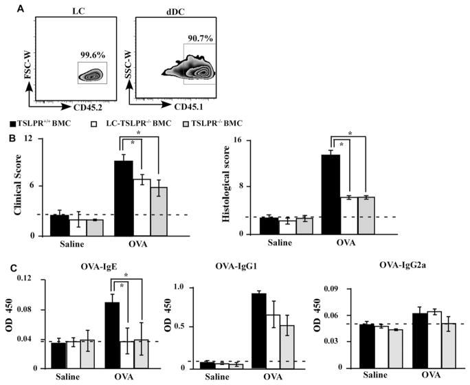
Methods: By using Langerin-diphtheria toxin receptor knock-in mice and human Langerin-diphtheria toxin A transgenic mice, we prepared mice deficient in LCs. We also prepared mice deficient in TSLP receptors in LCs by using TSLP receptor-deficient mice with bone marrow chimeric technique. We applied these mice to an ovalbumin (OVA)-induced epicutaneous sensitization model.
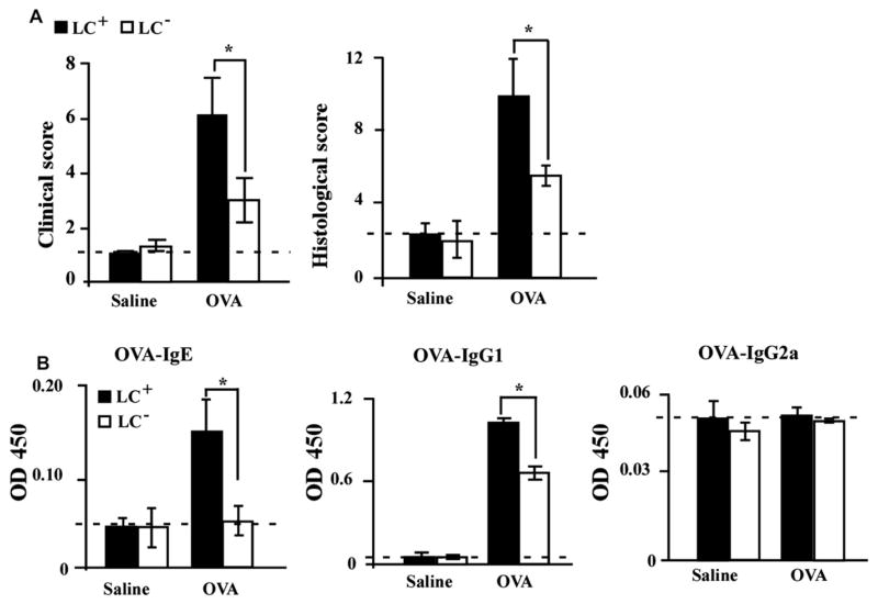
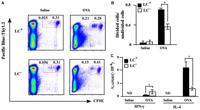
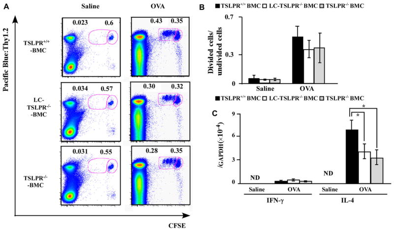
Results: Upon the epicutaneous application of OVA, conditional LC depletion attenuated the development of clinical manifestations as well as serum OVA-specific IgE increase, OVA-specific T-cell proliferation, and IL-4 mRNA expression in the draining lymph nodes. Consistently, even in the steady state, permanent LC depletion resulted in decreased serum IgE levels, suggesting that LCs mediate the T(H)2 local environment. In addition, mice deficient in TSLP receptors on LCs abrogated the induction of OVA-specific IgE levels upon epicutaneous OVA sensitization.
Conclusion: LCs initiate epicutaneous sensitization with protein antigens and induce T(H)2-type immune responses via TSLP signaling.
Copyright © 2012 American Academy of Allergy, Asthma & Immunology. Published by Mosby, Inc. All rights reserved.
Conflict of interest statement
Disclosure of potential conflict of interest: The authors declare that they have no relevant conflicts of interest.
Figures

Similar articles
-
Dual function of Langerhans cells in skin TSLP-promoted TFH differentiation in mouse atopic dermatitis.J Allergy Clin Immunol. 2021 May;147(5):1778-1794. doi: 10.1016/j.jaci.2020.10.006. Epub 2020 Oct 15. J Allergy Clin Immunol. 2021. PMID: 33068561
-
Thymic stromal lymphopoietin rather than IL-33 drives food allergy after epicutaneous sensitization to food allergen.J Allergy Clin Immunol. 2023 Jun;151(6):1660-1666.e4. doi: 10.1016/j.jaci.2023.02.025. Epub 2023 Mar 4. J Allergy Clin Immunol. 2023. PMID: 36878383 Free PMC article.
-
Selective AhR knockout in langerin-expressing cells abates Langerhans cells and polarizes Th2/Tr1 in epicutaneous protein sensitization.Proc Natl Acad Sci U S A. 2020 Jun 9;117(23):12980-12990. doi: 10.1073/pnas.1917479117. Epub 2020 May 27. Proc Natl Acad Sci U S A. 2020. PMID: 32461368 Free PMC article.
-
Thymic stromal lymphopoietin, OX40-ligand, and interleukin-25 in allergic responses.Clin Exp Allergy. 2009 Jun;39(6):798-806. doi: 10.1111/j.1365-2222.2009.03241.x. Epub 2009 Apr 7. Clin Exp Allergy. 2009. PMID: 19400908 Free PMC article. Review.
-
Thymic stromal lymphopoietin and OX40 ligand pathway in the initiation of dendritic cell-mediated allergic inflammation.J Allergy Clin Immunol. 2007 Aug;120(2):238-44; quiz 245-6. doi: 10.1016/j.jaci.2007.06.004. J Allergy Clin Immunol. 2007. PMID: 17666213 Review.
Cited by
-
TSLP Directly Interacts with Skin-Homing Th2 Cells Highly Expressing its Receptor to Enhance IL-4 Production in Atopic Dermatitis.J Invest Dermatol. 2015 Dec;135(12):3017-3024. doi: 10.1038/jid.2015.318. Epub 2015 Aug 19. J Invest Dermatol. 2015. PMID: 26288354
-
Novel Targeted Biological Agents for the Treatment of Atopic Dermatitis.BioDrugs. 2021 Jul;35(4):401-415. doi: 10.1007/s40259-021-00490-x. Epub 2021 Jul 2. BioDrugs. 2021. PMID: 34213742 Review.
-
Atopic Dermatitis: Pathophysiology.Adv Exp Med Biol. 2024;1447:21-35. doi: 10.1007/978-3-031-54513-9_3. Adv Exp Med Biol. 2024. PMID: 38724781 Review.
-
The Pathogenetic Effect of Natural and Bacterial Toxins on Atopic Dermatitis.Toxins (Basel). 2016 Dec 23;9(1):3. doi: 10.3390/toxins9010003. Toxins (Basel). 2016. PMID: 28025545 Free PMC article. Review.
-
Epidermal Overexpression of Xenobiotic Receptor PXR Impairs the Epidermal Barrier and Triggers Th2 Immune Response.J Invest Dermatol. 2018 Jan;138(1):109-120. doi: 10.1016/j.jid.2017.07.846. Epub 2017 Sep 18. J Invest Dermatol. 2018. PMID: 28927887 Free PMC article.
References
-
- Egawa G, Kabashima K. Skin as a peripheral lymphoid organ: revisiting the concept of skin-associated lymphoid tissues. J Invest Dermatol. 2011;131:2178–85. - PubMed
-
- Moniaga CS, Kabashima K. Filaggrin in atopic dermatitis: flaky tail mice as a novel model for developing drug targets in atopic dermatitis. Inflamm Allergy Drug Targets. 2011;10:477–85. - PubMed
-
- Werfel T. The role of leukocytes, keratinocytes, and allergen-specific IgE in the development of atopic dermatitis. J Invest Dermatol. 2009;129:1878–91. - PubMed
-
- Nickel R, Beck LA, Stellato C, Schleimer RP. Chemokines and allergic disease. J Allergy Clin Immunol. 1999;104:723–42. - PubMed
-
- Shimada Y, Takehara K, Sato S. Both Th2 and Th1 chemokines (TARC/CCL17, MDC/CCL22, and Mig/CXCL9) are elevated in sera from patients with atopic dermatitis. J Dermatol Sci. 2004;34:201–8. - PubMed
Publication types
MeSH terms
Substances
Grants and funding
LinkOut - more resources
Full Text Sources
Other Literature Sources
