. 2012 Mar 2;10(3):299-311.
doi: 10.1016/j.stem.2012.01.016.

Genetic regulators of a pluripotent adult stem cell system in planarians identified by RNAi and clonal analysis

Affiliations

Genetic regulators of a pluripotent adult stem cell system in planarians identified by RNAi and clonal analysis

Daniel E Wagner et al. Cell Stem Cell. .

Abstract

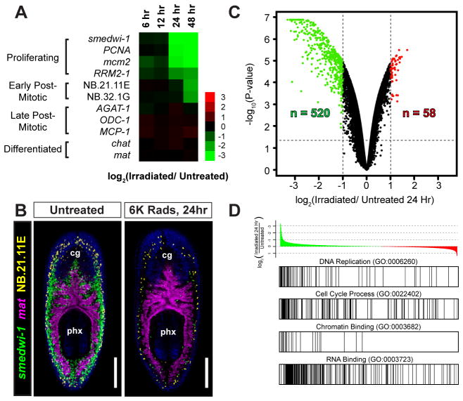
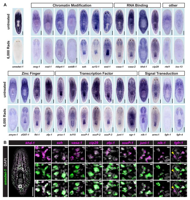
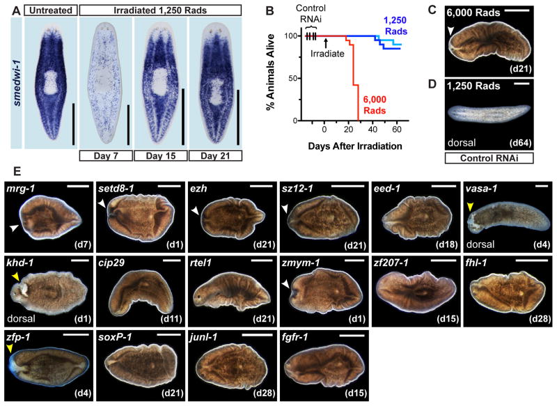
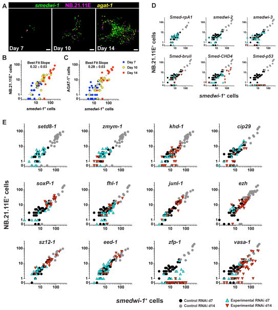
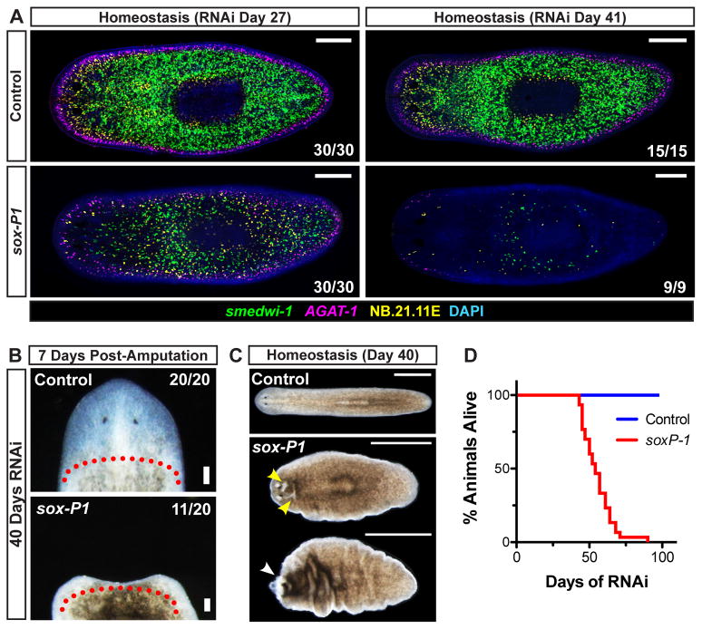
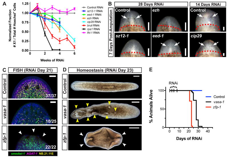
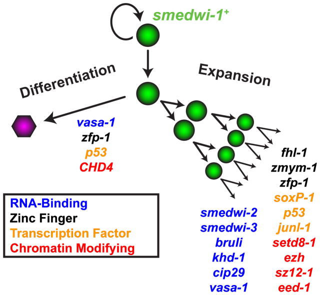
Pluripotency is a central, well-studied feature of embryonic development, but the role of pluripotent cell regulation in somatic tissue regeneration remains poorly understood. In planarians, regeneration of entire animals from tissue fragments is promoted by the activity of adult pluripotent stem cells (cNeoblasts). We utilized transcriptional profiling to identify planarian genes expressed in adult proliferating, regenerative cells (neoblasts). We also developed quantitative clonal analysis methods for expansion and differentiation of cNeoblast descendants that, together with RNAi, revealed gene roles in stem cell biology. Genes encoding two zinc finger proteins, Vasa, a LIM domain protein, Sox and Jun-like transcription factors, two candidate RNA-binding proteins, a Setd8-like protein, and PRC2 (Polycomb) were required for proliferative expansion and/or differentiation of cNeoblast-derived clones. These findings suggest that planarian stem cells utilize molecular mechanisms found in germ cells and other pluripotent cell types and identify genetic regulators of the planarian stem cell system.


Figures

Figure 1
Figure 1. Identification of Irradiation-Sensitive Transcripts in Adult Planarians by Microarrays
(A) Heat map illustrating mRNA depletion kinetics following 6,000 Rads γirradiation. Markers for proliferating cells (smedwi-1, PCNA, mcm2, RRM2-1), post-mitotic cells (NB.21.11E, NB.32.1G, AGAT-1, ODC-1, and MCP-1), neurons (chat), and intestine (mat) are shown. (B) Whole-mount triple-fluorescence in situ hybridization (FISH) shows the anatomical distribution of proliferative cells (neoblasts) with untreated and 24-hour irradiated animals. Shown are projections through z-stacks of multiple confocal planes in the interior of entire animals. Pharynx (px) and cephalic ganglia (cg) are indicated. Ventral views, anterior up. Scale bars, 200 μm. (C) Volcano plot showing transcripts depleted (green) or upregulated (red) 24 hours after irradiation. See Supplemental Table 1. (D) Gene set enrichment analysis (GSEA) with annotated gene list pre-ranked by log2 ratio (24-hour irradiated/untreated). Example gene sets enriched among irradiation-depleted transcripts are shown. See Supplemental Table 2.
Figure 2
Figure 2. Irradiation-Sensitive Transcripts are Expressed in smedwi-1+ Proliferating Cells
(A) Whole-mount in situ hybridization (ISH) in untreated animals and animals fixed 5 days after 6,000 Rads γ-irradiation. Genes were annotated by BLASTx and PFAM (See also Supplemental Table 3). (B) Expression of genes identified by microarray, analyzed by double FISH with a smedwi-1 RNA probe (proliferative cell marker). Zoomed images are single confocal planes from tail regions. Most cells detected by FISH co-expressed smedwi-1; cells with little/no smedwi-1 gene expression are labeled by arrowheads. Some transcripts (e.g., ezh and cip29) are expressed at low levels with background signal (scattered magenta dots) also visible. See also Supplemental Figure 2. Shown are representative ventral views, anterior up. Scale bars 200 μm, 10 μm (zoomed images).
Figure 3
Figure 3. Identification of RNAi Phenotypes after Sublethal Irradiation
(A) Proliferative cell repopulation detected by smedwi-1 ISH in animals fixed 7, 15, and 21 days after 1,250 Rads γ-irradiation. Ventral views, anterior up. (B) Survival curves of control RNAi animals exposed to 1,250 Rads (two independent experiments) and 6,000 Rads (n = 19–20 animals per sample). (C–E) Representative views of irradiated RNAi animals, anterior left. Images are ventral views unless otherwise noted. (C) 6,000 Rad-irradiated animals experienced head regression (white arrowheads) and ventral curling by day 21. (D) Most sublethally irradiated (1,250 Rad) animals, by contrast, were visibly normal on day 64. (E) Representative live images of animals with RNAi phenotypes after 1,250 rad exposure. Images are from the first day post-irradiation (noted in parenthesis) when tissue failure appeared; only cases where >50% of animals displayed defects are shown. White arrowheads: tissue regression. yellow arrowhead: epidermal lesions. Scale bar, 500 μm. See also Supplemental Figure 3 and Supplemental Table 4.
Figure 4
Figure 4. Genes Required for Proliferative Cell Expansion and Differentiation Identified by RNAi and Clonal Analysis
(A) Representative colonies from animals exposed to 1,750 Rads analyzed by triple FISH. Ventral views, adjacent to the pharynx, anterior left. Proliferating cells (smedwi-1+), and two post-mitotic cell types (NB.21.11E+ and AGAT-1+) are labeled. Scale bars, 50 μm. (B–C) Log-scale plots of raw cell count data. Each dot represents an individual colony. smedwi-1+ cell numbers per colony are plotted against numbers of NB.21.11E+ (B) and AGAT-1+ cells (C). (D–E) Animals irradiated 1,500 – 1,750 Rads fed one RNAi food dose displayed reduced numbers of smedwi-1+ and/or NB.21.11E+ cells per colony. RNAi was administered by feeding 4 days after irradiation except for junl-1, ezh, sz12-1, and eed-1, which were fed seven days prior to irradiation. See also Supplemental Figure 4. For statistical analysis of colony phenotypes, see Supplemental Table 5.
Figure 5
Figure 5. Smed-soxP-1 is Required for Maintenance of smedwi-1+ Cells, Tissue Homeostasis, and Regeneration
(A) Animals were assessed for proliferative (smedwi-1+) and post-mitotic (AGAT-1+ and NB.21.11E+) cell types after several weeks of RNAi. Shown are representative confocal planes, anterior left. (B–C) Regenerative ability and tissue homeostasis were assessed after 40 days of RNAi. (B) Representative live images of head regions 7 days post-amputation are shown, anterior up. Approximate amputation plane is indicated by dotted line. (C–D) Tissue maintenance and animal survival after continuous RNAi feedings every 3–4 days (n ≥ 30 animals/sample). (C) Representative live images of whole animals undergoing tissue failure, anterior left. Epidermal lesions and head regression are indicated by yellow and white arrowheads, respectively. (D) Survival curves. Scale bars 200 μm (A), 100μm (B), 1mm (C).
Figure 6
Figure 6. Genes Necessary for Colony Expansion and Differentiation are Required for Tissue Maintenance
(A) Animals fed RNAi food for several weeks were assessed for proliferative cell presence by flow cytometry. Numbers of proliferating X1 cells, represented as a fraction of total Hoechst+ cells, were normalized to internal RNAi controls. Three biological replicates were used per time point. Shown are means; error bars denote data range. (B) Regenerative ability after 14 or 28 days of continuous RNAi feeding. Shown are head regions, anterior up, seven days after decapitation. Dotted line, approximate amputation planes. Arrows, photoreceptors. (C–E) RNAi animals were assessed for tissue maintenance defects. (C) Confocal projections from animals after 21 days of RNAi. Dorsal views of head regions, anterior up. Arrows, tissue regression sites. (D–E) Tissue maintenance and animal survival after RNAi (n = 29–34 worms/sample). (D) Images after 23 days of RNAi. Anterior, left. Yellow arrows, lesions. White arrows, tissue regression and ventral curling. (E) Survival curves. Scale bars 100 μm (B–C), 500 μm (D).
Figure 7
Figure 7. RNA-Binding, Transcription, and Chromatin Modifying Factors Regulate Clonogenic Expansion and Differentiation of Planarian Stem Cells
Genes expressed within the proliferative cell compartment are required for expansion and differentiation activity associated with clonogenic cells (cNeoblasts). Several of these genes (e.g. p53, zfp-1, vasa-1) are involved in both proliferative cell expansion and differentiation.

Comment in

Similar articles

Cited by

References

    1. Avilion AA, Nicolis SK, Pevny LH, Perez L, Vivian N, Lovell-Badge R. Multipotent cell lineages in early mouse development depend on SOX2 function. Gene Dev. 2003;17:126–140. - PMC - PubMed
    1. Bardeen C, Baetjer F. The inhibitive action of the Roentgen rays on regeneration in planarians. J Exp Zool. 1904;1:191–195.
    1. Boyer L, Plath K, Zeitlinger J, Brambrink T, Medeiros L, Lee T, Levine S, Wernig M, Tajonar A, Ray M, et al. Polycomb complexes repress developmental regulators in murine embryonic stem cells. Nature. 2006;441:349–353. - PubMed
    1. Boyer LA, Lee TI, Cole MF, Johnstone SE, Levine SS, Zucker JP, Guenther MG, Kumar RM, Murray HL, Jenner RG, et al. Core transcriptional regulatory circuitry in human embryonic stem cells. Cell. 2005;122:947–956. - PMC - PubMed
    1. Chamberlain SJ, Yee D, Magnuson T. Polycomb repressive complex 2 is dispensable for maintenance of embryonic stem cell pluripotency. Stem Cells. 2008;26:1496–1505. - PMC - PubMed

Publication types

LinkOut - more resources