. 2012 Feb 22;102(4):802-9.
doi: 10.1016/j.bpj.2012.01.008. Epub 2012 Feb 21.

Single-molecule analysis of the microtubule cross-linking protein MAP65-1 reveals a molecular mechanism for contact-angle-dependent microtubule bundling

Affiliations

Single-molecule analysis of the microtubule cross-linking protein MAP65-1 reveals a molecular mechanism for contact-angle-dependent microtubule bundling

Amanda Tulin et al. Biophys J. .

Abstract

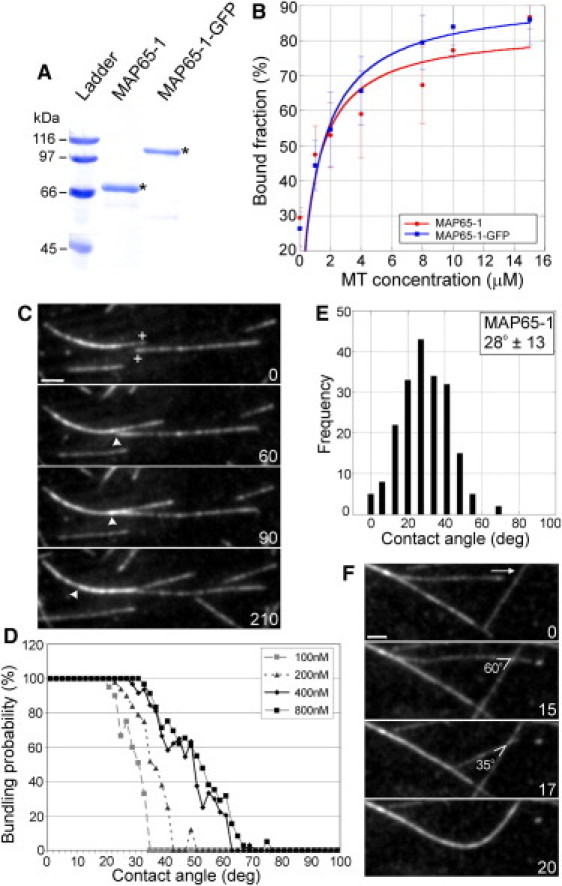
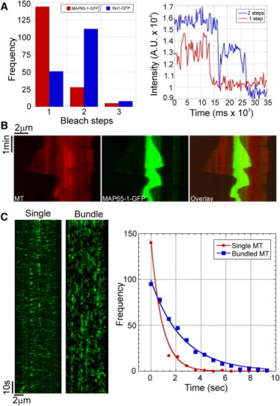
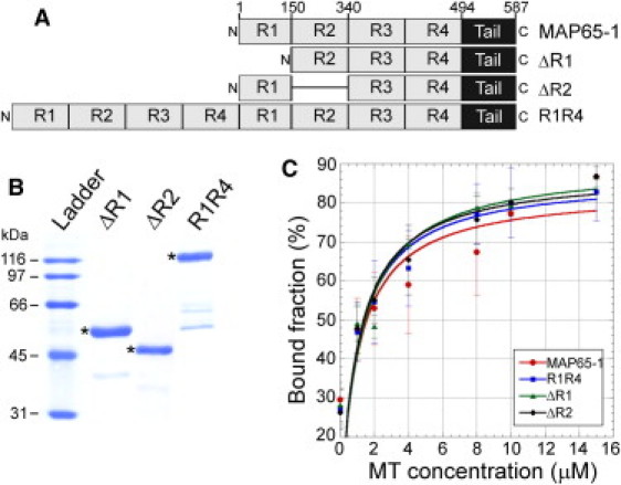
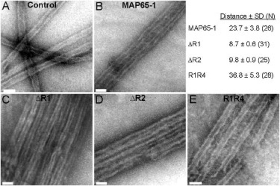
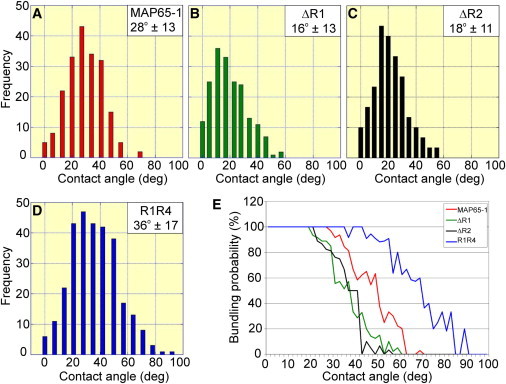
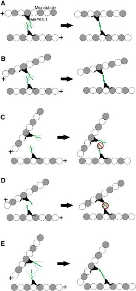
Bundling of microtubules (MTs) is critical for the formation of complex MT arrays. In land plants, the interphase cortical MTs form bundles specifically following shallow-angle encounters between them. To investigate how cells select particular MT contact angles for bundling, we used an in vitro reconstitution approach consisting of dynamic MTs and the MT-cross-linking protein MAP65-1. We found that MAP65-1 binds to MTs as monomers and inherently targets antiparallel MTs for bundling. Dwell-time analysis showed that the affinity of MAP65-1 for antiparallel overlapping MTs is about three times higher than its affinity for single MTs and parallel overlapping MTs. We also found that purified MAP65-1 exclusively selects shallow-angle MT encounters for bundling, indicating that this activity is an intrinsic property of MAP65-1. Reconstitution experiments with mutant MAP65-1 proteins with different numbers of spectrin repeats within the N-terminal rod domain showed that the length of the rod domain is a major determinant of the range of MT bundling angles. The length of the rod domain also determined the distance between MTs within a bundle. Together, our data show that the rod domain of MAP65-1 acts both as a spacer and as a structural element that specifies the MT encounter angles that are conducive for bundling.


Figures

Figure 1
Figure 1
MAP65-1 preferentially bundles antiparallel MTs after shallow-angle encounters. (A) Coomassie-stained gel of purified MAP65-1 and MAP65-1-GFP proteins. The expected protein sizes are marked by asterisks. (B) Binding curves with 1.5 μM MAP65-1 and MAP65-1-GFP proteins at increasing MT concentrations. Each data point represents the mean ± SD from at least three independent experiments. The data were fit to the Michaelis-Menten equation yielding Kd values of 1.03 ± 0.75 μM and 1.27 ± 0.68 μM for MAP65-1 and MAP65-1-GFP, respectively. (C) Montage showing antiparallel MT bundling by 400 nM MAP65-1. The plus-ends of the MTs of interest are indicated in the first frame. Arrowheads mark the position of the plus end within the MT bundle. (D) Plots showing the probability for MT bundling as a function of the encounter angle at various MAP65-1 concentrations. The bundling probability was calculated as a percentage of the number of MT encounters that resulted in MT bundling at a particular angle. The total number of MT encounters observed for 100 nM, 200 nM, 400 nM, and 800 nM of MAP65-1 are 245, 243, 323, and 311, respectively. (E) Distribution of the frequency of MT bundling at various encounter angles in the presence of 400 nM MAP65-1 (N = 199 events). The mean MT bundling angle is 28 ± 13°. (F) MT bundling after a decrease in the crossover angle from 60° to 35°. The arrow indicates the direction of the growing plus end of the MT of interest. Numbers in C and F indicate time in seconds. Scale bars, 2 μm.
Figure 2
Figure 2
MAP65-1 binds to MTs as a monomer and preferentially localizes to regions of MT overlap. (A) Bar graph of the number of bleaching steps for MAP65-1-GFP and Kinesin1-GFP molecules bound to taxol-stabilized MTs (N = 177 and 171 for MAP65-1-GFP and Kinesin1-GFP, respectively). Examples of fluorescence intensity traces showing one and two bleaching steps are shown at right. (B) Kymograph showing the localization of 400 nM MAP65-1-GFP in an antiparallel MT bundle. MAP65-1-GFP specifically tracks the region of MT overlap and is barely detectable along stretches with a single MT. (C) To the left are kymographs showing the binding of 8 nM MAP65-1-GFP to a single MT and an antiparallel MT bundle. To the right are the distributions of dwell-times of single binding events of MAP65-1-GFP on single MTs (N = 257) and bundled MTs (N = 384). Exponential fits to the data yielded half-times of 0.62 ± 0.07 s and 1.82 ± 0.01 s, respectively.
Figure 3
Figure 3
Purification and MT binding of MAP65-1 mutants. (A) Schematic of the domain architecture of MAP65-1 and the various mutants used in this study. The four predicted spectrin repeats are labeled R1–R4. Tail refers to the unstructured domain at the C-terminus of MAP65-1. (B) Coomassie-stained gel of purified ΔR1, ΔR2, and R1R4 proteins. The expected protein sizes are marked by asterisks. (C) Binding curves with 1.5 μM ΔR1, ΔR2, and R1R4 proteins at increasing MT concentrations. Each data point represents the mean ± SD from at least three independent experiments. The data were fit to the Michaelis-Menten equation, yielding Kd values of 1.17 ± 0.73 μM, 1.04 ± 0.61 μM, and 1.04 ± 0.63 μM for ΔR1, ΔR2, and R1R4, respectively. The binding curve for MAP65-1 is reproduced from Fig. 1B for comparison to the mutant proteins.
Figure 4
Figure 4
Length of the rod domain of MAP65-1 determines the distance between MTs in a bundle. Negative-stain electron microscopy of 1 μM MTs alone (A) or 1 μM MTs coincubated with 1 μM of MAP65-1 (B), ΔR1 (C), ΔR2 (D), and R1R4 (E), respectively. The mean ± SD of the distance (nm) is shown in the figure. The number of independent measurements between separate MTs is shown in parentheses. Scale bars, 50 nm.
Figure 5
Figure 5
Length of the rod domain of MAP65-1 determines the MT bundling angle. (AD) Distribution of the frequency of MT bundling at various encounter angles in the presence of 400 nM MAP65-1 (A), 800 nM ΔR1 (B), 400 nM ΔR2 (C), and 400 nM R1R4 (D). The data for MAP65-1 are reproduced from Fig. 1E for comparison to the mutant proteins. N = 199, 189, 231, and 295 for MAP65-1, ΔR1, ΔR2, and R1R4, respectively. The means ± SD of the bundling angle are shown in the figure. (E) Plots showing the probability for MT bundling as a function of the encounter angle in the presence of 400 nM MAP65-1, 800 nM ΔR1, 400 nM ΔR2, and 400 nM R1R4.
Figure 6
Figure 6
Model for encounter-angle-dependent MT bundling by MAP65-1. MAP65-1 monomers are shown bound to MTs (a single MT protofilament is shown for simplicity). The plus sign indicates the MT plus end. The N-terminal rod domain of MAP65-1 is shown projecting from the MT surface, and its conformational flexibility is represented by its multiple positions. If two MTs encounter each other in a nearly parallel orientation (A) or at a shallow-angle (B), the MAP65-1 monomers are able to dimerize and form a stable cross-link, thus resulting in MT bundling. In contrast, if two MTs encounter each other at a steep angle, the MAP65-1 monomers are unable to dimerize, because their rod domains cannot interact productively at these angles (C). Consequently, these MTs do not bundle. In the case of the ΔR1 and ΔR2 mutants, their shorter rod domains are probably stiffer, thus requiring even shallower encounter angles for dimer formation (D). In contrast, the R1R4 mutant has a longer rod domain that is likely to be more flexible than the rod domain of wild-type MAP65-1, which allows dimer formation and MT bundling even at steep encounter angles (E).

References

    1. Wasteneys G.O., Ambrose J.C. Spatial organization of plant cortical microtubules: close encounters of the 2D kind. Trends Cell Biol. 2009;19:62–71. - PubMed
    1. Shaw S.L., Kamyar R., Ehrhardt D.W. Sustained microtubule treadmilling in Arabidopsis cortical arrays. Science. 2003;300:1715–1718. - PubMed
    1. Chan J., Sambade A., Lloyd C. Arabidopsis cortical microtubules are initiated along, as well as branching from, existing microtubules. Plant Cell. 2009;21:2298–2306. - PMC - PubMed
    1. Nakamura M., Ehrhardt D.W., Hashimoto T. Microtubule and katanin-dependent dynamics of microtubule nucleation complexes in the acentrosomal Arabidopsis cortical array. Nat. Cell Biol. 2010;12:1064–1070. - PubMed
    1. Ambrose J.C., Wasteneys G.O. CLASP modulates microtubule-cortex interaction during self-organization of acentrosomal microtubules. Mol. Biol. Cell. 2008;19:4730–4737. - PMC - PubMed

Publication types

MeSH terms

Substances