. 2012 Feb 22;102(4):916-26.
doi: 10.1016/j.bpj.2011.12.010. Epub 2012 Feb 21.

A delicate interplay of structure, dynamics, and thermodynamics for function: a high pressure NMR study of outer surface protein A

Affiliations

A delicate interplay of structure, dynamics, and thermodynamics for function: a high pressure NMR study of outer surface protein A

Ryo Kitahara et al. Biophys J. .

Abstract

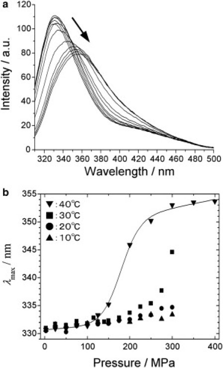
Outer surface protein A (OspA) is a crucial protein in the infection of Borrelia burgdorferi causing Lyme disease. We studied conformational fluctuations of OspA with high-pressure (15)N/(1)H two-dimensional NMR along with high-pressure fluorescence spectroscopy. We found evidence within folded, native OspA for rapid local fluctuations of the polypeptide backbone in the nonglobular single layer β-sheet connecting the N- and C-terminal domains with τ << ms, which may give the two domains certain independence in mobility and thermodynamic stability. Furthermore, we found that folded, native OspA is in equilibrium (τ >> ms) with a minor conformer I, which is almost fully disordered and hydrated for the entire C-terminal part of the polypeptide chain from β8 to the C-terminus. Conformer I is characterized with ΔG(0) = 32 ± 9 kJ/mol and ΔV(0) = -140 ± 40 mL/mol, populating only ∼0.001% at 40°C at 0.1 MPa, pH 5.9. Because in the folded conformer the receptor binding epitope of OspA is buried in the C-terminal domain, its transition into conformer I under in vivo conditions may be critical for the infection of B. burgdorferi. The formation and stability of the peculiar conformer I are apparently supported by a large packing defect or cavity located in the C-terminal domain.


Figures

Figure 1
Figure 1
Schematic representation of the secondary structure elements of OspA. (Sticks) Side chains of Leu-109, Val-199, and Trp-216. Strands 8–10 comprising the single layer β-sheet are labeled. The internal cavities (shaded spheres) of the protein (x-ray structure, PDB:1OSP) are shown with the program MOLMOL (43), as water inaccessible volumes.
Figure 2
Figure 2
Variable-pressure fluorescence spectroscopy of OspA. (a) Tryptophan fluorescence spectra of OspA at 40°C at different pressures from 0.1 to 400 MPa. The protein concentration was 22 μM in 10 mM phosphate buffer and 50 mM NaCl with a pH of 5.9. (b) Plots of the maximum wavelength of emission at 10, 20, 30, and 40°C as a function of pressure.
Figure 3
Figure 3
15N TROSY spectra of the OspA at 3, 50, 100, 150, 200, and 250 MPa at 40°C. (Inset) Region details the change in chemical shift of Val-199 that lies outside the plot.
Figure 4
Figure 4
(a and b) Changes in 1H (a) and 15N (b) chemical shifts between 3 and 150 MPa at 40°C. The average changes in 1H and15N shifts between 3 and 150 MPa are 0.05 ± 0.06 ppm and 0.4 ± 0.3 ppm, respectively. (Solid line) Average value. (First and second broken lines) Average values of average ± RMSD and ± 2 × RMSD, respectively. (c and d) Residues showing anomalous 1H (c) or 15N (d) chemical shift changes are marked. (Red) Residues with the deviation from the average larger than 2|RMSD|; (yellow) deviation between |RMSD| and 2|RMSD|.
Figure 5
Figure 5
(a) Changes in resonance intensities versus pressure. (Red) Residues unperturbed by pressure; (blue) residues losing intensity; (green) residues showing a mountain-like transition. (Solid circles) Averages for the three groups. ΔG0 and ΔV0 values are obtained from the average transition curves. (b) Resonance intensities relative to those at atmospheric pressure as a function of residue number at 225 and 250 MPa. (Green stars) Residues showing mountain-like transitions with pressure. (c) Mapping of the blue and red residues onto a crystal structure of OspA. (Dark-blue spheres) Cavities. (d) Protection factors PF versus residue number. Errors were estimated from the standard deviation of noise on the base plane.
Figure 6
Figure 6
1H one-dimensional NMR spectra of 1.5 mM OspA at pressures ranging from 3 MPa to 250 MPa. Two methyl peaks corresponding to residues L109 (a) and V199 (b) can be seen upfield of 0 ppm.
Figure 7
Figure 7
Conformational equilibrium of OspA depicted from high-pressure NMR. The folded conformer (N, left) has relatively large and rapid local fluctuations (τ < ms) in the nonglobular part of the protein consisting of a single-layer β-sheet (strands 8–10) connecting the N- and C-terminal domains. The folded conformer (N) also undergoes a large-amplitude fluctuation (ms << τ) into an intermediate conformer (I, middle) and the unfolded conformer (U, right). In the intermediate, approximately two-thirds of the polypeptide chain in the C-terminal side (β8-C-terminus) are disordered and hydrated, whereas the N-terminal side (β1–β8) remains largely intact. The overall equilibrium (with populations estimated from high-pressure NMR experiments at 40°C, pH 5.9, 0.1 MPa) is N (∼99.999%) ⇌ I (∼0.001%) ⇌ U (<<0.001%).

Similar articles

Cited by

References

    1. Akasaka K., Yamada H. On-line cell high-pressure nuclear magnetic resonance technique: application to protein studies. Methods Enzymol. 2001;338:134–158. - PubMed
    1. Akasaka K. Probing conformational fluctuation of proteins by pressure perturbation. Chem. Rev. 2006;106:1814–1835. - PubMed
    1. Kitahara R., Akasaka K. Close identity of a pressure-stabilized intermediate with a kinetic intermediate in protein folding. Proc. Natl. Acad. Sci. USA. 2003;100:3167–3172. - PMC - PubMed
    1. Kitahara R., Sareth S., Akasaka K. High pressure NMR reveals active-site hinge motion of folate-bound Escherichia coli dihydrofolate reductase. Biochemistry. 2000;39:12789–12795. - PubMed
    1. Kitahara R., Yokoyama S., Akasaka K. NMR snapshots of a fluctuating protein structure: ubiquitin at 30 bar-3 kbar. J. Mol. Biol. 2005;347:277–285. - PubMed

Publication types

MeSH terms

LinkOut - more resources