. 2012 Mar 30;149(1):88-100.
doi: 10.1016/j.cell.2012.02.018. Epub 2012 Mar 1.

Coding region polyadenylation generates a truncated tRNA synthetase that counters translation repression

Affiliations

Coding region polyadenylation generates a truncated tRNA synthetase that counters translation repression

Peng Yao et al. Cell. .

Abstract

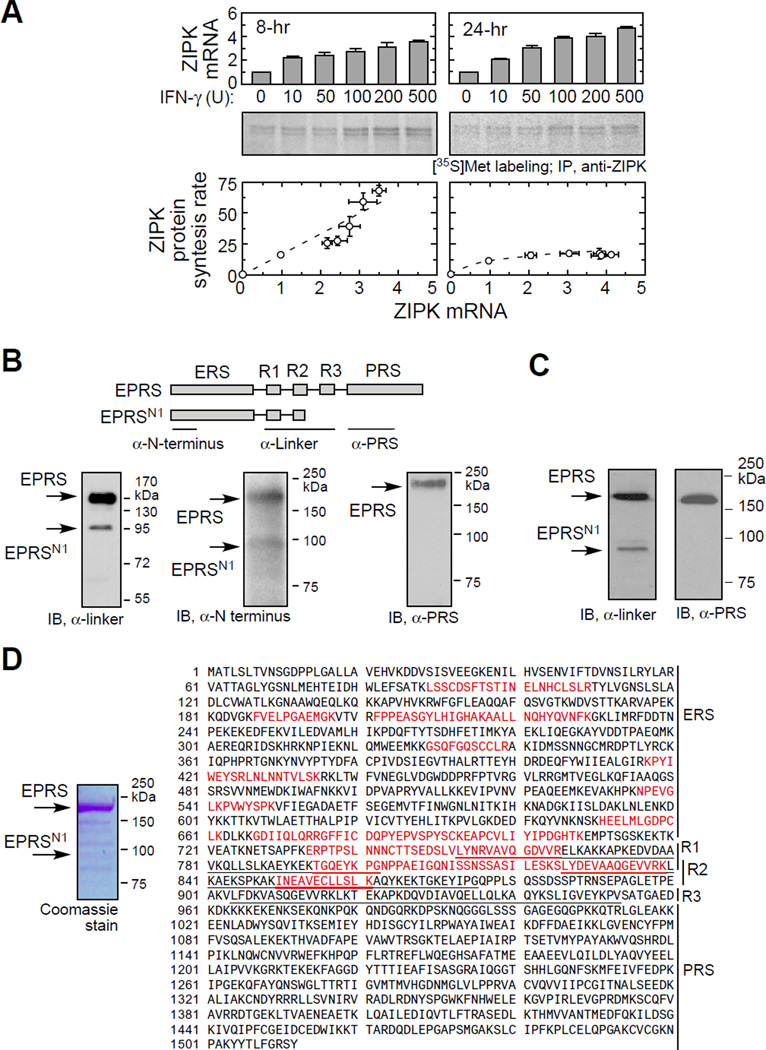
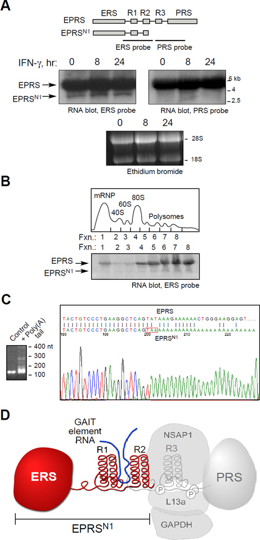
Posttranscriptional regulatory mechanisms superimpose "fine-tuning" control upon "on-off" switches characteristic of gene transcription. We have exploited computational modeling with experimental validation to resolve an anomalous relationship between mRNA expression and protein synthesis. The GAIT (gamma-interferon-activated inhibitor of translation) complex repressed VEGF-A synthesis to a low, constant rate independent of VEGF-A mRNA expression levels. Dynamic model simulations predicted an inhibitory GAIT-element-interacting factor to account for this relationship and led to the identification of a truncated form of glutamyl-prolyl tRNA synthetase (EPRS), a GAIT constituent that mediates binding to target transcripts. The truncated protein, EPRS(N1), shields GAIT-element-bearing transcripts from the inhibitory GAIT complex, thereby dictating a "translational trickle" of GAIT target proteins. EPRS(N1) mRNA is generated by polyadenylation-directed conversion of a Tyr codon in the EPRS-coding sequence to a stop codon (PAY(∗)). Genome-wide analysis revealed multiple candidate PAY(∗) targets, including the authenticated target RRM1, suggesting a general mechanism for production of C terminus-truncated regulatory proteins.


Figures

Figure 1
Figure 1. Differential Regulation of VEGF-A mRNA and Protein by the GAIT Pathway
(A) Schematic of GAIT pathway of transcript-selective translational control. (B) IFN-γ-stimulated monocytic cells maintain low-level expression of VEGF-A. U937 cells were treated with IFN-γ at up to 500 U/ml for 8 (left) or 24 (right) hr. VEGF-A mRNA was determined by qRT-PCR (top), and VEGF-A protein and β-actin in cell lysates by immunoblot analysis (middle panels). VEGF-A protein as function of VEGF-A mRNA is shown (mean ± standard error, n = 3) (bottom).
Figure 2
Figure 2. Dynamic Modeling of GAIT System Indicates a GAIT Element-Interacting Factor is Responsible for the “Translational Trickle” of VEGF-A Expression
(A) Dynamic modeling of GAIT system. Key model features are: Gi, inactive (or unformed) GAIT complex; G, active GAIT complex; M, GAIT element-bearing mRNA; GM, GAIT complex-bound mRNA, P, newly synthesized GAIT target protein. Dashed compartment contains: F, GEIF; FM, GEIF-bound mRNA. Rate constants are indicated within triangles. (B) Model simulations without GEIF. Rate of protein synthesis versus total mRNA before (8-hr) and after (24-hr) translational silencing using the initial model without GEIF (dashed lines); model simulations were done for range of IFN-γ concentrations (left). Model simulations at best-fit ratio of GAIT complex to target mRNA (ratio = 15) as determined by non-linear regression (right). (C) Model simulations with GEIF. Simulations (dashed lines and curves) in presence of GEIF based on experimentally determined parameters (left), and in presence of a 2-fold higher or 75% lower amount of GEIF (right). See also Figure S1 and Tables S1 and S2.
Figure 3
Figure 3. Monocytic Cells Contain a C-terminal Truncated EPRS, EPRSN1
(A) IFN-γ-stimulated monocytic cells maintain low-level synthesis of ZIPK. U937 cells were treated with IFN-γ as in (A). ZIPK mRNA was determined by qRT-PCR (top) and ZIPK synthesis determined by metabolic labeling with [35S]Met followed by immunoprecipitation (IP) with anti-ZIPK antibody (middle). ZIPK protein synthesis as function of ZIPK mRNA is shown (mean ± standard error, n = 3) (bottom). (B) Detection of EPRSN1 protein in U937 cells with domain-specific antibodies. Cytosolic lysates from U937 cells were subjected to immunoblot analysis with antibodies directed against: EPRS linker domain, EPRS-N-terminal peptide, and PRS. (C) Presence of EPRSN1 in human PBM. Immunoblot analysis with antibodies directed against the EPRS linker and PRS domains. (D) Mass spectrometric analysis indicates EPRSN1 contains ERS, R1, and R2 domains, but lacks R3 and PRS domains. EPRSN1 was isolated by immunoprecipitation with anti-EPRS linker antibody and SDS-PAGE, and detected with Coomassie stain (left). Peptides detected by mass spectrometric analysis of 95-kDa EPRSN1 band (red) and WHEP domains (underlined) are shown (right).
Figure 4
Figure 4. Monocytic Cells Express EPRSN1 mRNA
(A) EPRSN1 mRNA is expressed constitutively. Total RNA from U937 cells treated with IFN-γ was subjected to RNA blot analysis using ERS- and PRS-specific probes. (B) EPRSN1 mRNA is translatable. U937 cell lysates were fractionated on a sucrose gradient and extracted RNA in each fraction was subjected to RNA blot analysis with ERS-specific probe. (C) Nucleotide sequence of EPRSN1 mRNA. G/I stretches were added to poly(A)-tailed mRNA, RT-PCR was done using oligo-C and gene-specific upstream primers, and amplified fragment was cloned into T-vector and sequenced. (D) Schematic of EPRSN1 structure in context of full-length EPRS and its phosphorylation sites and its binding partners.
Figure 5
Figure 5. EPRSN1 Binding to GAIT Element RNA Blocks Translational Repression
(A) EPRSN1 exists as a free protein outside the MSC or GAIT complex. U937 cells were transfected with pcDNA3-EPRSN1-Myc plasmid and treated with IFN-γ for up to 24 hr. Cell lysates were immunoprecipitated with anti-KRS and anti-NSAP1 antibodies, and then subjected to immunoblot with anti-EPRS linker and anti-Myc tag antibodies. (B) High-affinity binding of EPRSN1 to GAIT RNA element. Biotinylated, 29-nt Cp GAIT element RNA was immobilized on a streptavidin sensor chip. Binding of human EPRSN1 (left) and full-length rat EPRS (right) was determined by SPR and expressed as resonance units (RU). (C) EPRSN1 interacts with VEGF-A mRNA in vivo. U937 cells were transfected with pcDNA3-EPRSN1-Myc or Myc vector control, and lysates immunoprecipitated (IP) with anti-Myc antibody. Extracted RNA was subjected to RT-PCR using primers specific for VEGF-A or β-actin mRNA. (D) EPRSN1 inhibits GAIT complex binding to VEGF-A mRNA. U937 cells were transfected with pcDNA3-EPRSN1-Myc or vector control, and GAIT complex in lysates was immunoprecipitated with anti-L13a antibody. Extracted RNA was subjected to RTPCR as in (C). (E) Recombinant EPRSN1 restores in vitro translation of GAIT element-bearing reporter. In vitro translation of Fluc reporter bearing the VEGF-A GAIT element (and Rluc control RNA) was determined in a rabbit reticulocyte lysate (RRL) in the presence of [35S]Met, cytosolic extracts from IFN-γ-treated U937 cells, and recombinant EPRSN1. (F) EPRSN1 restores translation of endogenous GAIT element-bearing mRNAs. Lysates from U937 cells transfected with pcDNA3-EPRSN1-Myc were fractionated on a sucrose gradient, and total RNA subjected to RT-PCR with VEGF-A- and GAPDH-specific primers. (G) EPRSN1 restores expression of GAIT element-bearing mRNAs. U937 cells were transfected with pcDNA3-EPRSN1-Myc and treated with IFN-γ for up to 24 hr. Cell lysates were subjected to immunoblot (left) and qRT-PCR (right) analyses as shown. See also Figure S2.
Figure 6
Figure 6. EPRSN1 mRNA is Generated from EPRS mRNA by the PAY* mechanism
(A) EPRS mRNA cassette containing upstream (UE) and downstream (DE) APA signal elements and cleavage site (CS). (B) In vitro cleavage assay for analysis of polyadenylation signal elements. [32P]UTP internal-labeled RNA cassettes from −109 to +131 relative to the cleavage site, containing the putative APA elements and mutants were generated by in vitro transcription and subjected to in vitro cleavage assay in presence of nuclear extracts from U937 cells. (C) In vitro polyadenylation of EPRS mRNA. A pre-cleaved, [32P]UTP internal-labeled, 109-nt RNA was generated from the polyadenylation cassette by in vitro transcription, and used in an in vitro polyadenylation assay in which substrate RNA is mixed with nuclear extract and ATP. (D) In vivo polyadenylation of EPRSN1 APA cassette. U937 cells were transfected with Renilla luciferase (Rluc) reporter plasmids containing wild-type and mutant APA cassettes and firefly luciferase (Fluc) reporter plasmid as transfection efficiency control. After 24 hr, luciferase activity of cell extracts was determined. (E) Reduction of EPRSN1 expression diminishes the translational trickle of VEGF-A. U937 cells transfected with antisense morpholino oligomer targeting the cleavage site were treated with IFN-γ. EPRS and EPRSN1 mRNA were determined by RNA blot with ERS-specific probe, and protein by immunoblot with anti-linker antibody. VEGF-A and β-actin in cell lysates were determined by immunoblot. (F) Truncated form of RRM1 mRNA is produced by in-CDS alternative polyadenylation mechanism. Total RNA from U937 cells was subjected to two-round nested RT-PCR using gene-specific primers (GSP). mRNA expression was determined by RT-PCR. (G) Detection of RRM1N1 mRNA. Total RNA from U937 cells was subjected to RNA blot analysis using 5’-and 3’-specific RRM1 probes. (H) Truncated mRNA of RRM1 is actively translatable. U937 cell lysates were fractionated by polysome profiling. The total RNA from various fractions was extracted and subjected to northern blot analysis with RRM1 5’-specific probe. (I) Detection of RRM1N1 protein. Western blot was performed by using N-terminus and C-terminus-specific antibody against RRM1, respectively. See also Figure S3.
Figure 7
Figure 7. Schematics of EPRSN1 Generation by PAY* and Function in Gene Expression
(A) Pathways of EPRSN1 expression by PAY* (left) and EPRS expression by 3’-UTR-based polyadenylation (right). Exon segments of EPRSN1 (green) and EPRS (brown) upstream of the cleavage sites (underlined) are indicated. (B) EPRSN1 function in maintaining a “translational trickle” of inflammatory gene expression in myeloid cells. See also Figure S4.

Comment in

References

    1. Ahmed YF, Gilmartin GM, Hanly SM, Nevins JR, Greene WC. The HTLV-I Rex response element mediates a novel form of mRNA polyadenylation. Cell. 1991;64:727–737. - PubMed
    1. Anderson S, Bankier AT, Barrell BG, de Bruijn MH, Coulson AR, Drouin J, Eperon IC, Nierlich DP, Roe BA, Sanger F, et al. Sequence and organization of the human mitochondrial genome. Nature. 1981;290:457–465. - PubMed
    1. Arif A, Jia J, Moodt RA, DiCorleto PE, Fox PL. Phosphorylation of glutamyl-prolyl tRNA synthetase by cyclin-dependent kinase 5 dictates transcriptselective translational control. Proc Natl Acad Sci U S A. 2011;108:1415–1420. - PMC - PubMed
    1. Arif A, Jia J, Mukhopadhyay R, Willard B, Kinter M, Fox PL. Twosite phosphorylation of EPRS coordinates multimodal regulation of noncanonical translational control activity. Mol Cell. 2009;35:164–180. - PMC - PubMed
    1. Bar-Shira A, Panet A, Honigman A. An RNA secondary structure juxtaposes two remote genetic signals for human T-cell leukemia virus type I RNA 3'- end processing. J Virol. 1991;65:5165–5173. - PMC - PubMed

Publication types

Substances