

Vorinostat: a potent agent to prevent and treat laser-induced corneal haze
- PMID: 22386369
- PMCID: PMC4508025
- DOI: 10.3928/1081597X-20120210-01
Vorinostat: a potent agent to prevent and treat laser-induced corneal haze
Abstract
Purpose: This study investigated the efficacy and safety of vorinostat, a deacetylase (HDAC) inhibitor, in the treatment of laser-induced corneal haze following photorefractive keratectomy (PRK) in rabbits in vivo and transforming growth factor beta 1 (TGFβ1) -induced corneal fibrosis in vitro.
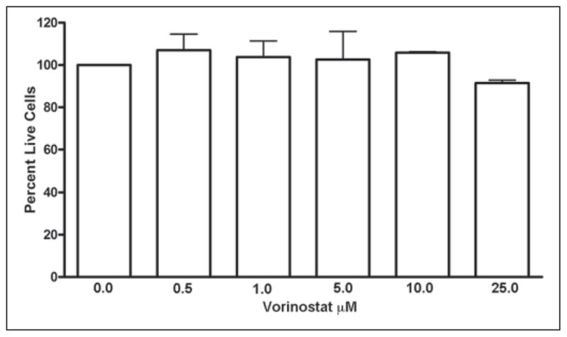
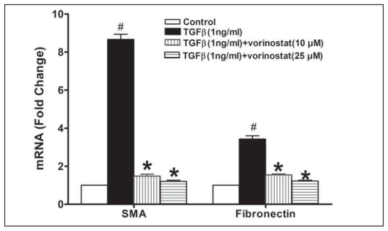
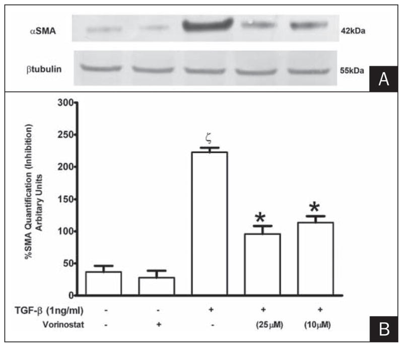
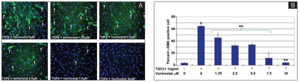
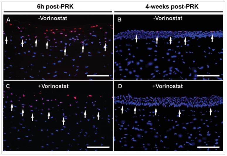
Methods: Corneal haze in rabbits was produced with -9.00 diopters (D) PRK. Fibrosis in cultured human and rabbit corneal fibroblasts was activated with TGFβ1. Vorinostat (25 μm) was topically applied once for 5 minutes on rabbit cornea immediately after PRK for in vivo studies. Vorinostat (0 to 25 μm) was given to human/rabbit corneal fibroblasts for 5 minutes or 48 hours for in vitro studies. Slit-lamp microscopy, TUNEL assay, and trypan blue were used to determined vorinostat toxicity, whereas real-time polymerase chain reaction, immunocytochemistry, and immunoblotting were used to measure its efficacy.
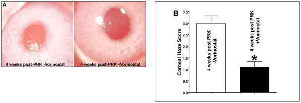
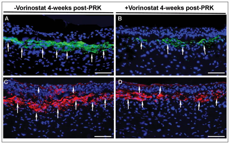
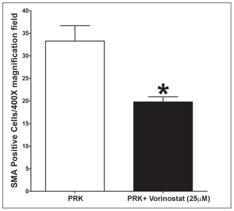
Results: Single 5-minute vorinostat (25 μm) topical application on the cornea following PRK significantly reduced corneal haze (P<.008) and fibrotic marker proteins (α-smooth muscle actin and f-actin; P<.001) without showing redness, swelling, or inflammation in rabbit eyes in vivo screened 4 weeks after PRK. Vorinostat reduced TGFβ1-induced fibrosis in human and rabbit corneas in vitro in a dose-dependent manner without altering cellular viability, phenotype, or proliferation.
Conclusions: Vorinostat is non-cytotoxic and safe for the eye and has potential to prevent laser-induced corneal haze in patients undergoing PRK for high myopia.
Copyright 2012, SLACK Incorporated.
Conflict of interest statement
The authors have no financial or proprietary interests in the materials presented herein.
Figures
Similar articles
-
Trichostatin a inhibits corneal haze in vitro and in vivo.Invest Ophthalmol Vis Sci. 2009 Jun;50(6):2695-701. doi: 10.1167/iovs.08-2919. Epub 2009 Jan 24. Invest Ophthalmol Vis Sci. 2009. PMID: 19168895 Free PMC article.
-
Efficacy and Safety Comparison Between Suberoylanilide Hydroxamic Acid and Mitomycin C in Reducing the Risk of Corneal Haze After PRK Treatment In Vivo.J Refract Surg. 2017 Dec 1;33(12):834-839. doi: 10.3928/1081597X-20170921-02. J Refract Surg. 2017. PMID: 29227512 Free PMC article.
-
Latrunculin B and substratum stiffness regulate corneal fibroblast to myofibroblast transformation.Exp Eye Res. 2018 May;170:101-107. doi: 10.1016/j.exer.2018.02.003. Epub 2018 Feb 6. Exp Eye Res. 2018. PMID: 29421383 Free PMC article.
-
Topical Losartan in the Management of Corneal Scarring Fibrosis: Update on Dosage, Efficacy, and Potential Epithelial Toxicity.J Ocul Pharmacol Ther. 2025 Jun;41(5):232-236. doi: 10.1089/jop.2024.0200. Epub 2025 Apr 16. J Ocul Pharmacol Ther. 2025. PMID: 40238717 Review.
-
Insights from animal studies exploring the efficacy and safety of topical losartan, in prophylaxis and treatment of corneal scarring fibrosis.Biomed Pharmacother. 2025 Feb;183:117857. doi: 10.1016/j.biopha.2025.117857. Epub 2025 Jan 17. Biomed Pharmacother. 2025. PMID: 39826357 Review.
Cited by
-
Histone Deacetylases Inhibitors in the Treatment of Retinal Degenerative Diseases: Overview and Perspectives.J Ophthalmol. 2015;2015:250812. doi: 10.1155/2015/250812. Epub 2015 Jun 2. J Ophthalmol. 2015. PMID: 26137316 Free PMC article. Review.
-
Attenuation of choroidal neovascularization by histone deacetylase inhibitor.PLoS One. 2015 Mar 25;10(3):e0120587. doi: 10.1371/journal.pone.0120587. eCollection 2015. PLoS One. 2015. PMID: 25807249 Free PMC article.
-
Collagen matrix perturbations in corneal stroma of Ossabaw mini pigs with type 2 diabetes.Mol Vis. 2021 Dec 7;27:666-678. eCollection 2021. Mol Vis. 2021. PMID: 35002212 Free PMC article.
-
Epigenetic regulation of anterior segment diseases and potential therapeutics.Ocul Surf. 2020 Jul;18(3):383-395. doi: 10.1016/j.jtos.2020.04.001. Epub 2020 Apr 25. Ocul Surf. 2020. PMID: 32344150 Free PMC article. Review.
-
Evaluation of CRISPR/Cas9 mediated TGIF gene editing to inhibit corneal fibrosis in vitro.Exp Eye Res. 2022 Jul;220:109113. doi: 10.1016/j.exer.2022.109113. Epub 2022 May 16. Exp Eye Res. 2022. PMID: 35588782 Free PMC article.
References
-
- Taneri S, Weisberg M, Azar DT. Surface ablation techniques. J Cataract Refract Surg. 2011;37(2):392–408. - PubMed
-
- Reynolds A, Moore JE, Naroo SA, Moore CB, Shah S. Excimer laser surface ablation - a review. Clin Experiment Ophthalmol. 2010;38(2):168–182. - PubMed
-
- Wilson SE, Mohan RR, Mohan RR, Ambrósio R, Jr, Hong J, Lee J. The corneal wound healing response: cytokine-mediated interaction of the epithelium, stroma, and inflammatory cells. Prog Retin Eye Res. 2001;20(5):625–637. - PubMed
Publication types
MeSH terms
Substances
Grants and funding
LinkOut - more resources
Full Text Sources
Other Literature Sources
Medical
