. 2012 May;143(2):188-99.
doi: 10.1016/j.clim.2012.01.019. Epub 2012 Feb 10.

Different signaling mechanisms regulating IL-6 expression by LPS between gingival fibroblasts and mononuclear cells: seeking the common target

Affiliations

Different signaling mechanisms regulating IL-6 expression by LPS between gingival fibroblasts and mononuclear cells: seeking the common target

Junfei Jin et al. Clin Immunol. 2012 May.

Abstract

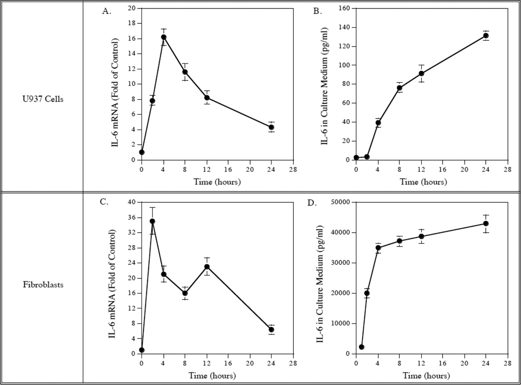
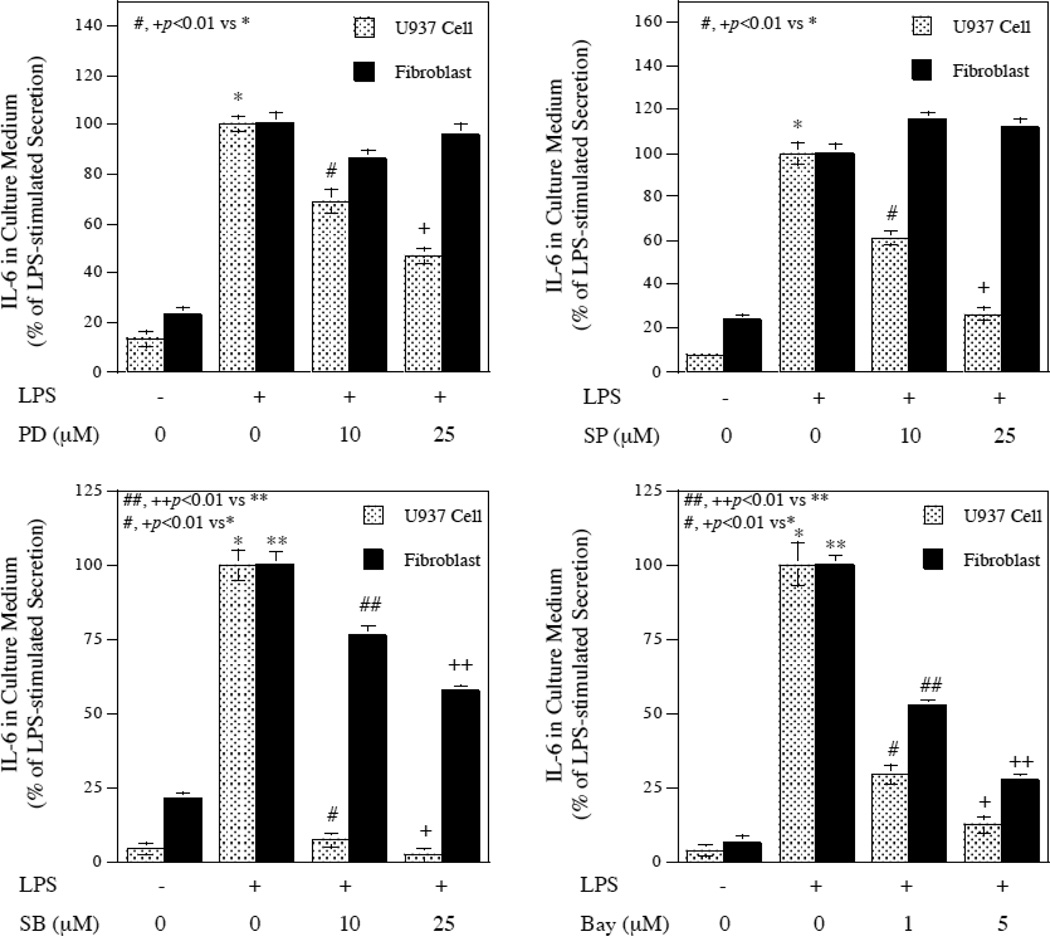
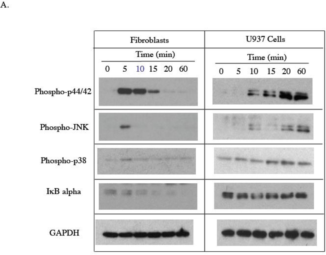
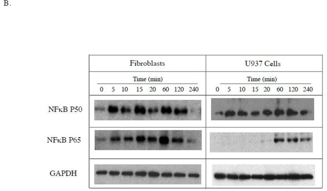
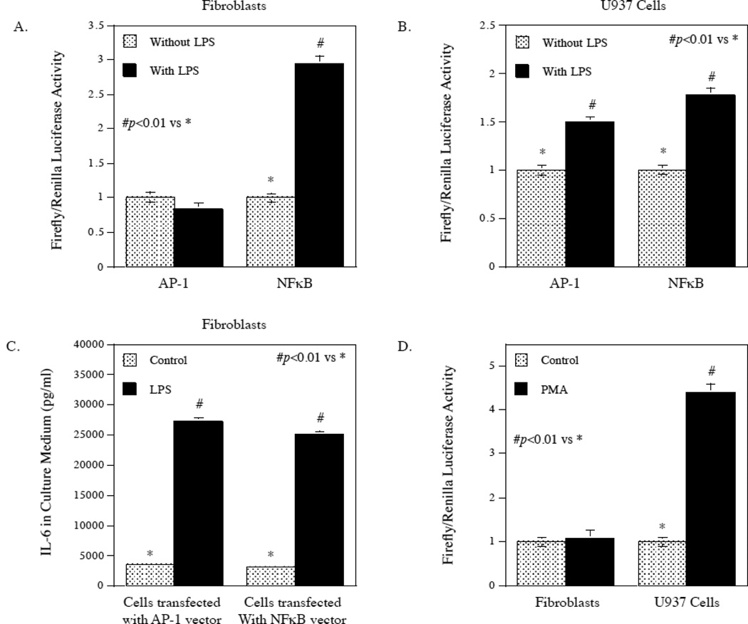
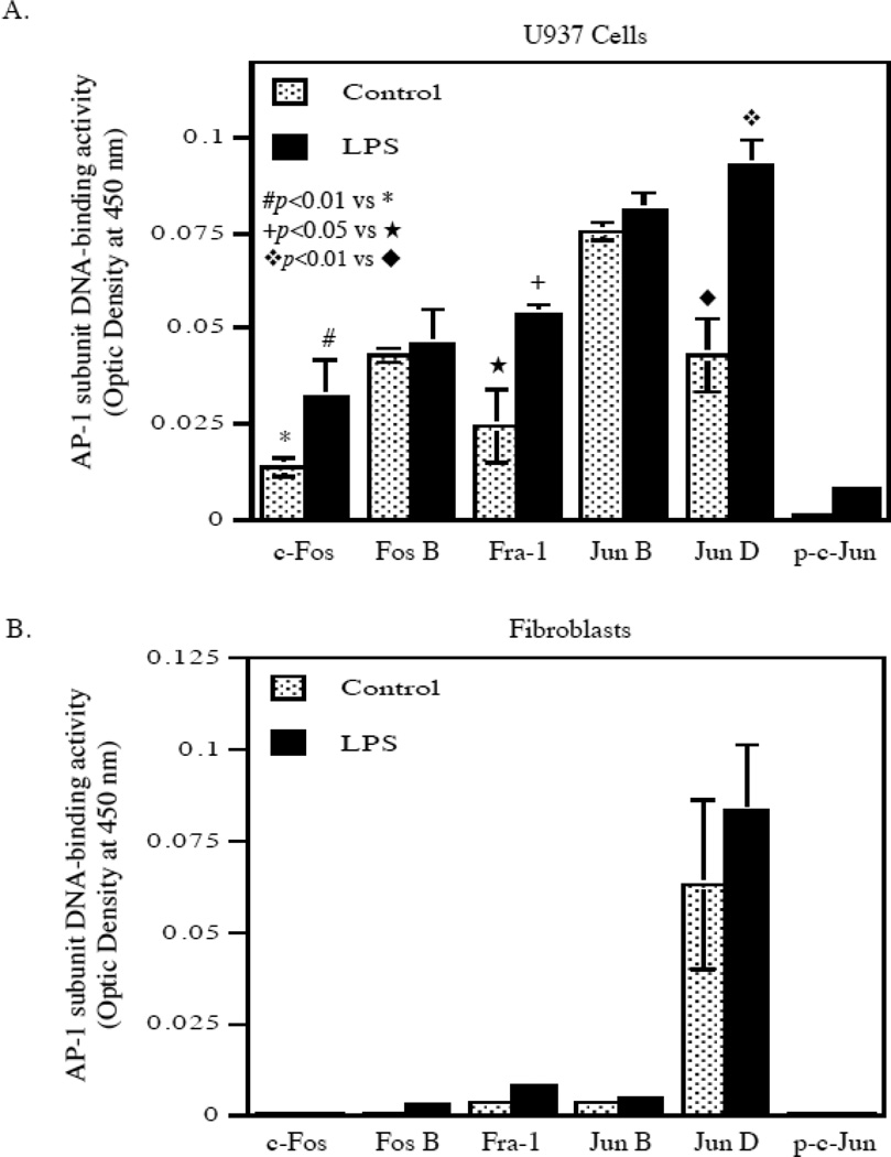
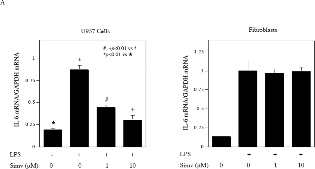
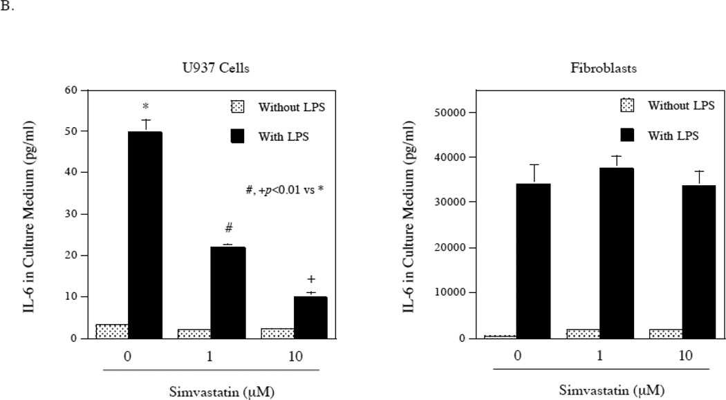
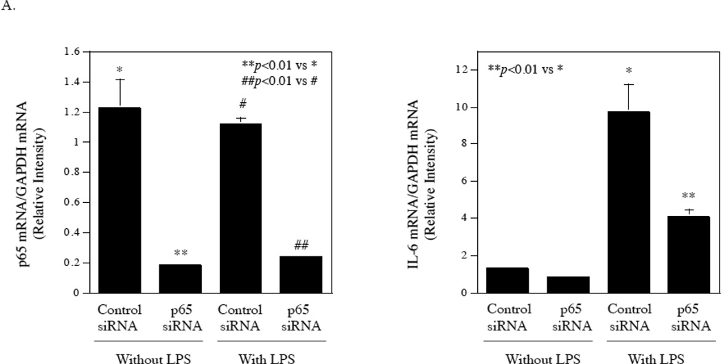
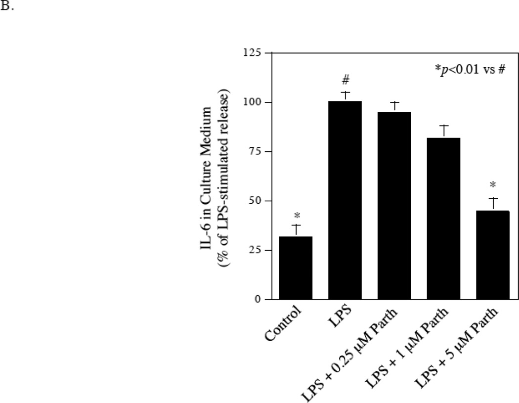
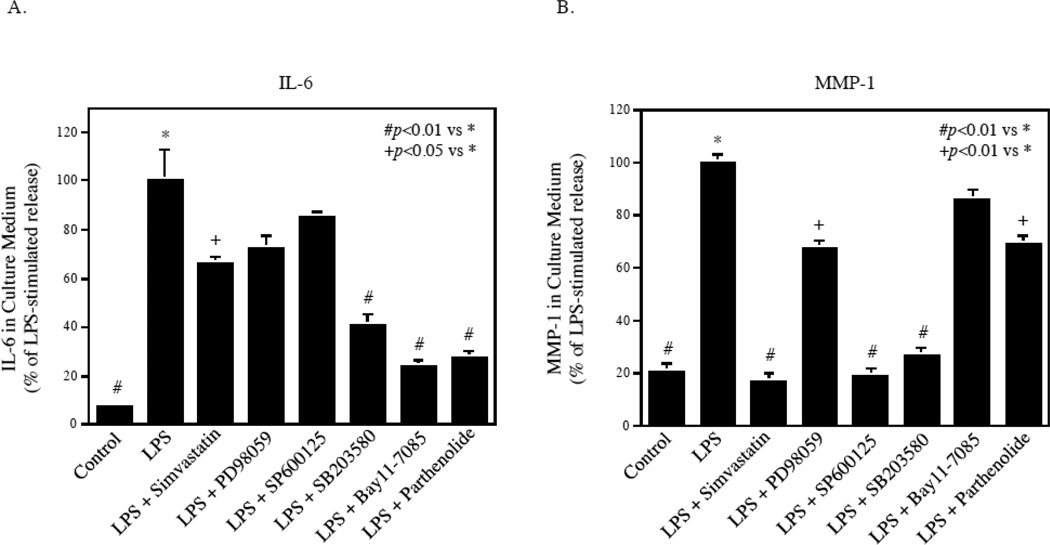
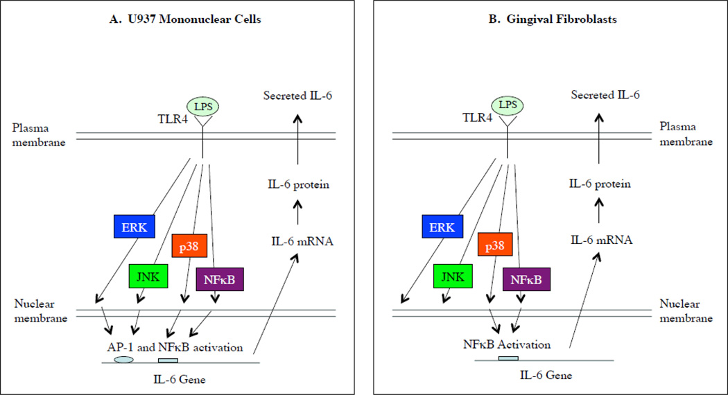
To reduce connective tissue IL-6 level stimulated by LPS, it is essential to control IL-6 expression in both mononuclear cells and fibroblasts. However, it is unclear whether the regulatory mechanisms for both cells are similar or not. In this study, we found that signaling pathways mediating LPS-stimulated IL-6 in mononuclear U937 cells and fibroblasts were different. Furthermore, our studies showed that while LPS activated AP-1 and NFκB in U937 cells, it only activated NFκB in fibroblasts. Analysis of nuclear AP-1 subunits showed that LPS stimulated c-Fos, Fra-1 and Jun D activities in U937 cells, but not fibroblasts. The lack of ERK involvement in LPS-stimulated IL-6 in fibroblasts was further supported by the observations that simvastatin, which is known to target ERK-AP-1, failed to inhibit LPS-stimulated IL-6 by fibroblasts. Finally, we showed that targeting NFκB pathway was highly effective in inhibition of LPS-stimulated IL-6 in coculture of U937 cells and fibroblasts.


Figures

Figure 1
Figure 1
Kinetics of IL-6 mRNA expression and protein secretion in response to LPS by U937 cells and gingival fibroblasts. U937 cells (A and B) or gingival fibroblasts (C and D) were treated with 100 ng/ml of LPS for different times as indicated and culture medium was collected and RNA was isolated at each time point for quantification of IL-6 mRNA using real-time PCR (A and C) and IL-6 protein using ELISA (B and D). The data presented are from one of three independent experiments with similar results.
Figure 2
Figure 2
Different effects of pharmacological inhibitors on LPS-stimulated IL-6 secretion between U937 cells and gingival fibroblasts. U937 cells or gingival fibroblasts were treated with or without 100 ng/ml of LPS in the absence or presence of different concentrations of PD98059, SP600125, SB203580, or Bay117085 as indicated for 24 h. After the treatment, IL-6 in culture medium was quantified using ELISA. The data presented are from one of two independent experiments with similar results.
Figure 3
Figure 3
Stimulation of signaling pathways by LPS in gingival fibroblasts and U937 cells. A. Gingival fibroblasts or U937 cells were treated with 100 ng/ml of LPS for different times as indicated. At each time point, cellular phosphorylated p44/p42 (phospho-p44/42), JNK (phosphor-JNK), p38 (phosphor-p38) as well as IκB alpha and GAPDH were detected using immunoblotting as described in Experimental Procedure. B. Gingival fibroblasts or U937 cells were treated with 100 ng/ml of LPS for different times as indicated. At each time point, nuclear NFκB p50 and NFκB p65, and cellular GAPDH were detected using immunoblotting as described in Experimental Procedure. The graphs presented are from one of two independent experiments with similar results.
Figure 3
Figure 3
Stimulation of signaling pathways by LPS in gingival fibroblasts and U937 cells. A. Gingival fibroblasts or U937 cells were treated with 100 ng/ml of LPS for different times as indicated. At each time point, cellular phosphorylated p44/p42 (phospho-p44/42), JNK (phosphor-JNK), p38 (phosphor-p38) as well as IκB alpha and GAPDH were detected using immunoblotting as described in Experimental Procedure. B. Gingival fibroblasts or U937 cells were treated with 100 ng/ml of LPS for different times as indicated. At each time point, nuclear NFκB p50 and NFκB p65, and cellular GAPDH were detected using immunoblotting as described in Experimental Procedure. The graphs presented are from one of two independent experiments with similar results.
Figure 4
Figure 4
Different effects of LPS on AP-1 transcriptional activation in gingival fibroblasts and U937 cells. A and B. Gingival fibroblasts (A) or U937 cells (B) were transfected with the AP-1 or NFκB-responsive luciferase constructs encoding the Firefly luciferase reporter gene for 24 h. After the transfection, cells were treated with or without 100 ng/ml of LPS for 24 h and Firefly luciferase activity was then determined and normalized to Renilla luciferase activity. C. The stimulation of IL-6 secretion by LPS from gingival fibroblasts transfected with AP-1 or NFκB-responsive luciferase constructs. The culture medium from the above experiment (A) was collected for IL-6 quantification using ELISA. D. The effect of PMA on AP-1 transcriptional activity in gingival fibroblasts and U937 cells. Gingival fibroblasts or U937 cells were transfected with AP-1-responsive luciferase constructs for 24 h and then treated with or without 10 ng/ml of PMA for 24 h. Firefly luciferase activity was determined after the treatment and normalized to Renilla luciferase activity. The data presented are representative of 3 independent experiments with similar results.
Figure 5
Figure 5
The effect of LPS on nuclear activities of AP-1 subunits in U937 cells and gingival fibroblasts. U937 cells (A) or gingival fibroblasts (B) were treated with or without 100 ng/ml of LPS for 2 h. After the treatment, the nuclear proteins were isolated from cells and subjected to a transcription factor activity assay system in which AP-1-binding element-containing oligonucleotides were immobilized on the surface of wells. After incubating the wells with nuclear proteins, the AP-1 subunits bound to the oligonucleotides were identified by specific antibodies. The bound levels of c-Fos, Fos B, Fra-1, Jun B, Jun D and phosphorylated c-Jun (p-c-Jun) were determined. The data presented are representative of two experiments with similar results.
Figure 6
Figure 6
Lack of inhibition by simvastatin on LPS-stimulated IL-6 secretion by gingival fibroblasts. A. The effect of simvastatin (Simv) on LPS-stimulated IL-6 mRNA expression by U937 cells and gingival fibroblasts. U937 cells and gingival fibroblasts were treated with 100 ng/ml of LPS in the absence or presence of 1 or 10 µM of simvastatin for 4 h for U937 cells and 2 h for fibroblasts and IL-6 mRNA was quantified using real-time PCR. B. The effect of simvastatin on LPS-stimulated IL-6 secretion by U937 cells and gingival fibroblasts. U937 cells or gingival fibroblasts were treated with or without 100 ng/ml of LPS in the absence or presence of 1 or 10 µM of simvastatin for 24 h. After the treatment, IL-6 in culture medium was quantified using ELISA. The data (mean ± SD) presented are representative of three independent experiments with similar results.
Figure 6
Figure 6
Lack of inhibition by simvastatin on LPS-stimulated IL-6 secretion by gingival fibroblasts. A. The effect of simvastatin (Simv) on LPS-stimulated IL-6 mRNA expression by U937 cells and gingival fibroblasts. U937 cells and gingival fibroblasts were treated with 100 ng/ml of LPS in the absence or presence of 1 or 10 µM of simvastatin for 4 h for U937 cells and 2 h for fibroblasts and IL-6 mRNA was quantified using real-time PCR. B. The effect of simvastatin on LPS-stimulated IL-6 secretion by U937 cells and gingival fibroblasts. U937 cells or gingival fibroblasts were treated with or without 100 ng/ml of LPS in the absence or presence of 1 or 10 µM of simvastatin for 24 h. After the treatment, IL-6 in culture medium was quantified using ELISA. The data (mean ± SD) presented are representative of three independent experiments with similar results.
Figure 7
Figure 7
Inhibition of LPS-stimulated IL-6 secretion from fibroblasts by knocking down NFκB p65 or pharmacological inhibitor. A. Gingival fibroblasts were transfected with p65 siRNA or control siRNA for 48 h and then treated with or without 100 ng/ml of LPS for 24 h. After the treatment, p65 and IL-6 mRNA were quantified using real-time PCR and normalized to GAPDH mRNA. B. Inhibition of LPS-stimulated IL-6 secretion from gingival fibroblasts by NFκB inhibitor parthenolide. Gingival fibroblasts were treated with 100 ng/ml of LPS in the absence or presence of different concentrations of parthenolide as indicated for 24 h. After the treatment, IL-6 in culture medium was quantified using ELISA. The data (mean ± SD) presented are representative of two independent experiments with similar results.
Figure 7
Figure 7
Inhibition of LPS-stimulated IL-6 secretion from fibroblasts by knocking down NFκB p65 or pharmacological inhibitor. A. Gingival fibroblasts were transfected with p65 siRNA or control siRNA for 48 h and then treated with or without 100 ng/ml of LPS for 24 h. After the treatment, p65 and IL-6 mRNA were quantified using real-time PCR and normalized to GAPDH mRNA. B. Inhibition of LPS-stimulated IL-6 secretion from gingival fibroblasts by NFκB inhibitor parthenolide. Gingival fibroblasts were treated with 100 ng/ml of LPS in the absence or presence of different concentrations of parthenolide as indicated for 24 h. After the treatment, IL-6 in culture medium was quantified using ELISA. The data (mean ± SD) presented are representative of two independent experiments with similar results.
Figure 8
Figure 8
The effect of simvastatin and different pharmacological inhibitors on LPS-stimulated IL-6 secretion by coculture of gingival fibroblasts and U937 cells. Gingival fibroblasts and U937 cells were co-cultured in the Transwell system and treated with or without 100 ng/ml of LPS in the absence or presence of 10 µM of simvastatin, 10 µM of PD98059, 10 µM of SP600125, 10 µM of SB203580, 5 µM of Bay117085 or 5 µM of parthenolide for 24 h. After the treatment, IL-6 (A) and MMP-1 (B) in culture medium were quantified using ELISA.
Figure 9
Figure 9
Illustration of different signaling mechanisms regulating IL-6 expression by LPS in U937 mononuclear cells and gingival fibroblasts. A. In U937 cells, LPS stimulates IL-6 expression via ERK, JNK, p38 and NFκB pathways and subsequent AP-1 and NFκB activation. B. In gingival fibroblasts, although LPS stimulates ERK, JNK, p38, NFκB pathways, it only activates NFκB. Only p38 and NFκB pathways are involved in LPS-stimulated IL-6 expression in gingival fibroblasts.

Similar articles

Cited by

References

    1. Iacopino AM. Periodontitis and diabetes interrelationships: role of inflammation. Annals of periodontology / the American Academy of Periodontology. 2001;6:125–137. - PubMed
    1. Nichols TC, Fischer TH, Deliargyris EN, Baldwin AS., Jr Role of nuclear factor-kappa B (NF-kappa B) in inflammation, periodontitis, and atherogenesis. Annals of periodontology / the American Academy of Periodontology. 2001;6:20–29. - PubMed
    1. Ridker PM, Silvertown JD. Inflammation, C-reactive protein, and atherothrombosis. Journal of periodontology. 2008;79:1544–1551. - PubMed
    1. Page RC. The role of inflammatory mediators in the pathogenesis of periodontal disease. Journal of periodontal research. 1991;26:230–242. - PubMed
    1. Ishihara K, Hirano T. IL-6 in autoimmune disease and chronic inflammatory proliferative disease. Cytokine Growth Factor Rev. 2002;13:357–368. - PubMed

Publication types

MeSH terms