

Attentional control: temporal relationships within the fronto-parietal network
- PMID: 22386880
- PMCID: PMC3348362
- DOI: 10.1016/j.neuropsychologia.2012.02.009
Attentional control: temporal relationships within the fronto-parietal network
Abstract
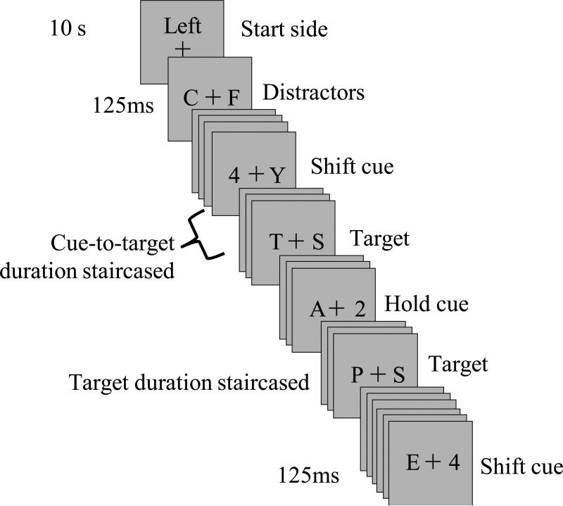
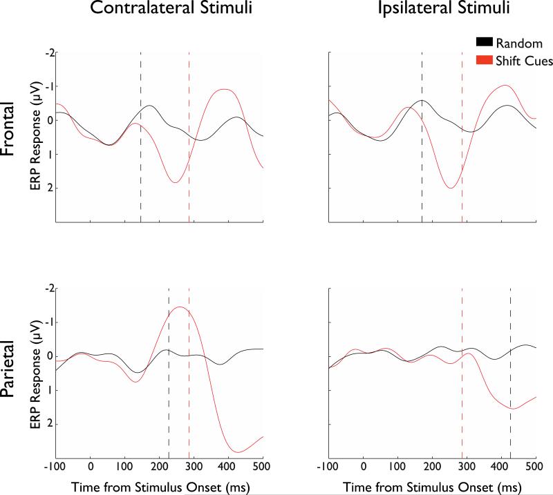
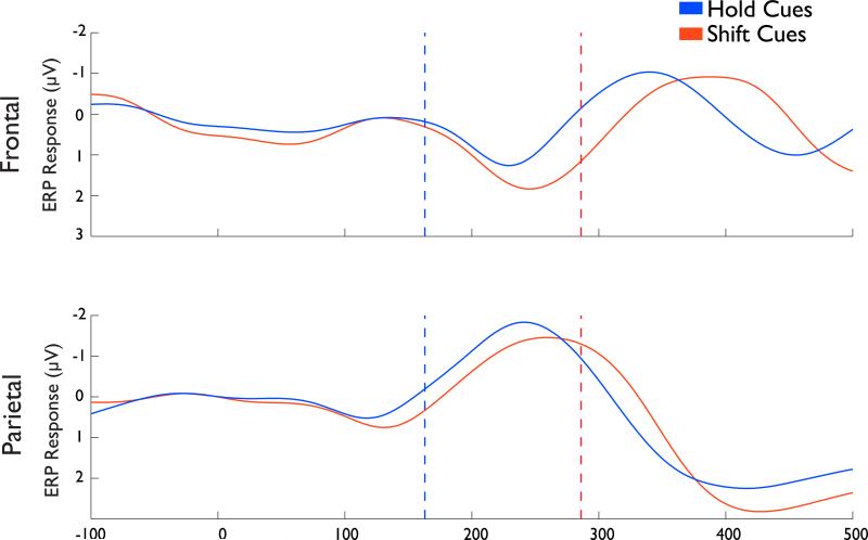
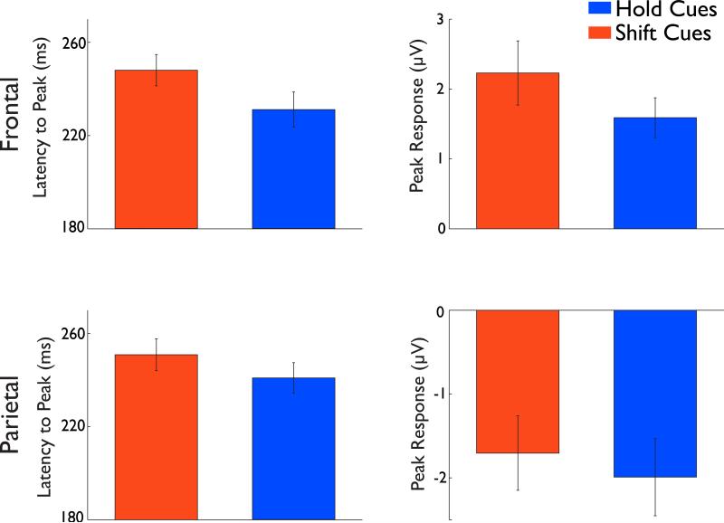
Selective attention to particular aspects of incoming sensory information is enabled by a network of neural areas that includes frontal cortex, posterior parietal cortex, and, in the visual domain, visual sensory regions. Although progress has been made in understanding the relative contribution of these different regions to the process of visual attentional selection, primarily through studies using neuroimaging, rather little is known about the temporal relationships between these disparate regions. To examine this, participants viewed two rapid serial visual presentation (RSVP) streams of letters positioned to the left and right of fixation point. Before each run, attention was directed to either the left or the right stream. Occasionally, a digit appeared within the attended stream indicating whether attention was to be maintained within the same stream ('hold' condition) or to be shifted to the previously ignored stream ('shift' condition). By titrating the temporal parameters of the time taken to shift attention for each participant using a fine-grained psychophysics paradigm, we measured event-related potentials time-locked to the initiation of spatial shifts of attention. The results revealed that shifts of attention were evident earlier in the response recorded over frontal than over parietal electrodes and, importantly, that the early activity over frontal electrodes was associated with a successful shift of attention. We conclude that frontal areas are engaged early for the purpose of executing an attentional shift, likely triggering a cascade through the fronto-parietal network ultimately, resulting in the attentional modulation of sensory events in posterior cortices.
Copyright © 2012 Elsevier Ltd. All rights reserved.
Figures
Similar articles
-
A matter of hand: Causal links between hand dominance, structural organization of fronto-parietal attention networks, and variability in behavioural responses to transcranial magnetic stimulation.Cortex. 2017 Jan;86:230-246. doi: 10.1016/j.cortex.2016.06.015. Epub 2016 Jun 25. Cortex. 2017. PMID: 27405259
-
Dynamic changes in phase-amplitude coupling facilitate spatial attention control in fronto-parietal cortex.PLoS Biol. 2014 Aug 26;12(8):e1001936. doi: 10.1371/journal.pbio.1001936. eCollection 2014 Aug. PLoS Biol. 2014. PMID: 25157678 Free PMC article.
-
Isolating event-related potential components associated with voluntary control of visuo-spatial attention.Brain Res. 2008 Aug 28;1227:96-109. doi: 10.1016/j.brainres.2008.06.034. Epub 2008 Jun 20. Brain Res. 2008. PMID: 18621037 Clinical Trial.
-
The timing of neural activity during shifts of spatial attention.J Cogn Neurosci. 2009 Dec;21(12):2369-83. doi: 10.1162/jocn.2008.21176. J Cogn Neurosci. 2009. PMID: 19199414
-
The hybrid model of attentional control: New insights into hemispheric asymmetries inferred from TMS research.Neuropsychologia. 2015 Jul;74:21-9. doi: 10.1016/j.neuropsychologia.2014.11.023. Epub 2014 Nov 20. Neuropsychologia. 2015. PMID: 25451041 Review.
Cited by
-
Task Performance Changes the Amplitude and Timing of the BOLD Signal.Transl Neurosci. 2017 Dec 19;8:182-190. doi: 10.1515/tnsci-2017-0025. eCollection 2017. Transl Neurosci. 2017. PMID: 29318035 Free PMC article.
-
Impaired fear recognition and attentional set-shifting is associated with brain structural changes in alcoholic patients.Addict Biol. 2014 Nov;19(6):1041-54. doi: 10.1111/adb.12175. Epub 2014 Aug 6. Addict Biol. 2014. PMID: 25123156 Free PMC article.
-
Broad domain generality in focal regions of frontal and parietal cortex.Proc Natl Acad Sci U S A. 2013 Oct 8;110(41):16616-21. doi: 10.1073/pnas.1315235110. Epub 2013 Sep 23. Proc Natl Acad Sci U S A. 2013. PMID: 24062451 Free PMC article.
-
Transient Distraction and Attentional Control during a Sustained Selective Attention Task.J Cogn Neurosci. 2016 Jul;28(7):935-47. doi: 10.1162/jocn_a_00949. Epub 2016 Mar 11. J Cogn Neurosci. 2016. PMID: 26967946 Free PMC article.
-
Functional dissociation of the inferior frontal junction from the dorsal attention network in top-down attentional control.J Neurophysiol. 2018 Nov 1;120(5):2498-2512. doi: 10.1152/jn.00506.2018. Epub 2018 Aug 29. J Neurophysiol. 2018. PMID: 30156458 Free PMC article.
References
-
- Bartolomeo P, Thiebaut de Schotten M, Doricchi F. Left unilateral neglect as a disconnection syndrome. Cerebral cortex. 2007;17:2479–2490. - PubMed
-
- Bisley JW, Goldberg ME. Neuronal activity in the lateral intraparietal area and spatial attention. Science. 2003;299:81–86. - PubMed
-
- Brignani D, Lepsien J, Rushworth MF, Nobre AC. The timing of neural activity during shifts of spatial attention. Journal of cognitive neuroscience. 2009;21:2369–2383. - PubMed
Publication types
MeSH terms
Grants and funding
LinkOut - more resources
Full Text Sources
