

An MRM-based workflow for quantifying cardiac mitochondrial protein phosphorylation in murine and human tissue
- PMID: 22387130
- PMCID: PMC3405178
- DOI: 10.1016/j.jprot.2012.02.014
An MRM-based workflow for quantifying cardiac mitochondrial protein phosphorylation in murine and human tissue
Abstract
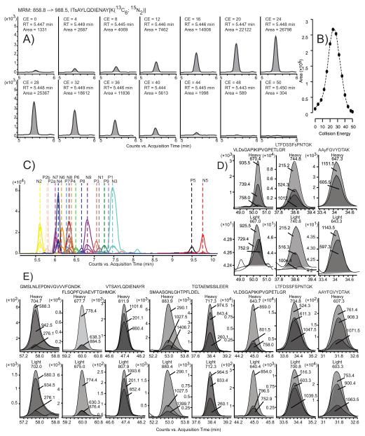
The regulation of mitochondrial function is essential for cardiomyocyte adaptation to cellular stress. While it has long been understood that phosphorylation regulates flux through metabolic pathways, novel phosphorylation sites are continually being discovered in all functionally distinct areas of the mitochondrial proteome. Extracting biologically meaningful information from these phosphorylation sites requires an adaptable, sensitive, specific and robust method for their quantification. Here we report a multiple reaction monitoring-based mass spectrometric workflow for quantifying site-specific phosphorylation of mitochondrial proteins. Specifically, chromatographic and mass spectrometric conditions for 68 transitions derived from 23 murine and human phosphopeptides, and their corresponding unmodified peptides, were optimized. These methods enabled the quantification of endogenous phosphopeptides from the outer mitochondrial membrane protein VDAC, and the inner membrane proteins ANT and ETC complexes I, III and V. The development of this quantitative workflow is a pivotal step for advancing our knowledge and understanding of the regulatory effects of mitochondrial protein phosphorylation in cardiac physiology and pathophysiology. This article is part of a Special Issue entitled: Translational Proteomics.
Copyright © 2012 Elsevier B.V. All rights reserved.
Figures

Similar articles
-
Site-specific quantitative analysis of cardiac mitochondrial protein phosphorylation.J Proteomics. 2013 Apr 9;81:15-23. doi: 10.1016/j.jprot.2012.09.015. Epub 2012 Sep 25. J Proteomics. 2013. PMID: 23022582 Free PMC article.
-
Phosphoproteomics of the developing heart identifies PERM1 - An outer mitochondrial membrane protein.J Mol Cell Cardiol. 2021 May;154:41-59. doi: 10.1016/j.yjmcc.2021.01.010. Epub 2021 Feb 5. J Mol Cell Cardiol. 2021. PMID: 33549681
-
Phosphoproteome analysis reveals regulatory sites in major pathways of cardiac mitochondria.Mol Cell Proteomics. 2011 Feb;10(2):M110.000117. doi: 10.1074/mcp.M110.000117. Epub 2010 May 22. Mol Cell Proteomics. 2011. PMID: 20495213 Free PMC article.
-
What can we learn about cardioprotection from the cardiac mitochondrial proteome?Cardiovasc Res. 2010 Nov 1;88(2):211-8. doi: 10.1093/cvr/cvq277. Epub 2010 Aug 30. Cardiovasc Res. 2010. PMID: 20805096 Free PMC article. Review.
-
Mitochondrial turnover in the heart.Biochim Biophys Acta. 2011 Jul;1813(7):1295-301. doi: 10.1016/j.bbamcr.2010.11.017. Epub 2010 Dec 13. Biochim Biophys Acta. 2011. PMID: 21147177 Free PMC article. Review.
Cited by
-
Proteomics of the heart.Physiol Rev. 2024 Jul 1;104(3):931-982. doi: 10.1152/physrev.00026.2023. Epub 2024 Feb 1. Physiol Rev. 2024. PMID: 38300522 Free PMC article. Review.
-
Regulation of cardiac proteasomes by ubiquitination, SUMOylation, and beyond.J Mol Cell Cardiol. 2014 Jun;71:32-42. doi: 10.1016/j.yjmcc.2013.10.008. Epub 2013 Oct 17. J Mol Cell Cardiol. 2014. PMID: 24140722 Free PMC article. Review.
-
Characterization and application of a disease-cell model for a neurodegenerative lysosomal disease.Mol Genet Metab. 2014 Feb;111(2):172-83. doi: 10.1016/j.ymgme.2013.09.011. Epub 2013 Sep 21. Mol Genet Metab. 2014. PMID: 24094551 Free PMC article.
-
Multiplexed Liquid Chromatography-Multiple Reaction Monitoring Mass Spectrometry Quantification of Cancer Signaling Proteins.Methods Mol Biol. 2017;1647:19-45. doi: 10.1007/978-1-4939-7201-2_2. Methods Mol Biol. 2017. PMID: 28808993 Free PMC article.
-
Application of targeted mass spectrometry in bottom-up proteomics for systems biology research.J Proteomics. 2018 Oct 30;189:75-90. doi: 10.1016/j.jprot.2018.02.008. Epub 2018 Feb 13. J Proteomics. 2018. PMID: 29452276 Free PMC article. Review.
References
-
- Bindoff L. Mitochondria and the heart. Eur Heart J. 2003;24:221–4. - PubMed
Publication types
MeSH terms
Substances
Grants and funding
LinkOut - more resources
Full Text Sources
Other Literature Sources
