

A locally aggressive solitary fibrous tumor of the leg: Case report and literature review
- PMID: 22387414
- PMCID: PMC3316767
- DOI: 10.1016/j.ijscr.2012.01.009
A locally aggressive solitary fibrous tumor of the leg: Case report and literature review
Abstract
Introduction: The solitary fibrous tumor (SFT) is a rare soft tissue tumor with a substantially benign clinical behavior. However, malignant neoplasms with local recurrence or distant metastases have been reported.
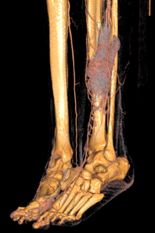
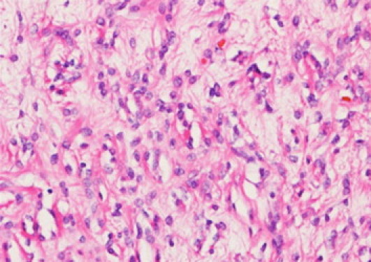
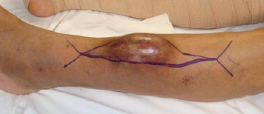
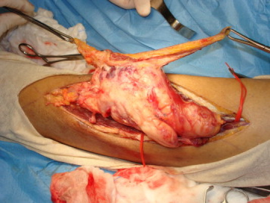
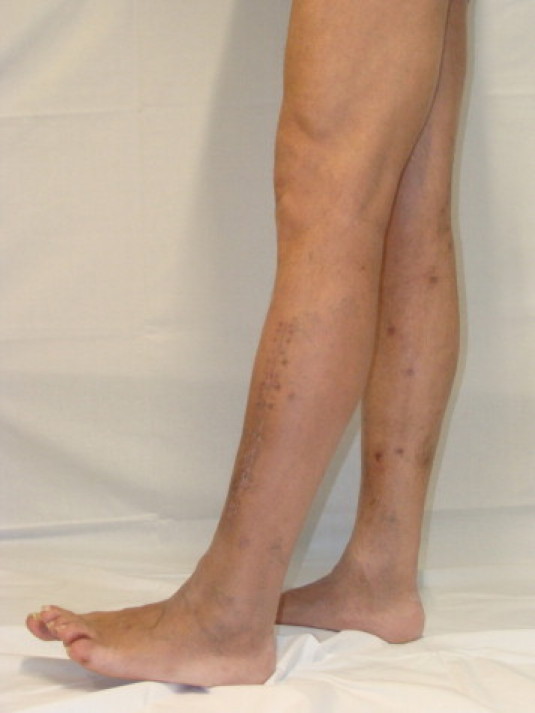
Presentation of the case: The authors present a case of an aggressive SFT of the leg, in a 55 years old Caucasian man. Radiological, histological and molecular findings are reported. The differential diagnosis, therapy and outcome of this rare tumor are also discussed.
Discussion: An extensive review of literature showed SFT's clinical behavior as substantially benign, anyway aggressive or malignant neoplasms have been described. The potential risk of local recurrence and distant metastasis thus suggests wide surgical resection and careful long-term follow-up. Differential diagnosis may be quite laborious as SFT can mimic a variety of benign and malignant mesenchymal tumors; immunohistochemical analysis for CD34, CD99, vimentin and bcl-2 is then mandatory.
Conclusion: Our clinical experience confirmed that SFT may have an aggressive behavior, however, conservative surgical treatment may be successful in the long term.
Copyright © 2012 Surgical Associates Ltd. Published by Elsevier Ltd. All rights reserved.
Figures
Similar articles
-
Solitary Fibrous Tumor of the Lower Leg: A Rare and Difficult Diagnosis.Plast Reconstr Surg Glob Open. 2015 Oct 1;3(10):e528. doi: 10.1097/GOX.0000000000000501. eCollection 2015 Oct. Plast Reconstr Surg Glob Open. 2015. PMID: 26579334 Free PMC article.
-
Clinicopathologic and Immunohistochemical Characteristics of Solitary Fibrous Tumor and Its Mimics: A Single-Center Experience.Clin Pathol. 2021 Jul 2;14:2632010X211028209. doi: 10.1177/2632010X211028209. eCollection 2021 Jan-Dec. Clin Pathol. 2021. PMID: 34278302 Free PMC article.
-
Aggressive Behavior Predictors in Solitary Fibrous Tumor: Demographic, Clinical, and Histopathologic Characteristics of 81 Cases.Ann Surg Oncol. 2021 Oct;28(11):6861-6867. doi: 10.1245/s10434-021-09592-w. Epub 2021 Jan 29. Ann Surg Oncol. 2021. PMID: 33512676
-
A review of solitary fibrous tumor/hemangiopericytoma tumor and a comparison of risk factors for recurrence, metastases, and death among patients with spinal and intracranial tumors.Neurosurg Rev. 2021 Jun;44(3):1299-1312. doi: 10.1007/s10143-020-01335-x. Epub 2020 Jun 18. Neurosurg Rev. 2021. PMID: 32556679
-
Solitary fibrous tumor of the orbit: is it rare? Report of a case series and review of the literature.Ophthalmology. 2003 Jul;110(7):1442-8. doi: 10.1016/S0161-6420(03)00459-7. Ophthalmology. 2003. PMID: 12867407 Review.
Cited by
-
Solitary Fibrous Tumor of the Lower Leg: A Rare and Difficult Diagnosis.Plast Reconstr Surg Glob Open. 2015 Oct 1;3(10):e528. doi: 10.1097/GOX.0000000000000501. eCollection 2015 Oct. Plast Reconstr Surg Glob Open. 2015. PMID: 26579334 Free PMC article.
-
Sinonasal and rhinopharyngeal solitary fibrous tumour: a case report and review of the literature.Acta Otorhinolaryngol Ital. 2015 Dec;35(6):455-8. doi: 10.14639/0392-100X-163813. Acta Otorhinolaryngol Ital. 2015. PMID: 26900253 Free PMC article. Review.
References
-
- Anders J.O., Aurich M., Lang T., Wagner A. Solitary fibrous tumor in the thigh: review of the literature. J Cancer Res Clin Oncol. 2006;132:69–75. - PubMed
-
- Abe S., Imamura T., Tateishi A., Park P., Nakano H., Harasawa A. Intramuscular solitary fibrous tumor: a clinicopathological case study. J Comp Assist Tomogr. 1999;23(3):458–462. - PubMed
-
- Gengler C., Guillou L. Solitary fibrous tumour and haemangiopericytoma: evolution of a concept. Histopathology. 2006;48(January (1)):63–74. - PubMed
-
- Wignall O.J., Moskovic E.C., Thway K., Thomas J.M. Solitary fibrous tumors of the soft tissues: review of the imaging and clinical features with histopathologic correlation. AJR Am J Roentgenol. 2010;195(July (1)):55–62. - PubMed
-
- Ramdial P.K., Madaree A. Aggressive CD34-positive fibrous scalp lesion of childhood: extrapulmonary solitary fibrous tumor. Pediatr Dev Pathol. 2001;4(May–June (3)):267–275. - PubMed
LinkOut - more resources
Full Text Sources
Research Materials
