. 2012 May;127(1):120-9.
doi: 10.1093/toxsci/kfs099. Epub 2012 Mar 2.

Poly(ADP-ribose) polymerase-1 inhibition by arsenite promotes the survival of cells with unrepaired DNA lesions induced by UV exposure

Affiliations

Poly(ADP-ribose) polymerase-1 inhibition by arsenite promotes the survival of cells with unrepaired DNA lesions induced by UV exposure

Xu-Jun Qin et al. Toxicol Sci. 2012 May.

Abstract

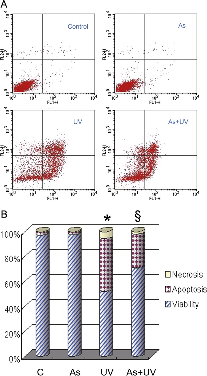
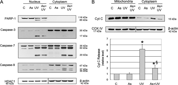
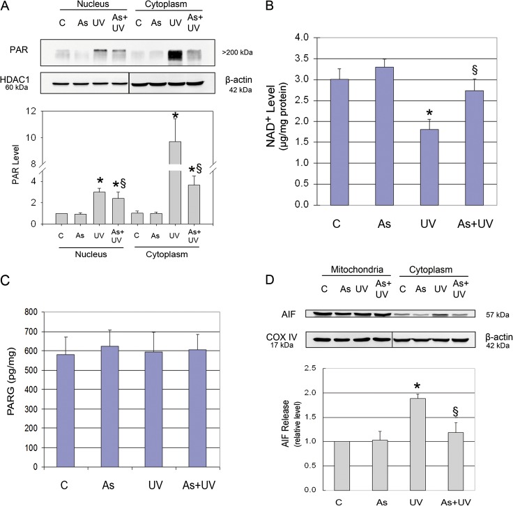
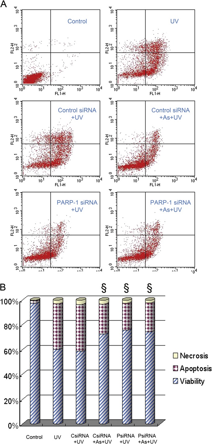
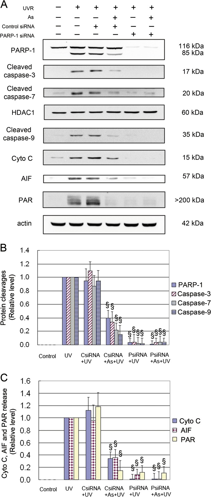
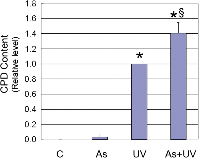
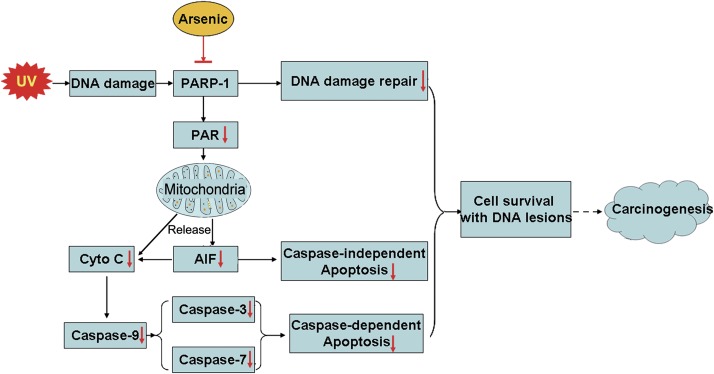
Human arsenic exposure is associated with increased risk of skin cancer, and arsenite greatly enhances ultraviolet (UV)-induced skin tumors in a mouse model of carcinogenesis. Inhibition of DNA repair is one proposed mechanism for the observed cocarcinogenicity. We have previously demonstrated that low concentrations of arsenite inhibit poly(ADP-ribose) polymerase (PARP)-1, thus interfering with DNA repair process triggered by UV radiation. Because overactivation of PARP-1 often leads to apoptotic cell death, and unrepaired DNA lesions promote genomic instability and carcinogenesis, we hypothesized that inhibition of PARP-1 by arsenic may promote the survival of potentially "initiated carcinogenic cells," i.e., cells with unrepaired DNA lesions. In the present study, we tested this hypothesis on UV-challenged HaCat cells. Cells were pretreated with 2μM arsenite for 24 h before UV exposure. Outcome parameters included apoptotic death rate, PARP-1 activation, apoptotic molecules, and retention of DNA lesions. UV exposure induced PARP-1 activation and associated poly(ADP-ribose) production, apoptosis-inducing factor release, cytochrome C release, and caspases activation, which led to apoptotic death in HaCat cells. Pretreatment with 2μM arsenite significantly inhibited UV-induced cell death as well as the associated molecular events. Notably, knockdown of PARP-1 with small interfering RNA completely abolished the antagonism of arsenite. Furthermore, arsenite pretreatment led to long-term retention of UV-induced cyclobutane pyrimidine dimers. Together, these results suggest that low concentration of arsenite reduces UV-induced apoptosis via inhibiting PARP-1, thus promoting the survival of cells with unrepaired DNA lesions, which may be an important mechanism underlying arsenic cocarcinogenic action.


Figures

FIG. 1.
FIG. 1.
Effect of arsenite on UV-induced apoptosis. HaCat cells were incubated with 2μM arsenite for 24 h before UV exposure. Twelve hours after UV radiation, cell apoptosis was measured by flow cytometry using the TACS Annexin V kit (A). Percentages of cell apoptosis, necrosis, and viability were calculated (B). *p < 0.05 versus control and §p < 0.05 versus UV.
FIG. 2.
FIG. 2.
Effect of arsenite on UV-induced cleavages of PARP-1, caspase-3, -7, and -9 and release of Cyto C. HaCat cells were incubated with 2μM arsenite for 24 h before UV exposure. Twelve hours after UV radiation, nuclear and cytoplasmic cell extracts were prepared. Protein levels of cleaved caspase-3, -7, and -9 were determined by Western blot (A). Six hours after UV radiation, mitochondrial and cytoplasmic extracts were prepared. The release of Cyto C from mitochondria to the cytoplasm was detected by Western blot (B). The band intensities were quantified by KODAK Molecular Imaging Software version 4.0. Data represent the mean ± SD of three independent experiments. *p < 0.05 versus control and §p < 0.05 versus UV.
FIG. 3.
FIG. 3.
Effect of arsenite on UV-induced PAR production. HaCat cells were incubated with 2μM arsenite for 24 h before UV exposure. Six hours after UV radiation, the PAR levels in nuclear and cytoplasmic cell extracts and the release of AIF from mitochondria to the cytoplasm were measured by Western blot. The band intensities were quantified by KODAK Molecular Imaging Software version 4.0 (A and D). The NAD+ level was measured using an NAD+/NADH quantification kit (B). PARG activity was measured by Trevigen's HT Colorimetric PARG Assay Kit (C). Data represent the mean ± SD of three independent experiments. *p < 0.05 versus control and §p < 0.05 versus UV.
FIG. 4.
FIG. 4.
Effect of PARP-1 knockdown on UV-induced apoptosis. HaCat cells were transfected with control or PARP-1 siRNA. Seventy-two hours after transfection, cells were incubated with 2μM arsenite for 24 h before UV exposure. Twelve hours after UV radiation, cell apoptosis was measured by flow cytometry using the TACS Annexin V kit (A). Percentages of cell apoptosis, necrosis, and viability were calculated (B). §p < 0.05 versus UV.
FIG. 5.
FIG. 5.
Effect of PARP-1 knockdown on UV-induced PARP-1, caspase cleavages, releases of Cyto C, and AIF and PAR formation. HaCat cells were transfected with control or PARP-1 siRNA. Seventy-two hours after transfection, cells were incubated with 2μM arsenite for 24 h before UV exposure. Cells were collected 6 h after UV radiation for assessment of PAR production and Cyto C and AIF release in cytosolic fraction. Cells were collected 12 h after UV radiation for detection of PARP-1 and caspase cleavages in nuclear extract by Western blot (A). The band intensities were quantified by KODAK Molecular Imaging Software version 4.0 (B and C). Data represent the mean ± SD of three independent experiments. §p < 0.05 versus UV.
FIG. 6.
FIG. 6.
Effect of arsenite on persistent UV-induced CPDs. Cells were incubated with 2μM arsenite for 24 h before UV exposure. After UV radiation, cells were cultured for 14 days before detection of CPDs by ELISA. Data represent the mean ± SD of three independent experiments. *p < 0.05 versus control and §p < 0.05 versus UV.
FIG. 7.
FIG. 7.
Scheme depicting the mode of action for the cocarcinogenicity of arsenite: promoting the survival of cells with unrepaired DNA lesion following UV radiation through inhibiting PARP-1.

Similar articles

Cited by

References

    1. Agency for Toxic Substances and Disease Registry (ATSDR) Toxicological Profile for Arsenic. Atlanta, GA: U.S. Department of Health and Human Services, Public Health Service; 2007. - PubMed
    1. Andrabi SA, Kim NS, Yu SW, Wang H, Koh DW, Sasaki M, Klaus JA, Otsuka T, Zhang Z, Koehler RC, et al. Poly(ADP-ribose) (PAR) polymer is a death signal. Proc. Natl. Acad. Sci. U.S.A. 2006;103:18308–18313. - PMC - PubMed
    1. Beyersmann D, Hartwig A. Carcinogenic metal compounds: Recent insight into molecular and cellular mechanisms. Arch. Toxicol. 2008;82:493–512. - PubMed
    1. Burns FJ, Uddin AN, Wu F, Nadas A, Rossman TG. Arsenic-induced enhancement of ultraviolet radiation carcinogenesis in mouse skin: A dose-response study. Environ. Health Perspect. 2004;112:599–603. - PMC - PubMed
    1. Chaitanya GV, Steven AJ, Babu PP. PARP-1 cleavage fragments: Signatures of cell-death proteases in neurodegeneration. Cell Commun. Signal. 2010;8:31. - PMC - PubMed

Publication types

MeSH terms