. 2012 Mar 4;9(4):379-84.
doi: 10.1038/nmeth.1904.

TULIPs: tunable, light-controlled interacting protein tags for cell biology

Affiliations

TULIPs: tunable, light-controlled interacting protein tags for cell biology

Devin Strickland et al. Nat Methods. .

Abstract

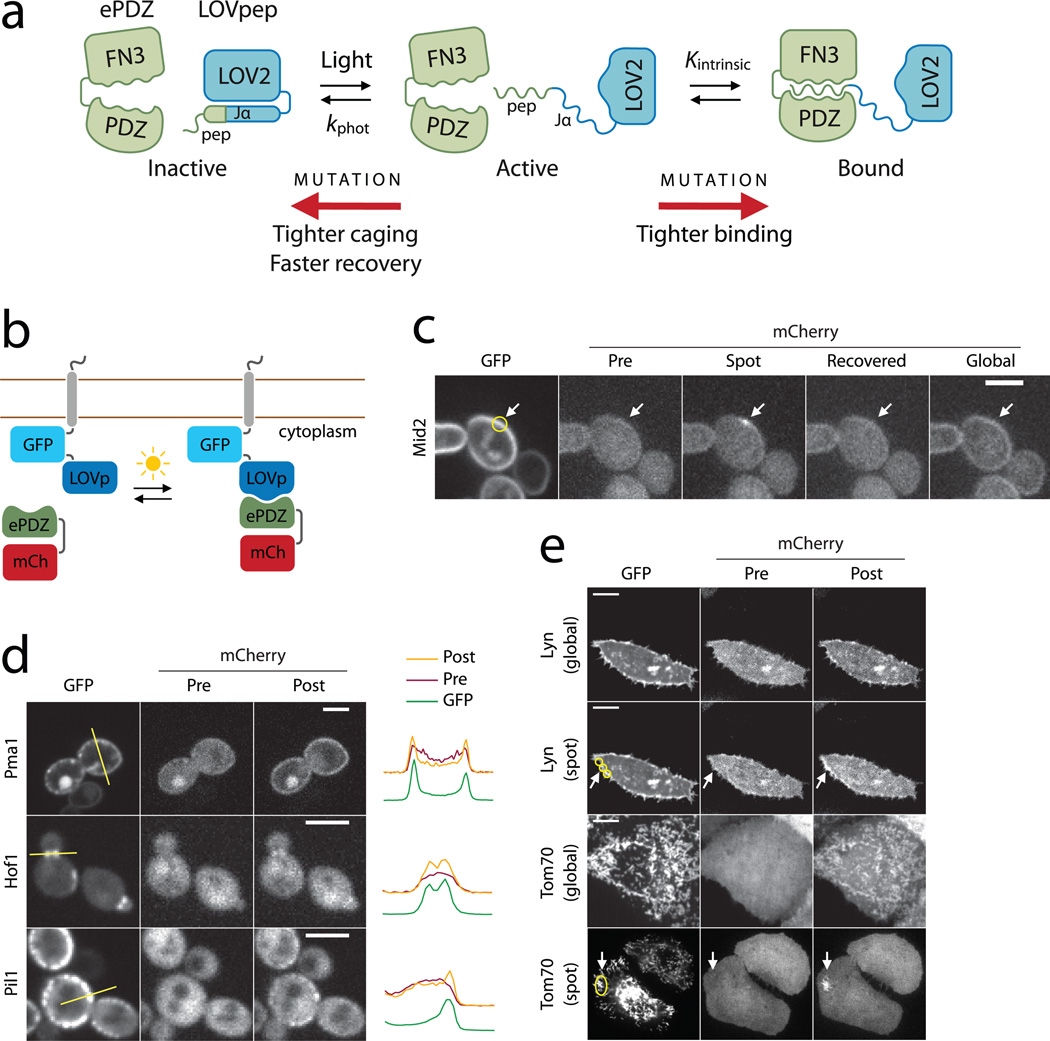
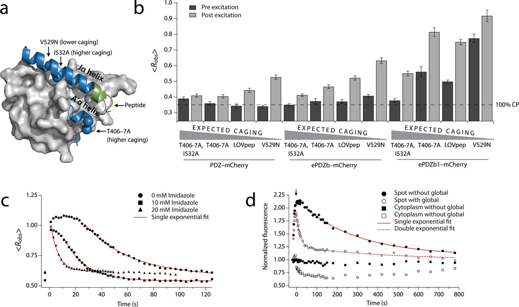
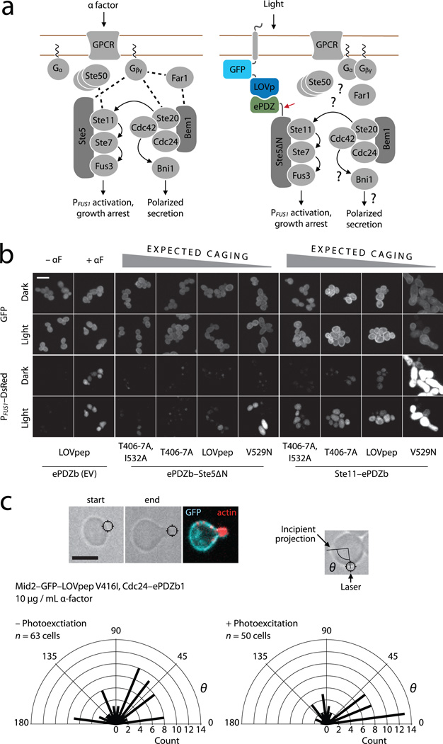
Naturally photoswitchable proteins offer a means of directly manipulating the formation of protein complexes that drive a diversity of cellular processes. We developed tunable light-inducible dimerization tags (TULIPs) based on a synthetic interaction between the LOV2 domain of Avena sativa phototropin 1 (AsLOV2) and an engineered PDZ domain (ePDZ). TULIPs can recruit proteins to diverse structures in living yeast and mammalian cells, either globally or with precise spatial control using a steerable laser. The equilibrium binding and kinetic parameters of the interaction are tunable by mutation, making TULIPs readily adaptable to signaling pathways with varying sensitivities and response times. We demonstrate the utility of TULIPs by conferring light sensitivity to functionally distinct components of the yeast mating pathway and by directing the site of cell polarization.


Figures

Figure 1
Figure 1
Design and characterization of TULIPs. (a) Schematic design of TULIPs. In the dark, a peptide epitope is caged by docking of the Jα helix to the LOV2 core (blue). Upon photoexcitation, the Jα helix undocks and exposes the peptide epitope for binding by ePDZ (green). The caging, intrinsic ePDZ–peptide affinity (Kintrinsic) and lifetime of the photoexcited state (kphot) can all be tuned by mutations. (b) Schematic of the assay used to measure ePDZ–LOVpep binding in living yeast. (c) Recruitment of ePDZb1–mCherry to the integral plasma membrane protein Mid2 in yeast using spot (arrow) and global photoexcitation. Scale bar, 5 µm. (d) Recruitment of ePDZb–mCherry to diverse subcellular markers in yeast by global photoexcitation. Scale bars, 5 µm. The plots depict pixel intensities measured along the yellow lines indicated in the GFP images. (e) Recruitment of ePDZb1–mCherry to the plasma membrane and mitochondria of HeLa cells by global and spot (arrow) photoexcitation. Scale bars, 10 µm.
Figure 2
Figure 2
Mutational and chemical control of binding. (a) AsLOV2 structure (Protein Data Bank: 2v0u) showing the location of the ePDZ epitope (green) and the caging mutations used in this study. (b) Lit- and dark-state <Robs> using LOVpep with caging mutations. Data are the means from a population (n ≥ 34) of cells; error bars show s.e.m. The dashed line represents <Robs> for ~100% cytoplasmic ePDZ–mCherry. (c) Kinetics of global recruitment and dissociation of ePDZb1–mCherry for LOVpep with wild-type dark-recovery kinetics. Imidazole is added to the media in the concentrations indicated. Data are the means from a population (n ≥ 8) of cells. Red lines are exponential fits of the dissociation phase (kobs). (d) Kinetics of spot recruitment and dissociation of ePDZb1–mCherry using slow-cycling (V416I) LOVpep. ePDZb1–mCherry is recruited to a spot as in Fig. 1c. For the filled symbols, the recruited molecules are allowed to recover without further illumination. For the open symbols, the cell is globally photoexcited at the time indicated by the arrow so as to deplete the unbound cytoplasmic pool (open squares) of ePDZb1–mCherry. Data are the means from a population (n ≥ 13) of cells. Red lines are exponential fits of the dissociation phase.
Figure 3
Figure 3
Optical control of MAPK activation and polarity establishment in yeast. (a) The wild-type mating pathway in budding yeast (left), and a scheme for light-dependent plasma membrane recruitment of Ste5∆N (right). The red arrow indicates the fusion between ePDZ and Ste5∆N. Dashed lines indicate wild-type binding interactions, many of which may be absent in Ste5∆N recruitment. (b) PFUS1 promoter activation, cell cycle arrest and polarized growth in light- and dark-grown cells. ePDZb–Ste5∆N or ePDZb–Ste11 are globally recruited to plasma membrane-tethered LOVpep variants as indicated. Scale bar, 10 µm. (c) Light-directed polarized growth. Cells are exposed to mating pheromone to induce cell cycle arrest, then stimulated with spot photoexcitation to recruit Cdc24–ePDZb1 to plasma membrane-tethered LOVpep. Radial plots show quantification of light-directed polarized growth. θ is the angle between the spot of laser photoexcitation, the center of the cell and the incipient projection. Radial bars depict the number of polarization events of angle θ in each 15° sector. “– Photoexcitation” denotes a negative control experiment in which the spot photoexcitation laser was switched off. P < 0.01 for a comparison of experimental and control distributions (two-sample Kolmogorov–Smirnov). Scale bar, 5 µm.

References

    1. Hartman NC, Groves JT. Signaling clusters in the cell membrane. Curr. Opin. Cell Biol. 2011;23:370–376. - PMC - PubMed
    1. Toettcher JE, Voigt CA, Weiner OD, Lim WA. The promise of optogenetics in cell biology: interrogating molecular circuits in space and time. Nat. Methods. 2011;8:35–38. - PMC - PubMed
    1. Wu YI, et al. A genetically encoded photoactivatable Rac controls the motility of living cells. Nature. 2009;461:104–108. - PMC - PubMed
    1. Yoo SK, et al. Differential regulation of protrusion and polarity by PI3K during neutrophil motility in live zebrafish. Dev. Cell. 2010;18:226–236. - PMC - PubMed
    1. Wang X, He L, Wu YI, Hahn KM, Montell DJ. Light-mediated activation reveals a key role for Rac in collective guidance of cell movement in vivo. Nat. Cell Biol. 2010;12:591–597. - PMC - PubMed

Publication types

MeSH terms