Mdm2 controls CREB-dependent transactivation and initiation of adipocyte differentiation
- PMID: 22388350
- PMCID: PMC3392627
- DOI: 10.1038/cdd.2012.15
Mdm2 controls CREB-dependent transactivation and initiation of adipocyte differentiation
Abstract
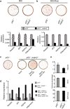
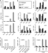
The role of the E3 ubiquitin ligase murine double minute 2 (Mdm2) in regulating the stability of the p53 tumor suppressor is well documented. By contrast, relatively little is known about p53-independent activities of Mdm2 and the role of Mdm2 in cellular differentiation. Here we report a novel role for Mdm2 in the initiation of adipocyte differentiation that is independent of its ability to regulate p53. We show that Mdm2 is required for cAMP-mediated induction of CCAAT/enhancer-binding protein δ (C/EBPδ) expression by facilitating recruitment of the cAMP regulatory element-binding protein (CREB) coactivator, CREB-regulated transcription coactivator (Crtc2)/TORC2, to the c/ebpδ promoter. Our findings reveal an unexpected role for Mdm2 in the regulation of CREB-dependent transactivation during the initiation of adipogenesis. As Mdm2 is able to promote adipogenesis in the myoblast cell line C2C12, it is conceivable that Mdm2 acts as a switch in cell fate determination.
Figures


Similar articles
-
MDM2 facilitates adipocyte differentiation through CRTC-mediated activation of STAT3.Cell Death Dis. 2016 Jun 30;7(6):e2289. doi: 10.1038/cddis.2016.188. Cell Death Dis. 2016. PMID: 27362806 Free PMC article.
-
CREB represses p53-dependent transactivation of MDM2 through the complex formation with p53 and contributes to p53-mediated apoptosis in response to glucose deprivation.Biochem Biophys Res Commun. 2011 Mar 4;406(1):79-84. doi: 10.1016/j.bbrc.2011.01.114. Epub 2011 Feb 3. Biochem Biophys Res Commun. 2011. PMID: 21295542
-
Long lasting inhibition of Mdm2-p53 interaction potentiates mesenchymal stem cell differentiation into osteoblasts.Biochim Biophys Acta Mol Cell Res. 2019 May;1866(5):737-749. doi: 10.1016/j.bbamcr.2019.01.012. Epub 2019 Jan 28. Biochim Biophys Acta Mol Cell Res. 2019. PMID: 30703414
-
Regulation of p53: a collaboration between Mdm2 and Mdmx.Oncotarget. 2012 Mar;3(3):228-35. doi: 10.18632/oncotarget.443. Oncotarget. 2012. PMID: 22410433 Free PMC article. Review.
-
A new twist in the feedback loop: stress-activated MDM2 destabilization is required for p53 activation.Cell Cycle. 2005 Mar;4(3):411-7. doi: 10.4161/cc.4.3.1522. Epub 2005 Mar 2. Cell Cycle. 2005. PMID: 15684615 Review.
Cited by
-
Adipose MDM2 regulates systemic insulin sensitivity.Sci Rep. 2021 Nov 8;11(1):21839. doi: 10.1038/s41598-021-01240-3. Sci Rep. 2021. PMID: 34750429 Free PMC article.
-
Dermal adipogenesis protects against neutrophilic skin inflammation during psoriasis pathogenesis.Cell Mol Immunol. 2025 Aug;22(8):901-917. doi: 10.1038/s41423-025-01296-5. Epub 2025 Jun 23. Cell Mol Immunol. 2025. PMID: 40550921
-
Mdm2 promotes myogenesis through the ubiquitination and degradation of CCAAT/enhancer-binding protein β.J Biol Chem. 2015 Apr 17;290(16):10200-7. doi: 10.1074/jbc.M115.638577. Epub 2015 Feb 26. J Biol Chem. 2015. PMID: 25720496 Free PMC article.
-
Involvement of tumor suppressors PTEN and p53 in the formation of multiple subtypes of liposarcoma.Cell Death Differ. 2015 Nov;22(11):1785-91. doi: 10.1038/cdd.2015.27. Epub 2015 Mar 27. Cell Death Differ. 2015. PMID: 25822339 Free PMC article.
-
MDM2 facilitates adipocyte differentiation through CRTC-mediated activation of STAT3.Cell Death Dis. 2016 Jun 30;7(6):e2289. doi: 10.1038/cddis.2016.188. Cell Death Dis. 2016. PMID: 27362806 Free PMC article.
References
-
- Zhang JW, Klemm DJ, Vinson C, Lane MD. Role of CREB in transcriptional regulation of CCAAT/enhancer-binding protein beta gene during adipogenesis. J Biol Chem. 2004;279:4471–4478. - PubMed
Publication types
MeSH terms
Substances
Grants and funding
LinkOut - more resources
Full Text Sources
Other Literature Sources
Molecular Biology Databases
Research Materials
Miscellaneous

