. 2012 Mar;4(3):187-201.
doi: 10.18632/aging.100440.

Prevention of β-amyloid degeneration of microglia by erythropoietin depends on Wnt1, the PI 3-K/mTOR pathway, Bad, and Bcl-xL

Affiliations

Prevention of β-amyloid degeneration of microglia by erythropoietin depends on Wnt1, the PI 3-K/mTOR pathway, Bad, and Bcl-xL

Yan Chen Shang et al. Aging (Albany NY). 2012 Mar.

Abstract

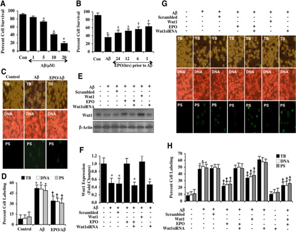
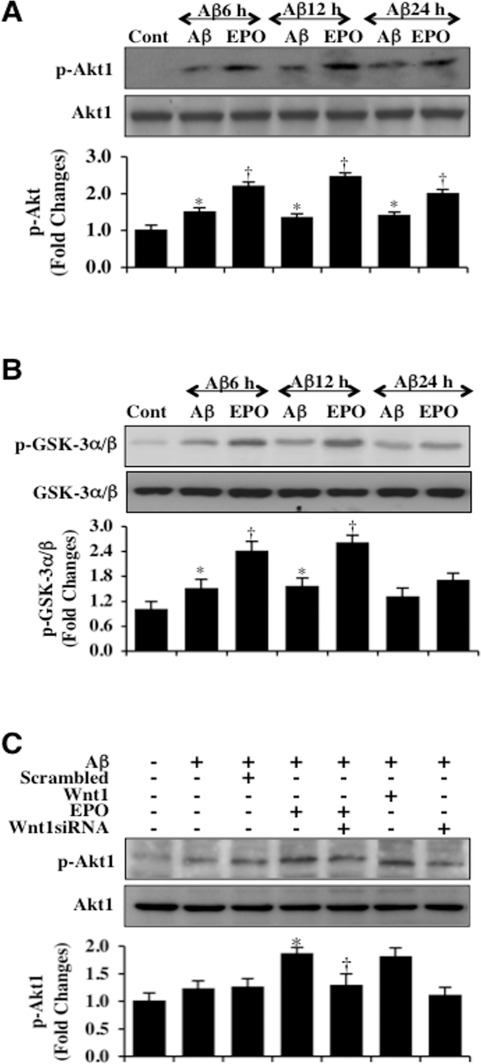
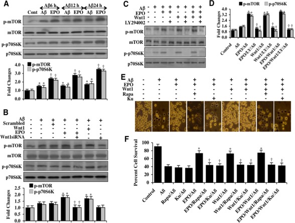
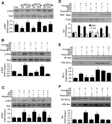
Central nervous system microglia promote neuronal regeneration and sequester toxic β-amyloid (Aβ) deposition during Alzheimer's disease. We show that the cytokine erythropoietin (EPO) decreases the toxic effect of Aβ on microgliain vitro. EPO up-regulates the cysteine-rich glycosylated wingless protein Wnt1 and activates the PI 3-K/Akt1/mTOR/ p70S6K pathway. This in turn increases phosphorylation and cytosol trafficking of Bad, reduces the Bad/Bcl-xL complex and increases the Bcl-xL/Bax complex, thus preventing caspase 1 and caspase 3 activation and apoptosis. Our data may foster development of novel strategies to use cytoprotectants such as EPO for Alzheimer's disease and other degenerative disorders.


Conflict of interest statement

The authors of this manuscript have no conflict of interest to declare.

Figures

Figure 1
Figure 1. EPO preserves microglia survival against Aβ through Wnt1
(A) Microglia were exposed to Aβ(10 μM) at the concentrations of 1, 5, 10, and 20 μM and cell survival was determined 24 hours after administration of Aβwith trypan blue dye exclusion method (*P< 0.01 vs. Control). Con = control = untreated microglia. Each data point represents the mean and SEM from 6 experiments. (B) EPO (10 ng/ml) was applied to microglial cultures at 24, 12, 6, or 1 hour prior to administration of Aβ (10 μM) and cell survival was determined 24 hours after Aβ administration with the trypan blue dye exclusion method (*P<0.01 vs. untreated control; P <0.05 vs. Aβ). Each data point represents the mean and SEM from 6 experiments. Con = control = untreated microglia. (C and D) EPO (10 ng/ml) was applied to microglial cultures 1 hour prior to the administration of Aβ (10 μM) and cell survival, DNA fragmentation, and PS exposure were determined 24 hours later. Representative images (C) and quantitative analysis (D) demonstrate that Aβ leads to a significant increase in trypan blue staining, DNA fragmentation, and membrane PS exposure in microglia 24 hours after Aβ exposure compared to untreated control cultures. EPO (10 ng/ml) applied 1 hour prior to Aβ exposure prevented microglial cell injury, DNA fragmentation, and membrane PS exposure(*P < 0.01 vs. Control; P <0.05 vs. Aβ). Each data point represents the mean and SEM from 6 experiments. (E and F) EPO (10 ng/ml) or Wnt1 (100 ng/ml) were applied 1 hour prior to Aβ (10 μM) administration with Wnt1 expression determined 6 hours following Aβ exposure. EPO (10 ng/ml) and Wnt1 (100 ng/ml) maintained the expression of Wnt1 that is otherwise down-regulated during Aβ exposure. Gene reduction ofWnt1with Wnt1 siRNA significantly reduced the expression of Wnt1 following a 6 hour period of Aβ exposure or treatment with EPO (10 ng/ml) during Aβ exposure. Non-specific scrambled siRNA did not alter Wnt1 expression during Aβ exposure (*P<0.01 vs. Control). (G and H) EPO was applied to microglial cultures 1 hour prior to the administration of Aβ and trypan blue dye exclusion, DNA fragmentation, and membrane PS exposure were determined 24 hours later. Representative images (G) and quantitative results (H) show that EPO (10 ng/ml) or Wnt1 (100 ng/ml) applied 1 hour prior to Aβ significantly reducedtrypan blue staining, DNA fragmentation, and membrane PS exposure in microglia 24 hours after Aβ exposure. Gene reduction of Wnt1 with transfection of Wnt1 siRNA prior to Aβ exposure prevented EPO (10 ng/ml) from blocking cell injury and resulted in increased trypan blue staining, DNA fragmentation, and membrane PS exposure in microglia 24 hours following Aβ exposure. Non-specific scrambled siRNA did not significantly alter microglial cell injury following Aβ exposure (*P<0.01 vs. untreated control; P < 0.05 vs. Aβ). Each data point represents the mean and SEM from 6 experiments.
Figure 2
Figure 2. EPO maintains Akt1 activation through Wnt1 during Aβ exposure
(A) Microglial protein extracts (50 μg/lane) were immunoblotted with phosphorylated Akt1 (p-Akt1) (active form) at 6, 12, and 24 hours following administration of Aβ (10 μM). Aβ resulted a mild increase in the expression of p-Akt1 over a 24 hour period. EPO (10 ng/ml) with a 1 hour pretreatment significantly increased the expression of p-Akt1 over a 24 hour period following Aβ exposure (*P < 0.01 vs. Control; P< 0.01 vs. Aβ of corresponding time point). In all cases, each data point represents the mean and SEM from 3 experiments. (B) Akt1 activity in microglia was determined with a GSK-3β fusion protein through assessment of p-GSK-3α/β expression following Aβ exposure. EPO (10 ng/ml) with a 1 hour pretreatment significantly increased the activity of Akt1 over 12 hours following Aβ exposure (*P <0.01 vs. Control; P<0.01 vs. Aβ of corresponding time point). In all cases, each data point represents the mean and SEM from 3 experiments. (C) Gene reduction of Wnt1 was performed with transfection of Wnt1 siRNA prior to Aβ exposure in microglia and p-Akt1 expression was determined at 6 hours following Aβ exposure. Loss of Wnt1 resulted in a decreased expression of p-Akt1 following a 6 hour period of Aβ exposure and significantly reduced EPO (10 ng/ml) expression of p-Akt1 during Aβ exposure. Non-specific scrambled siRNA did not alter p-Akt1 expression during Aβ exposure (*P < 0.01 vs. Aβ; P < 0.01 vs. EPO/Aβ).
Figure 3
Figure 3. EPO oversees Wnt1, mTOR and p70S6K and the PI 3-K/Akt1 pathway to protect microglia during Aβ exposure
(A) Microglial protein extracts (50 μg/lane) were immunoblotted with phosphorylated mTOR (p-mTOR, (Ser2448)) and phosphorylated p70S6K (p-p70S6K, Thr389)) antibodies at 6, 12, and 24 hours following exposure to Aβ (10 μM). Aβ resulted in a slight increase in the expression of p-mTOR and p-p70S6K. In contrast, EPO (10 ng/ml) with a 1 hour pretreatment significantly increased the expression of p-mTOR and p-p70S6K during Aβ exposure (*P <0.01 vs. Control; †P<0.01 vs. Aβ of corresponding exposure time). In all cases, each data point represents the mean and SEM from 3 experiments. (B) Gene reduction ofWnt1was performed with transfection of Wnt1 siRNA prior to Aβ (10 μM) exposure in microglia. Expression of p-mTOR and p-p70S6K were determined at 6 hours following Aβ exposure. EPO (10 ng/ml) or Wnt1 (100 ng/ml) applied 1 hour prior to Aβ exposure significantly increased the expression of p-mTOR and p-p70S6K. Transfection with Wnt1 siRNA significantly reduced the expression of p-mTOR and p-p70S6K following a 6 hour period of Aβ exposure and during EPO (10 ng/ml) administration with Aβ exposure. Non-specific scrambled siRNA did not alter the expression of p-mTOR and p-p70S6K during Aβ exposure (*P<0.01 vs. Aβ; P<0.01 vs. EPO/Aβ). (C and D) EPO (10 ng/ml), Wnt1 (100 ng/ml), or combined EPO with Wnt1 were applied to microglial cultures 1 hour prior to Aβ(10 μM) exposure and p-mTOR and p-p70S6K expression were determined 6 hours following Aβ exposure. EPO, Wnt1, or combined EPO with Wnt1 administration significantly increased the expression of p-mTOR and p-p70S6K to a similar levels 6 hours following Aβ exposure. Yet, combined application of the PI 3-K inhibitor LY294002 (10 μM, given 1.5 hours prior to Aβ) with EPO, Wnt1, or combined EPO with Wnt1 resulted in a significant decrease in expression of p-mTOR and p-p70S6K (*P<0.01 vs. Aβ; P<0.01 vs. EPO/Aβ, Wnt1/Aβ, or EPO/Wnt1/Aβ). (E and F) EPO (10 ng/ml), Wnt1 (100 ng/ml), or combined EPO with Wnt1 were applied to microglial cultures 1 hour prior to Aβ exposure and cell survival was determined by the using trypan blue dye exclusion method 24 hours later. Representative pictures (E) and quantitative results (F) indicated that EPO, Wnt1, or combined EPO with Wnt1 application significantly reduced trypan blue staining and increased cell survival to a similar level following Aβ(10 μM) exposure. Application of the mTOR specific inhibitors rapamycin (Rapa, 50 nM) or KU 0063794 (KU, 100 nM) 1.5 hours prior to Aβ administration prevented EPO, Wnt1, or combined EPO with Wnt1 to foster cell survival and resulted in an increased staining of trypan blue and a decrease in cell survival in microglia (*P<0.01 vs. Aβ; †P<0.01 vs. EPO/Aβ, Wnt1/Aβ, or EPO/Wnt1/Aβ). Each data point represents the mean and SEM from 6 experiments.
Figure 4
Figure 4. EPO through Wnt1 phosphorylates Bad, controls mitochondrial trafficking of Bad, and modulates Bad, Bcl-xL, and Bax binding
(A) Microglial protein extracts (50 μg/lane) were immunoblotted with phospho-rylated Bad (p-Bad, (Ser136)) antibody at 6, 12, and 24 hours following administration of Aβ (10 μM). Aβ exposure resulted in a significant decrease in the expression of p-Bad. EPO (10 ng/ml) with a 1 hour pretreatment significantly increased the expression of p-Bad 6 hours following Aβ exposure (*P <0.01 vs. Control; P<0.01 vs. Aβ of corresponding exposure time). In all cases, each data point represents the mean and SEM from 3 experiments. (B) Gene reduction of Wnt1 was performed with transfection of Wnt1 siRNA prior to Aβ (10 μM) administration in microglia. The expression of p-Bad was determined at 6 hours following Aβ exposure. EPO (10 ng/ml) or Wnt1 (100 ng/ml) with 1 hour pretreatments significantly increased the expression of p-Bad 6 hours following Aβ exposure. Yet, Wnt1 siRNA transfection prior to Aβ exposure prevented EPO from significantly phosphorylating Bad. Non-specific scrambled siRNA did not significantly alter p-Bad expression during Aβ exposure (*P < 0.01 vs. Aβ; P<0.01 vs. EPO/Aβ). (C) EPO (10 ng/ml), Wnt1 (100 ng/ml) or combined EPO with Wnt1 with 1 hour pretreatments significantly increased the expression of p-Bad 6 hours following Aβ (10 μM) exposure. Application of the mTOR inhibitor rapamycin (50 nM) with EPO (10 ng/ml), Wnt1 (100 ng/ml) or combined EPO with Wnt1 1.5 hour prior to Aβ exposure resulted in the loss of the ability of EPO, Wnt1, or combined EPO with Wnt1 to increase in the expression of p-Bad during Aβ exposure (*P < 0.01 vs. Aβ; P<0.01 vs. EPO/Aβ, Wnt1/Aβ or EPO/Wnt1/Aβ). (D) Gene reduction of Wnt1 was performed with transfection of Wnt1 siRNA prior to Aβ (10 μM) administration in microglia and the expression of Bad in both cytosolic and mitochondrial fractions was determined at 6 hours following Aβ exposure. EPO (10 ng/ml) or Wnt1 (100 ng/ml) with 1 hour pretreatments significantly reduced mitochondrial expression of Bad and increased the cytosolic expression of Bad following Aβ exposure. Yet, gene reduction of Wnt1 with Wnt1 siRNA transfection led to the loss of the ability of EPO to promote the release of Bad for the mitochondria to the cytosol during Aβ exposure. Non-specific scrambled siRNA did not significantly change the translocation of Bad during Aβ exposure (*P < 0.01 vs. Aβ; P < 0.01 vs. EPO/Aβ). (E) Gene reduction of Wnt1 was performed in microglia with transfection of Wnt1 siRNA prior to Aβ (10 μM) administration. Protein extracts were immunoprecipitated using Bad antibody 6 hours following Aβ exposure. Western blot for Bcl-xL expression in the precipitates was performed. EPO (10 ng/ml) or Wnt1 (100 ng/ml) with 1 hour pretreatments decreased binding of Bcl-xL to Bad during Aβ exposure. Gene reduction of Wnt1 with Wnt1 siRNA transfection prevented EPO to decrease the binding of Bcl-xL to Bad during Aβ exposure. Non-specific scrambled siRNA did not significantly alter the binding of Bad to Bcl-xL during Aβ exposure (*P < 0.01 vs. Aβ; P<0.01 vs. EPO/Aβ). (F) Gene reduction of Wnt1 was performed with transfection of Wnt1 siRNA prior to Aβ (10 μM) administration in microglia. Protein extracts were immunoprecipitated using Bcl-xL antibody 6 hours following Aβ exposure. Western blot for Bax expression in the precipitates was performed. EPO (10 ng/ml) or Wnt1 (100 ng/ml) with 1 hour pretreatments increased the binding of Bcl-xL to Bax during Aβ exposure. Gene reduction of Wnt1 with Wnt1 siRNA transfection prevented EPO to increase the binding of Bcl-xL to Bax during Aβ exposure. Non-specific scrambled siRNA did not significantly alter the binding of Bcl-xL with Bax during Aβ exposure (*P < 0.01 vs. Aβ; P<0.01 vs. EPO/Aβ).
Figure 5
Figure 5. EPO and Wnt1 control mitochondrial membrane potential, block early and late apoptotic microglial Aβdegeneration, and prevent caspase 1 and 3 activation through Bcl-xL
(A) Representative images and quantitative results from JC-1 staining illustrate that Aβ (10 μM)results in a significant decrease in the red/green fluorescence intensity ratio of mitochondria within 6 hours when compared with untreated control cultures, demonstrating that Aβ exposure leads to significant mitochondrial membrane depolarization. EPO (10 ng/ml) or Wnt1 (100 ng/ml) with 1 hour pretreatments significantly increase the red/green fluorescence intensity of mitochondria in microglia, demonstrating that mitochondrial membrane potential was restored. In contrast, gene reduction of Wnt1 with transfection of Wnt1 siRNA increased mitochondrial membrane depolarization to a greater degree than Aβ exposure alone and prevented the ability of EPO to maintain mitochondrial membrane potential during Aβ exposure. The relative ratio of red/green fluorescent intensity of mitochondrial staining was measured in 6 independent experiments with analysis performed using the public domain NIH Image program (http://rsb.info.nih.gov/nih-image) (*P<0.01 vs. Aβ; †P <0.01 vs. EPO/Aβ). (B) Microglial cell protein extracts (50 μg/lane) were immunoblotted with cleaved caspase 1 (active) and cleaved caspase 3 (active) antibodies 6 hours following Aβ(10 μM) exposure.Aβ(10 μM) exposure significantly increased caspase 1 and caspase 3 activities. In contrast, EPO (10 ng/ml) or Wnt1 (100 ng/ml) administration significantly decreased the expression of cleaved (active) caspase 1 and caspase 3 at 6 hours following Aβ(10 μM) exposure. Gene reduction of Wnt1 with transfection with Wnt1 siRNA abrogated the ability of EPO to prevent caspase activation (*P<0.01 vs. Aβ; †P <0.01 vs. EPO/Aβ). Non-specific scrambled siRNA did not significantly change the expression of cleaved caspase 1 and caspase 3 during Aβ exposure. Each data point represents the mean and SEM from 3 experiments. Quantification of western band intensity from 3 experiments was performed using the public domain NIH Image program (http://rsb.info.nih.gov/nih-image). (C) Microglial cell protein extracts (50 μg/lane) were immunoblotted with cleaved caspase 1 (active) and cleaved caspase 3 (active) antibodies 6 hours following Aβ(10 μM) exposure.Aβ(10 μM) exposure significantly increased caspase 1 and caspase 3 activities. In contrast, EPO (10 ng/ml) or Wnt1 (100 ng/ml) administration significantly decreased the expression of cleaved (active) caspase 1 and caspase 3 at 6 hours following Aβ(10 μM) exposure. Gene reduction of Bcl-xLwith transfection with Bcl-xL siRNA abrogated the ability of EPO to prevent caspase activation (*P<0.01 vs. Aβ; †P <0.01 vs. EPO/Aβ). Non-specific scrambled siRNA did not significantly change the expression of cleaved caspase 1 and caspase 3 during Aβ exposure. Each data point represents the mean and SEM from 3 experiments. Quantification of western band intensity from 6 experiments was performed using the public domain NIH Image program (http://rsb.info.nih.gov/nih-image). (D and E)EPO (10 ng/ml) or Wnt1 (100 ng/ml) were administered to microglial cultures 1 hour prior to the Aβ (10 μM) exposure and trypan blue dye exclusion, DNA fragmentation, and membrane PS exposure were determined 24 hours later.Representative images (B) and quantitative analysis (C) demonstrate that Aβ(10 μM) results in a significant increase in trypan blue staining, DNA fragmentation, and membrane PS exposure in microglia 24 hours after Aβ exposure compared to untreated control cultures. In contrast, EPO (10 ng/ml), Wnt1 (100 ng/ml), or combined EPO and Wnt1 applied 1 hour prior to Aβ significantly reducedtrypan blue staining, DNA fragmentation, and membrane PS exposure in microglia 24 hours after Aβ exposure. Gene reduction of Bcl-xLwith transfection of Bcl-xL siRNA prior to Aβ exposure prevented EPO (10 ng/ml), Wnt1 (100 ng/ml), or combined EPO and Wnt1 from blocking cell injury and resulted in increased trypan blue staining, DNA fragmentation, and membrane PS exposure in microglia 24 hours following Aβ exposure. Non-specific scrambled siRNA did not significantly change cell injury during Aβ exposure (*P < 0.01 vs. Aβ; P <0.01 vs. EPO/Aβ, Wnt1/Aβ, or EPO/Wnt1/Aβ). Each data point represents the mean and SEM from 6 experiments.

Similar articles

Cited by

References

    1. Erol A. Unraveling the Molecular Mechanisms Behind the Metabolic Basis of Sporadic Alzheimer's Disease. J Alzheimers Dis. 2009 - PubMed
    1. Maiese K, Chong ZZ, Hou J, Shang YC. New strategies for Alzheimer's disease and cognitive impairment. Oxid Med Cell Longev. 2009;2:279–289. - PMC - PubMed
    1. Chong ZZ, Li F, Maiese K. Erythropoietin requires NF-kappaB and its nuclear translocation to prevent early and late apoptotic neuronal injury during beta-amyloid toxicity. Curr Neurovasc Res. 2005;2:387–399. - PMC - PubMed
    1. Chong ZZ, Li F, Maiese K. Cellular demise and inflammatory microglial activation during beta-amyloid toxicity are governed by Wnt1 and canonical signaling pathways. Cell Signal. 2007;19:1150–1162. - PMC - PubMed
    1. Salminen A, Kaarniranta K, Haapasalo A, Soininen H, Hiltunen M. AMP-activated protein kinase: a potential player in Alzheimer's disease. J Neurochem. 2011;118:460–474. - PubMed

Publication types

MeSH terms