. 2012 Apr 18;31(8):1961-74.
doi: 10.1038/emboj.2012.52. Epub 2012 Mar 2.

Multiple factors dictate target selection by Hfq-binding small RNAs

Affiliations

Multiple factors dictate target selection by Hfq-binding small RNAs

Chase L Beisel et al. EMBO J. .

Abstract

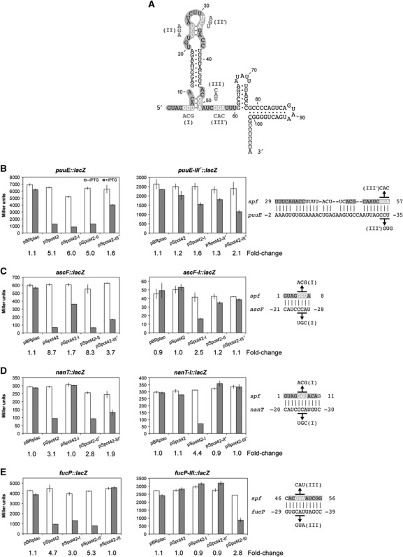
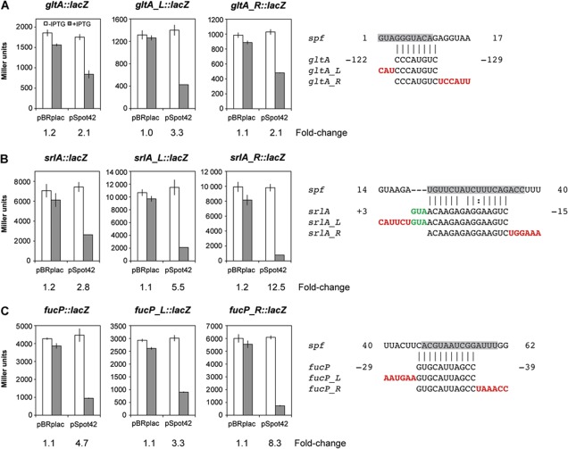
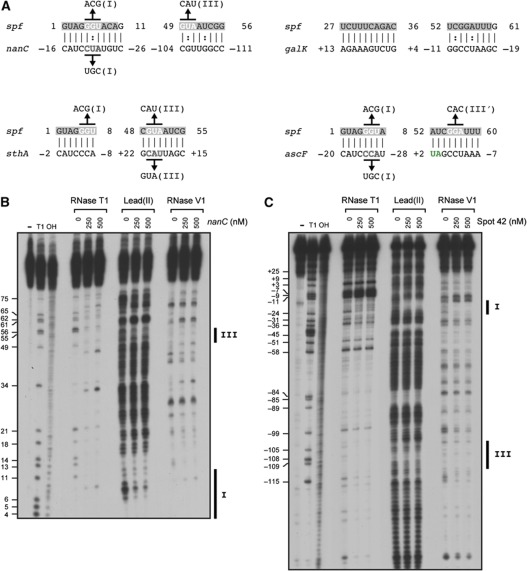
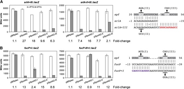
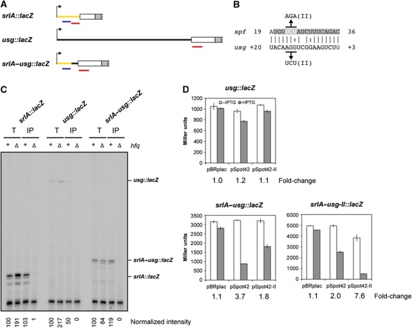
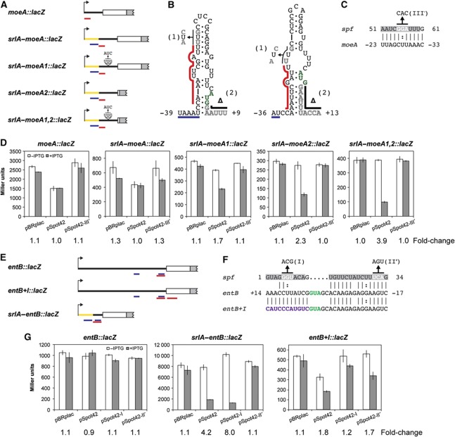
Hfq-binding small RNAs (sRNAs) in bacteria modulate the stability and translational efficiency of target mRNAs through limited base-pairing interactions. While these sRNAs are known to regulate numerous mRNAs as part of stress responses, what distinguishes targets and non-targets among the mRNAs predicted to base pair with Hfq-binding sRNAs is poorly understood. Using the Hfq-binding sRNA Spot 42 of Escherichia coli as a model, we found that predictions using only the three unstructured regions of Spot 42 substantially improved the identification of previously known and novel Spot 42 targets. Furthermore, increasing the extent of base-pairing in single or multiple base-pairing regions improved the strength of regulation, but only for the unstructured regions of Spot 42. We also found that non-targets predicted to base pair with Spot 42 lacked an Hfq-binding site, folded into a secondary structure that occluded the Spot 42 targeting site, or had overlapping Hfq-binding and targeting sites. By modifying these features, we could impart Spot 42 regulation on non-target mRNAs. Our results thus provide valuable insights into the requirements for target selection by sRNAs.


Conflict of interest statement

The authors declare that they have no conflict of interest.

Figures

Figure 1
Figure 1
Mutational analysis of base-pairing interactions between Spot 42 and selected target mRNAs. (A) Secondary structure of Spot 42 supported by in vitro structural probing data (Møller et al, 2002). The three unstructured regions (I–III) are highlighted in grey, while Spot 42 mutations are shown in white. The base pairs between nucleotides 7–9 and nucleotides 23–25 likely are unpaired most of the time. (BE) β-Galactosidase assay results for lacZ translational fusion strains carrying the empty vector pBRplac or different Spot 42 expression plasmids. Genes tested as lacZ fusions are (B) puuE, (C) ascF, (D) nanT, and (E) fucP. See Supplementary Table S1 for the sequences of the gene fragment included in each lacZ fusion. Strains harbouring each plasmid were induced with 0.2% L-arabinose with or without 1 mM IPTG for 1 h before assaying each culture. The fold-change is the ratio of β-galactosidase activity of cells grown in the absence and presence of IPTG. Error bars represent the standard deviation from measurements of three independent colonies. Base-pairing interactions predicted by TargetRNA are on the right. Nucleotides in the unstructured regions of Spot 42 are highlighted in grey. The start codon of the fusion is in green, where the number of nucleotides upstream (−) and downstream (+) of the start codon is indicated. Sequences above the predicted base-pairing interactions correspond to the indicated pSpot42 mutations, while sequences below the predicted base-pairing interactions correspond to the compensatory mutations in the target gene fusions.
Figure 2
Figure 2
Improved regulation with extended base-pairing interactions in the unstructured regions of Spot 42. Up to six nucleotides were inserted immediately downstream (L) or upstream (R) of each targeting site in the lacZ fusions with (A) gltA, (B) srlA, and (C) fucP. Inserted nucleotides (in red) were designed to extend base-pairing between Spot 42 and each fusion for all three regions of Spot 42 (gltA, region I; srlA, region II; fucP, region III). See Figure 1 for a description of the β-galactosidase assay conditions, fold-change, and colouring and numbering of nucleotides in the predicted base-pairing interactions. Error bars represent the standard deviation from measurements of three independent colonies.
Figure 3
Figure 3
Base-pairing between multiple unstructured regions of Spot 42 and individual target mRNAs. Four Spot 42 target genes that met the requirements for multi-site pairing described in the main text were identified: nanC, galK, sthA, and ascF. (A) Predicted base-pairing interactions between Spot 42 and each target mRNA. See Figure 1 for a description of the colouring and numbering of the predicted base-pairing interactions. Results of β-galactosidase assays for fusions and Spot 42 variants containing the indicated mutations are available either in this work (ascF; Figure 1B) or in our previous work (nanC, sthA) (Beisel and Storz, 2011a). (B) In vitro structural probing of labelled Spot 42 hybridized with the unlabelled nanC mRNA. 5′ Radiolabelled Spot 42 (20 nM) was incubated at 37°C with 0.1 μg/μl yeast RNA and various concentrations of an unlabelled 192-nucleotide portion of the nanC mRNA (0, 250, 500 nM). Concentrations of unlabelled nanC mRNAs were selected based on gel shift assays (Supplementary Figure S6A). Incubated RNAs were treated with RNase T1 (cleavage of single-stranded G residues), lead acetate (cleavage of single-stranded nucleotides), or RNase V1 (cleavage of double-stranded and stacked single-stranded nucleotides) and resolved by denaturing PAGE. Untreated Spot 42 (−), denatured Spot 42 treated with RNase T1 (T1), and alkaline hydrolysis of Spot 42 (OH) were resolved as references. Regions I and III of Spot 42 are indicated by black bars to the right of the gel image. Numbering to the left of the gel image indicates the position relative to the 5′ G in Spot 42. (C) In vitro structural probing of labelled nanC mRNA hybridized to unlabelled Spot 42. 5′ 32P-radiolabelled nanC mRNA (20 nM) was incubated as described in (B) with various concentrations of unlabelled Spot 42 (0, 250, 500 nM). Concentrations of unlabelled Spot 42 were selected based on gel shift assays (Supplementary Figure S6B). Predicted targeting sites for regions I and III of Spot 42 are indicated by black bars to the right of the gel image. Numbering to the left of the gel image indicates the position upstream (−) and downstream (+) of the start codon. The RNase V1 cleavage products have a 3′-hydroxyl, which results in slightly reduced mobility compared with cleavage products with a 3′-phosphate resulting from RNase T1 digestion and alkaline hydrolysis. Figure source data can be found in Supplementary data.
Figure 4
Figure 4
Improved regulation with base-pairing through multiple unstructured regions of Spot 42. Eleven nucleotides were either inserted (in red) or mutated (in purple) in the (A) srlA or (B) fucP fusions to create a new targeting site for Spot 42 (srlA+III::lacZ, fucP+I::lacZ). Indicated mutations were introduced at site II in srlA+III::lacZ and site III in fucP+I::lacZ (to give srlA-II+III::lacZ and fucP-III+I::lacZ, respectively). See Figure 1 for a description of the β-galactosidase assay conditions, fold-change, and colouring and numbering of the predicted base-pairing interactions. Error bars represent the standard deviation from measurements of three independent colonies.
Figure 5
Figure 5
Gene regulation by Spot 42 conferred through insertion of an Hfq-binding site. (A) Schematic representation of the srlA and usg fusions. Putative Hfq-binding sites are in blue, Spot 42 targeting sites are in red, the coding region of each gene included in the fusion is a white box, the 5′ portion of lacZ is a grey box, and the 5′ untranslated region of srlA included in each fusion is coloured yellow. To generate srlA–usg::lacZ, the first 26 nucleotides of the srlA mRNA were fused immediately upstream of the ribosome-binding site in usg::lacZ. (B) Base-pairing interactions between Spot 42 and the usg mRNA predicted by TargetRNA. The indicated mutations are contained in pSpot42-II (above) and srlA–usg-II::lacZ (below). (C) Primer extension analysis of srlA, usg, and srlA–usg fusion mRNAs co-immunoprecipitated with Hfq. Total RNA (T, 2 μg) or RNA eluted from Hfq immunoprecipitated with α-Hfq antibodies (IP, 0.2 μg) from wild-type (+) or hfq-deletion (Δ) strains were reverse-transcribed with a 5′ radiolabelled lacZ-specific primer and resolved by denaturing PAGE. The anticipated primer extension product for each strain is indicated on the right. The intensity of bands at these locations were quantified using a phosphorimager and normalized to the band for total RNA from the wild-type strain following subtraction of background intensity. Similar results were obtained using gene-specific primers. The non-specific bands at the bottom of the gel indicate equal loading of each sample. (D) β-Galactosidase assay results for the usg fusions. See Figure 1 for a description of the β-galactosidase assay conditions and fold-change. Error bars represent the standard deviation from measurements of three independent colonies. Figure source data can be found in Supplementary data.
Figure 6
Figure 6
Gene regulation by Spot 42 conferred by freeing the Spot 42 targeting site or separating the Hfq-binding site and Spot 42 targeting site. (A) Schematic representation of the moeA fusions. See Figure 5A for a description of the colouring. moeA::lacZ was generated by fusing lacZ to the 14th codon of moeA, srlA–moeA::lacZ was generated by fusing the 5′ end of srlA to the transcriptional start site of moeA::lacZ, srlA–moeA1::lacZ was generated by introduction of two point mutations in the 5′ untranslated region of moeA, and srlA–moeA2::lacZ was generated by fusing lacZ to the srlA–moeA start codon. (B) Secondary structures of srlA–moeA predicted by NUPACK and mfold. The mutations contained within srlA–moeA1::lacZ (1), srlA–moeA2::lacZ (2), or srlA–moeA1,2::lacZ (both 1 and 2) are bordered by black lines. Nucleotides bordered by a red line are the Spot 42 targeting site, nucleotides bordered by a blue line are the Hfq-binding site, and nucleotides in green are the start codon. Indicated is the number of nucleotides upstream (–) and downstream (+) of the start codon. (C) Base-pairing interactions between Spot 42 and the moeA mRNA predicted by TargetRNA. See Figure 1 for a description of the colouring and numbering of the predicted base-pairing interactions. (D) β-Galactosidase assay results for the moeA fusions. See Figure 1 for a description of the β-galactosidase assay conditions and fold-change. (E) Schematic representation of the entB fusions. See Figure 5A for a description of colouring. (F) Predicted base-pairing interactions between Spot 42 and either entB or entB mutated to include a targeting site for region I of Spot 42 (entB+I). See Figure 1 for a description of the colouring and numbering of the predicted base-pairing interactions. Mutated nucleotides are in purple. (G) β-Galactosidase assay results for the entB fusions. See Figure 1 for a description of the β-galactosidase assay conditions and fold-change. Error bars in (D, G) represent the standard deviation from measurements of three independent colonies. Different fusions showed differing levels of basal expression, which for moeA and srlA–moeA (D) as well as entB and srlA–entB (G) are reflected in differing mRNA levels (Supplementary Figure S7). Although there also was some variation in the basal levels between experiments, the fold-change was very consistent.

Similar articles

Cited by

References

    1. Beisel CL, Storz G (2011a) The base-pairing RNA Spot 42 participates in a multioutput feedforward loop to help enact catabolite repression in Escherichia coli. Mol Cell 41: 286–297 - PMC - PubMed
    1. Beisel CL, Storz G (2011b) Discriminating tastes: physiological contributions of the Hfq-binding small RNA Spot 42 to catabolite repression. RNA Biol 8: 766–770 - PMC - PubMed
    1. Bouvier M, Sharma CM, Mika F, Nierhaus KH, Vogel J (2008) Small RNA binding to 5′ mRNA coding region inhibits translational initiation. Mol Cell 32: 827–837 - PubMed
    1. Buchet A, Nasser W, Eichler K, Mandrand-Berthelot MA (1999) Positive co-regulation of the Escherichia coli carnitine pathway cai and fix operons by CRP and the CaiF activator. Mol Microbiol 34: 562–575 - PubMed
    1. Busch A, Richter AS, Backofen R (2008) IntaRNA: efficient prediction of bacterial sRNA targets incorporating target site accessibility and seed regions. Bioinformatics 24: 2849–2856 - PMC - PubMed

Publication types