

doi: 10.1038/nsmb.2250.
Structure of the human metapneumovirus fusion protein with neutralizing antibody identifies a pneumovirus antigenic site
Affiliations
- PMID: 22388735
- PMCID: PMC3546531
- DOI: 10.1038/nsmb.2250
Item in Clipboard
Structure of the human metapneumovirus fusion protein with neutralizing antibody identifies a pneumovirus antigenic site
Nat Struct Mol Biol.
.
Abstract
Human metapneumovirus and respiratory syncytial virus cause lower respiratory tract infections. The virus fusion (F) glycoprotein promotes membrane fusion by refolding from a metastable pre-fusion to a stable post-fusion conformation. F is also a major target of the neutralizing antibody response. Here we show that a potently neutralizing anti-human metapneumovirus antibody (DS7) binds a structurally invariant domain of F, revealing a new epitope that could be targeted in vaccine development.
Figures
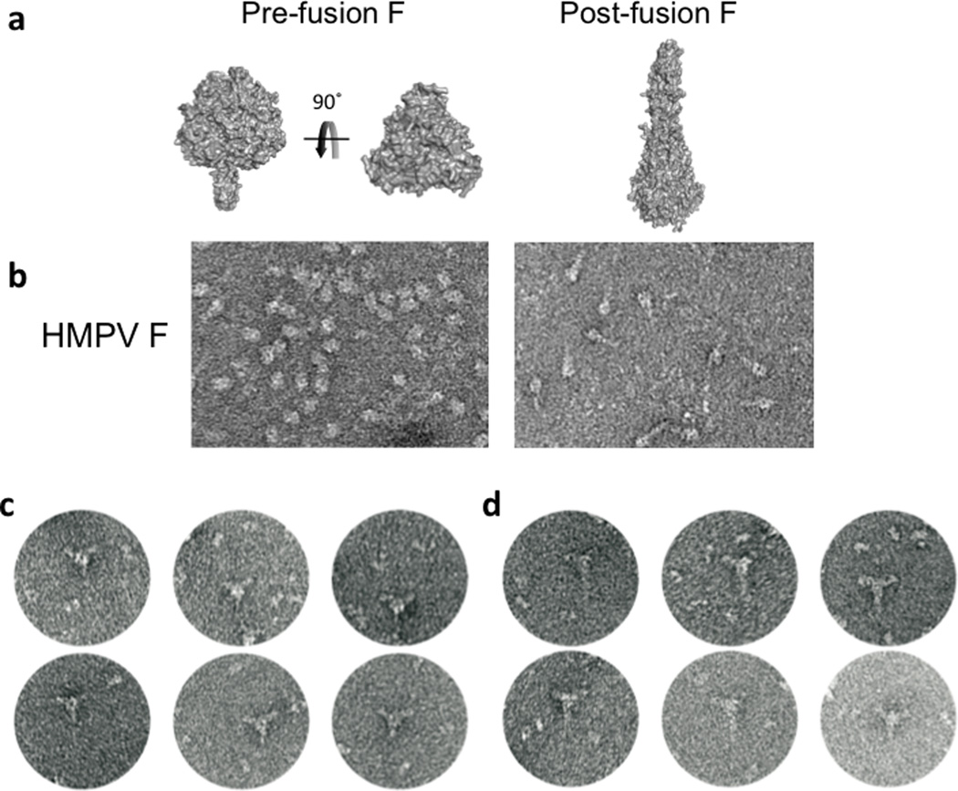
(a) The PIV5 F (pre-fusion) structure is shown in two views, rotated 90° from each other. One orientation shows the pre-fusion HRB stalk, and the second is oriented along the trimer axis and shows only the head region. The RSV F (post-fusion) trimer is shown, oriented perpendicular to the trimer axis. (b) EM images of HMPV F that appear consistent with the pre-fusion (left panel) and post-fusion (right panel) conformational states. (c, d) EM images of DS7 Fab bound to HMPV F in putative pre-fusion (c) and post-fusion (d) conformations.

(a, b) The DS7 Fab engages a single F fragment consisting of DI (yellow), DII (red) and DIII (magenta) domains. The DS7 heavy chain (light blue) binds a hydrophobic pocket in DI. The DS7 light chain (pale green) buries residues in both DI and a loop in DII. (b) The complex is rotated 90° from the orientation in (a). (c) Residues in the DS7 binding site interactions are shown in stick format, with carbon atoms colored by domain as in (a). CDR3H inserts into a hydrophobic pocket on the broad side of DI, formed by Leu24, Ile31, Pro282, and Trp284, flanked by charged residues Lys20, Glu33, Lys312, Arg348 and Glu349. The light chain buries residues at the DI N-terminus and three residues in DII.

(a) Major antigenic sites in HMPV and RSV F are mapped onto the HMPV F structure, with the DS7 buried surface area shown as a transparent yellow surface. The view is from the perspective of the DS7 Fab, rotated ~90° from Figure 2a. Escape mutant sites are shown as side chains with dark blue (HMPV F) or magenta (RSV F) spheres. The individual HMPV residues are labeled, along with the HMPV antigenic sites 2–6 and the RSV antigenic sites I-VI. The RSV antigenic sites correspond to the following residues: Ag Site I: 357 (RSV - 389); Ag Site II: 232, 238, 242, 245 (RSV - 262, 268, 272, 275), Ag Sites IV, V, VI: 397, 400, 401, 404, 415 (RSV – 429, 432, 433, 436, 447). (b) The DS7 epitope surface is shown colored by atom type (yellow:carbon; blue:nitrogen; red:oxygen), with underlying residues in stick format. Asterisks mark residues conserved in HMPV and RSV F. A dotted line circumscribes a predominantly hydrophobic pocket that binds DS7 CDR3H. (c) The RSV F surface corresponding to the DS7 epitope is shown as in panel (b). The dotted line highlights the partially conserved hydrophobic pocket with Trp314 at its base.
References
-
- Lamb RA, Parks GD. Paramyxoviridae: The viruses and their replication. In: Knipe DM, Howley PM, editors. Fields Virology. Vol. 1. Wolters Kluwer: Lippincott WIlliams & Wilkins; 2007. pp. 1449–1496.
-
- Collins PL, Crowe JE., Jr . Respiratory Syncytial Virus and Metapneumovirus. In: Knipe DM, Howley PM, editors. Fields Virology. Vol. 2. Wolters Kluwer: Lippincott WIlliams & Wilkins; 2007. pp. 1601–1646.
-
- Papenburg J, Boivin G. Rev Med Virol. 2011;20:245–260. - PubMed
Publication types
MeSH terms
Substances
Associated data
- Actions
Grants and funding
- AI-072414/AI/NIAID NIH HHS/United States
- AI-23173/AI/NIAID NIH HHS/United States
- R01 AI072414/AI/NIAID NIH HHS/United States
- R01 AI085062/AI/NIAID NIH HHS/United States
- R21 AI073697/AI/NIAID NIH HHS/United States
- P30 CA060553/CA/NCI NIH HHS/United States
- AI-073697/AI/NIAID NIH HHS/United States
- R01 AI023173/AI/NIAID NIH HHS/United States
- GM-61050/GM/NIGMS NIH HHS/United States
- AI-085062/AI/NIAID NIH HHS/United States
- HHMI/Howard Hughes Medical Institute/United States
- R01 GM061050/GM/NIGMS NIH HHS/United States
LinkOut - more resources
Full Text Sources
Other Literature Sources
Molecular Biology Databases
