. 2012 Mar 4;19(4):424-9.
doi: 10.1038/nsmb.2255.

A sensor-adaptor mechanism for enterovirus uncoating from structures of EV71

Affiliations

A sensor-adaptor mechanism for enterovirus uncoating from structures of EV71

Xiangxi Wang et al. Nat Struct Mol Biol. .

Abstract

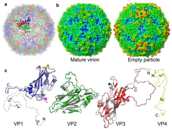
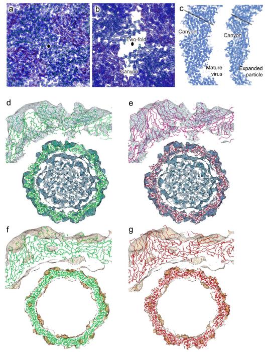
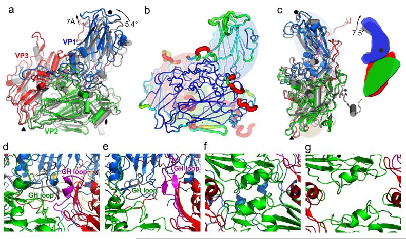
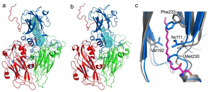
Enterovirus 71 (EV71) is a major agent of hand, foot and mouth disease in children that can cause severe central nervous system disease and death. No vaccine or antiviral therapy is available. High-resolution structural analysis of the mature virus and natural empty particles shows that the mature virus is structurally similar to other enteroviruses. In contrast, the empty particles are markedly expanded and resemble elusive enterovirus-uncoating intermediates not previously characterized in atomic detail. Hydrophobic pockets in the EV71 capsid are collapsed in this expanded particle, providing a detailed explanation of the mechanism for receptor-binding triggered virus uncoating. These structures provide a model for enterovirus uncoating in which the VP1 GH loop acts as an adaptor-sensor for cellular receptor attachment, converting heterologous inputs to a generic uncoating mechanism, highlighting new opportunities for therapeutic intervention.


Figures

Figure 1
Figure 1
Overall structures. (a), Cartoon of the mature EV71 virion, looking down an icosahedral 2-fold axis; VP1, VP2, VP3 and VP4 are drawn in blue, green, red and yellow, respectively. A single icosahedral protomer is drawn more brightly. (b), Radius colored surface representation of EV71 mature and empty particles. The surfaces for both are colored from blue to red according to their distances from the particle center. (c), Structures of the EV71 capsid proteins. VP0-3 (we treat VP0 and VP2 interchangeably) are shown in a similar orientation to the brighter protomer in (a); proteins from the expanded empty particle are coloured as in (a), whereas the corresponding chains from the mature virion are grey. VP4 of the mature virion is shown in yellow. The proteins have been superimposed as rigid bodies (216, 227, 220 Cαs superpose with rms deviations of 1.6, 0.9 and 1.3Å for VP1, VP0(2) and VP3 respectively). Residues 1-297 of VP1, 10-254 of VP2, 1-242 of VP3 and 12-69 of VP4 have well defined electron density in the mature virion, while residues 73-210 and 219-297 of VP1, 82-319 of VP0 and 1-238 of VP3 in the empty particle are modeled. Major surface exposed loops are marked with coloured washers: VP1 BC (yellow), VP1 DE (orange), VP1 HI (red), VP1 GH (magenta), VP2 EF (light blue), VP3 GH (black).
Figure 2
Figure 2
Electron density. (a) and (b): averaged 2|F|o-|Fc| maps in the vicinity of an icosahedral 2-fold axis (marked as black ellipse) for the mature virus (a) and expanded particle (b), showing the well defined electron density and demonstrating the perforations in the expanded particle. Cα traces, coloured as in Fig. 1 are also shown. (c), Equivalent equatorial slices through the two electron density maps shown in (a) and (b). The black lines mark the approximate position of an icosahedral 5-fold axis. (d-g) Comparisons of the fit of our crystallographic EV71 expanded particle coordinates and modeled poliovirus into the 10 Å resolution cryo-EM reconstructions for 135S and 82S poliovirus particles,. The density was displayed and correlation coefficients calculated using the program URO. (d) and (e) show the fit of EV71 (green) and poliovirus (red) into the density for the 135S particle (correlation coefficient 0.66 for both EV71 and the deposited poliovirus coordinates). (f) and (g) show the corresponding fits into the cryo-EM density for the late 80S poliovirus (correlation coefficients 0.63 for EV71 and 0.64 for poliovirus).
Figure 3
Figure 3
Protomers of mature virion and expanded particle compared. (a), protomeric units shown with respect to the icosahedral axes of the particles (i.e. superposing whole particles). The mature virus is grey and the expanded particle coloured as in Figure 1c. The orientation is similar to the bright protomer in Figure 1a, icoshedral symmetry axes are drawn in black. (b), Superposed protomers. Structural differences are mapped onto the protomer of the mature virion, the thickness and colour of the worm representation reflects the local deviation between the structures (ramped blue 0 to orange 8Å). Regions missing in the expanded particle are shown in red (VP4 is omitted). (c), the change in the jackknife structure of VP1 on capsid expansion. The superposition is based on VP0(2), VP3 and the 5-fold distal portion of VP1. Black dot in the right diagram marks the point about which VP1 flexes. (d)-(g) show the regions where perforations appear in the expanded particle (the base of the canyon in (d) and (e) and the 2-fold region in (f) and (g), the mature virus is on the left and expanded particle on the right). (c)-(g) coloured as in Figure 1c, with the GH loops of VP1 and VP3 highlighted in grey and magenta, respectively. Residues 136-141 of VP3 and 227-250 and 48-52 of VP2 form a bridge separating the perforations.
Figure 4
Figure 4
Pocket factor binding site. (a), VP1 of EV71 mature virus harbours a hydrophobic pocket (blue mesh) similar to that seen in other enteroviruses, which is occupied by a natural lipid, probably sphingosine (magenta). The protein cartoon is coloured as for Figure 1a. (b), The expanded particle, showing the collapsed pocket. (c), Comparison of the VP1 pockets between the mature virus (grey, with pocket factor shown in magenta) and empty particles (blue). During pocket collapse residues 110-114, 152- 160, 190-194 and 228-234 move inwards taking some internal hydrophobic residues with them (including I111, F135 and F155).

Comment in

Similar articles

Cited by

References

    1. Brown BA, Pallansch MA. Complete nucleotide sequence of enterovirus 71 is distinct from poliovirus. Virus Res. 1995;39:195–205. - PubMed
    1. McMinn PC. An overview of the evolution of enterovirus 71 and its clinical and public health significance. FEMS Microbiol Rev. 2002;26:91–107. - PubMed
    1. Wu Y, et al. Structures of EV71 RNA-dependent RNA polymerase in complex with substrate and analogue provide a drug target against the hand-foot-and- mouth disease pandemic in China. Protein Cell. 2010;1:491–500. - PMC - PubMed
    1. Basavappa R, et al. Role and mechanism of the maturation cleavage of VP0 in poliovirus assembly: structure of the empty capsid assembly intermediate at 2.9 A resolution. Protein Sci. 1994;3:1651–69. - PMC - PubMed
    1. Curry S, et al. Dissecting the roles of VP0 cleavage and RNA packaging in picornavirus capsid stabilization: the structure of empty capsids of foot-and- mouth disease virus. J Virol. 1997;71:9743–52. - PMC - PubMed

Publication types

Substances