. 2012 Jun 1;21(11):2559-71.
doi: 10.1093/hmg/dds082. Epub 2012 Mar 2.

Rescue of severely affected dystrophin/utrophin-deficient mice through scAAV-U7snRNA-mediated exon skipping

Affiliations

Rescue of severely affected dystrophin/utrophin-deficient mice through scAAV-U7snRNA-mediated exon skipping

Aurélie Goyenvalle et al. Hum Mol Genet. .

Abstract

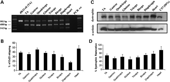
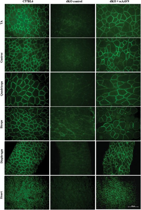
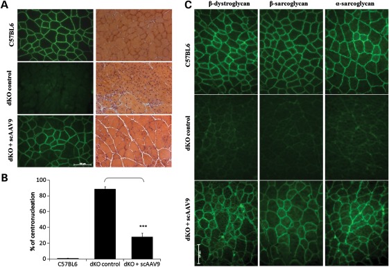
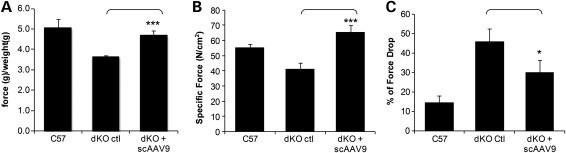
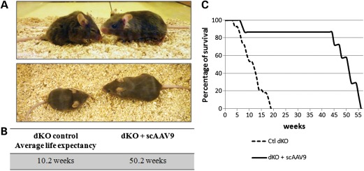
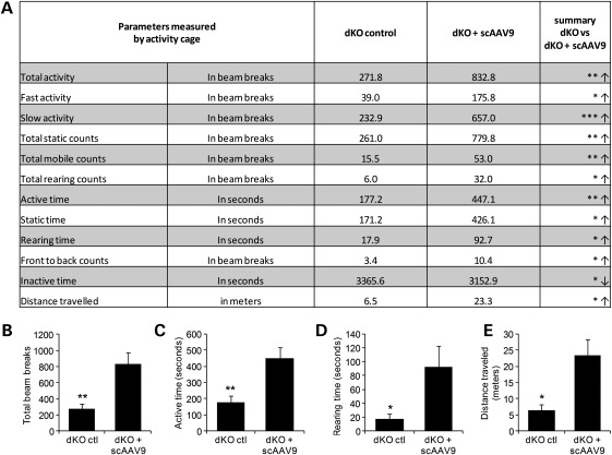
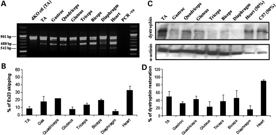
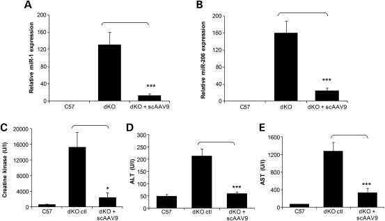
Duchenne muscular dystrophy (DMD) is a severe neuromuscular disorder caused by mutations in the dystrophin gene that result in the absence of functional protein. Antisense-mediated exon skipping is one of the most promising approaches for the treatment of DMD and recent clinical trials have demonstrated encouraging results. However, antisense oligonucleotide-mediated exon skipping for DMD still faces major hurdles such as extremely low efficacy in the cardiac muscle, poor cellular uptake and relatively rapid clearance from circulation, which means that repeated administrations are required to achieve some therapeutic efficacy. To overcome these limitations, we previously proposed the use of small nuclear RNAs (snRNAs), especially U7snRNA to shuttle the antisense sequences after vectorization into adeno-associated virus (AAV) vectors. In this study, we report for the first time the efficiency of the AAV-mediated exon skipping approach in the utrophin/dystrophin double-knockout (dKO) mouse which is a very severe and progressive mouse model of DMD. Following a single intravenous injection of scAAV9-U7ex23 in dKO mice, near-normal levels of dystrophin expression were restored in all muscles examined, including the heart. This resulted in a considerable improvement of their muscle function and dystrophic pathology as well as a remarkable extension of the dKO mice lifespan. These findings suggest great potential for AAV-U7 in systemic treatment of the DMD phenotype.


Figures

Figure 1.
Figure 1.
Exon-skipping and dystrophin expression in tissues from dKO mice following one i.v. injection of scAAV9-U7ex23. (A) RT–PCR analysis to detect exon 23 skipping efficiency at the RNA level. The 901 bp product represents the full-length transcript, and the products of 688 and 542 bp represent transcripts that exclude exon 23 and exons 22 and 23, respectively. (B) Quantification of exon 23 skipping measured by Taqman qPCR. Exon 23 skipping is expressed as a percentage of total dystrophin, measured by the exon 4–5 expression level, after normalization with an endogenous control (n = 6). (C) Western blot to detect dystrophin expression in tissues from scAAV9-treated dKO mice, compared with C57BL6 control mice (top gel). Equal loading of 100 µg protein is shown for each sample with α-actinin expression detected as a loading control except for the C57BL6 samples for which only 50 µg was loaded (bottom gel). (D) Quantification of levels of dystrophin protein restored in various muscles. Membranes were converted to numerical pictures by scanning, and band intensities were analysed using the ImageJ 1.33a software. Dystrophin levels are expressed as percentage compared with levels in wild-type tissue.
Figure 2.
Figure 2.
Dystrophin restoration in treated dKO muscles. Immunostaining of muscle tissue cross-sections to detect dystrophin expression and localization in C57BL6 normal control mice (left panel), untreated dKO mice (middle panel) and scAAV9-U7ex23-treated mice (right panel). Tissues analysed were from TA, gastrocnemius, quadriceps, biceps, diaphragm and heart muscles (scale bar = 100 µm).
Figure 3.
Figure 3.
scAAV-U7ex23 treatment improves muscle histopathology in dKO mice. (A) The left panel shows immunofluorescent staining for dystrophin (green) of tibialis anterior cross-sections from C57BL6 normal control mice (up), untreated dKO mice (middle) and peptide conjugated PMO treated mice (bottom). The lower panel shows haematoxylin and eosin staining of the same muscles. (B) Percentage of myofibres with centronucleation (n = 600–1800 myofibres/cohort). ***P < 0.001 when compared with untreated dKO mice. (C) Expression of dystrophin restores the DAPC to the sarcolemma in the scAAV9-treated dKO mice. DAPC components α- and β-sarcoglycan and β-dystroglycan were detected by immunostaining in tissue cross-section of TA muscles from scAAV9-treated dKO mice (lower panel), compared with C57BL6 normal mice (upper panel) and untreated dKO mice (middle panel) (scale bar = 100 µm).
Figure 4.
Figure 4.
Improvement of muscle function in scAAV9-treated mice. (A) Forelimb muscle function assessment shows the physical improvement of scAAV-treated dKO mice compared with untreated dKO mice and C57BL6 normal control mice (n = 6 per cohort). ***P < 0.005 compared with untreated dKO mice. (B) EDL muscles of scAAV-treated dKO mice were analysed for their maximal force (peak force) producing capacity compared with untreated dKO mice and C57BL6 control mice. Maximal force was also normalized for cross-sectional area to assess specific force. ***P < 0.005 compared with untreated dKO mice. Specific force is not significantly different between treated dKO and C57Bl6 (P > 0.09). (C) The percentage of force drop is assessed by measuring the force deficit following a series of five eccentric contractions. *P < 0.05 compared with untreated dKO mice. Error bars are shown as mean ± SEM (n = 6 per cohort).
Figure 5.
Figure 5.
scAAV9-U7ex23 treatment averts the onset of dystrophic pathology in the dKO mice. (A) Photograph of an untreated dKO mouse at 12 weeks of age (left), displaying a strong kyphosis and joint contractures compared with an scAAV9-U7ex23-treated mouse at 24 weeks of age (right), looking healthy. (B) Average lifespan of untreated dKO and scAAV9-U7ex23-treated dKO mice. (C) Survival curves of untreated dKO (dash line) and scAAV9-U7ex23-treated dKO mice (continuous line).
Figure 6.
Figure 6.
Improvement of physical activity and behaviour following scAAV9-U7ex23 injection. Mice were analysed at 10 weeks of age with open-field behavioural activity cages. (A) The 12 most relevant out of 22 recorded parameters are reported. Arrows indicate an increase (↑) or a decrease (↓) in the value of the measured parameter for the treated dKO mice compared with untreated controls. (B) Total activity, (C) total active time, (D) rearing time and (E) distance travelled are represented as graphs. *P < 0.05, **P < 0.01 and ***P < 0.005 compared with untreated dKO mice. Error bars are shown as mean ± SEM (n = 6 per cohort).
Figure 7.
Figure 7.
Analysis of exon-skipping and dystrophin expression at an endpoint in scAAV9-U7ex23-treated dKO mice. (A) RT–PCR analysis to detect exon 23 skipping efficiency at the RNA level. The 901 bp product represents the full-length transcript, and the products of 688 and 542 bp represent transcripts that exclude exon 23 and exons 22 and 23, respectively. (B) Quantification of exon 23 skipping measured by Taqman qPCR. Exon 23 skipping is expressed as a percentage of total dystrophin, measured by the exon 4–5 expression level, after normalization with an endogenous control. (C) Western blot to detect dystrophin expression in tissues from scAAV9-treated dKO mice, compared with untreated dKO and C57BL6 control mice (top gel). Equal loading of 100 µg protein is shown for each sample with α-actinin expression detected as a loading control, except for the heart and the C57BL6 control, where only 50% were loaded (bottom gel). (D) Quantification of levels of dystrophin protein restored in various muscles. Membranes were converted to numerical pictures by scanning, and band intensities were analysed using the ImageJ 1.33a software. Dystrophin levels are expressed as percentage compared with levels in wild-type tissue.
Figure 8.
Figure 8.
scAAV9-U7ex23 injection leads to a decrease in circulating biomarkers levels. (A and B) dKO mice were treated with a single i.v. injection of scAAV9-U7ex23 at 3 weeks of age, and blood serum was sampled at 40 weeks of age. Total RNA was extracted from serum and qPCR was utilized to calculate the relative expression of miR-1 and miR-206 in untreated and treated mice to healthy controls. miR-223 expression was used as an endogenous control for ΔΔCt analysis as described previously (29). Data indicate the mean and standard deviation of the expression levels of a minimum of five mice for each treatment. ***P < 0.001. (C) Measurement of serum CK levels as an index of ongoing muscle membrane instability in scAAV9-U7ex23-treated dKO mice compared with untreated dKO mice (*P < 0.005) and C57BL6 normal control mice. (D and E) Measurement of serum ALT and AST scAAV9-U7ex23-treated dKO mice compared with untreated dKO mice (***P < 0.001) and C57BL6 normal control mice.

Similar articles

Cited by

References

    1. Monaco A.P., Bertelson C.J., Liechti-Gallati S., Moser H., Kunkel L.M. An explanation for the phenotypic differences between patients bearing partial deletions of the DMD locus. Genomics. 1988;2:90–95. - PubMed
    1. Koenig M., Beggs A.H., Moyer M., Scherpf S., Heindrich K., Bettecken T., Meng G., Muller C.R., Lindlof M., Kaariainen H., et al. The molecular basis for Duchenne versus Becker muscular dystrophy: correlation of severity with type of deletion. Am. J. Hum. Genet. 1989;45:498–506. - PMC - PubMed
    1. Aartsma-Rus A., Janson A.A., Heemskerk J.A., De Winter C.L., Van Ommen G.J., Van Deutekom J.C. Therapeutic modulation of DMD splicing by blocking exonic splicing enhancer sites with antisense oligonucleotides. Ann. N. Y. Acad. Sci. 2006;1082:74–76. - PubMed
    1. Mann C.J., Honeyman K., Cheng A.J., Ly T., Lloyd F., Fletcher S., Morgan J.E., Partridge T.A., Wilton S.D. Antisense-induced exon skipping and synthesis of dystrophin in the mdx mouse. Proc. Natl Acad. Sci. USA. 2001;98:42–47. - PMC - PubMed
    1. van Deutekom J.C., Janson A.A., Ginjaar I.B., Frankhuizen W.S., Aartsma-Rus A., Bremmer-Bout M., den Dunnen J.T., Koop K., van der Kooi A.J., Goemans N.M., et al. Local dystrophin restoration with antisense oligonucleotide PRO051. N. Engl. J. Med. 2007;357:2677–2686. - PubMed

Publication types