

Inflammation assessment in patients with arthritis using a novel in vivo fluorescence optical imaging technology
- PMID: 22388997
- PMCID: PMC3298665
- DOI: 10.1136/annrheumdis-2010-148288
Inflammation assessment in patients with arthritis using a novel in vivo fluorescence optical imaging technology
Erratum in
- Ann Rheum Dis. 2012 Oct;71(10):1756
- Ann Rheum Dis. 2013 Jan;72(1):156
Abstract
Background: Indocyanine green (ICG)-enhanced fluorescence optical imaging (FOI) is an established technology for imaging of inflammation in animal models. In experimental models of arthritis, FOI findings corresponded to histologically proven synovitis. This is the first comparative study of FOI with other imaging modalities in humans with arthritis.
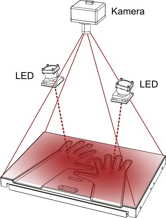
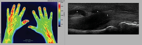
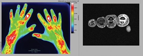
Methods: 252 FOI examinations (Xiralite system, mivenion GmbH, Berlin, Germany; ICG bolus of 0.1 mg/kg/body weight, sequence of 360 images, one image per second) were compared with clinical examination (CE), ultrasonography (US) and MRI of patients with arthritis of the hands.
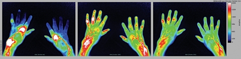
Results: In an FOI sequence, three phases could be distinguished (P1-P3). With MRI as reference, FOI had a sensitivity of 76% and a specificity of 54%, while the specificity of phase 1 was 94%. FOI had agreement rates up to 88% versus CE, 64% versus greyscale US, 88% versus power Doppler US and 83% versus MRI, depending on the compared phase and parameter. FOI showed a higher rate of positive results compared to CE, US and MRI. In individual patients, FOI correlated significantly (p<0.05) with disease activity (Disease Activity Score 28, r=0.41), US (r=0.40) and RAMRIS (Rheumatoid Arthritis MRI Score) (r=0.56). FOI was normal in 97.8% of joints of controls.
Conclusion: ICG-enhanced FOI is a new technology offering sensitive imaging detection of inflammatory changes in subjects with arthritis. FOI was more sensitive than CE and had good agreement with CE, US in power Doppler mode and MRI, while showing more positive results than these. An adequate interpretation of an FOI sequence requires a separate evaluation of all phases. For the detection of synovitis and tenosynovitis, FOI appears to be as informative as 1.5 T MRI and US.
Conflict of interest statement
Figures
References
-
- Saag KG, Teng GG, Patkar NM, et al. American College of Rheumatology 2008 recommendations for the use of nonbiologic and biologic disease-modifying antirheumatic drugs in rheumatoid arthritis. Arthritis Rheum 2008;59:762–84 - PubMed
-
- Brown AK, Quinn MA, Karim Z, et al. Presence of significant synovitis in rheumatoid arthritis patients with disease-modifying antirheumatic drug-induced clinical remission: evidence from an imaging study may explain structural progression. Arthritis Rheum 2006;54:3761–73 - PubMed
-
- Brown AK, Conaghan PG, Karim Z, et al. An explanation for the apparent dissociation between clinical remission and continued structural deterioration in rheumatoid arthritis. Arthritis Rheum 2008;58:2958–67 - PubMed
-
- Taylor PC, Steuer A, Gruber J, et al. Comparison of ultrasonographic assessment of synovitis and joint vascularity with radiographic evaluation in a randomized, placebo-controlled study of infliximab therapy in early rheumatoid arthritis. Arthritis Rheum 2004;50:1107–16 - PubMed
Publication types
MeSH terms
Substances
LinkOut - more resources
Full Text Sources
Other Literature Sources
Medical
