. 2012 Feb 1;125(Pt 3):724-34.
doi: 10.1242/jcs.092726.

N-WASP-mediated invadopodium formation is involved in intravasation and lung metastasis of mammary tumors

Affiliations

N-WASP-mediated invadopodium formation is involved in intravasation and lung metastasis of mammary tumors

Bojana Gligorijevic et al. J Cell Sci. .

Abstract

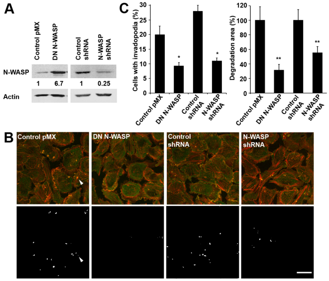
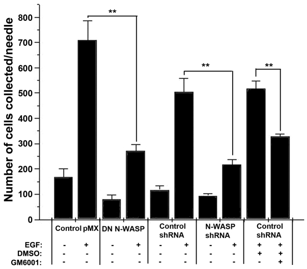
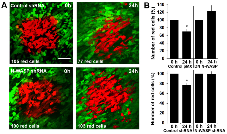
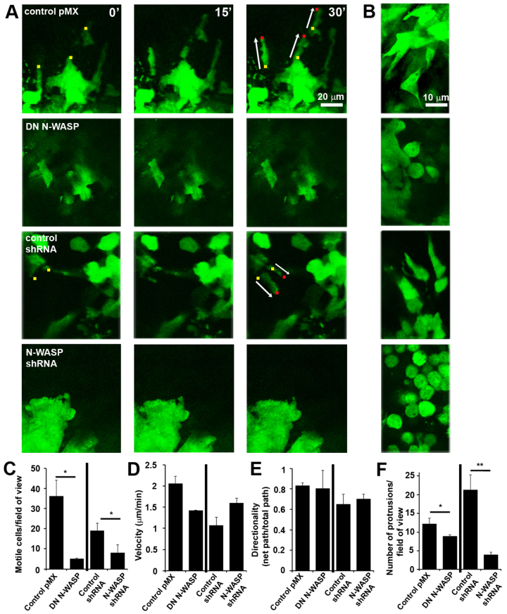
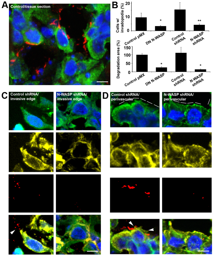
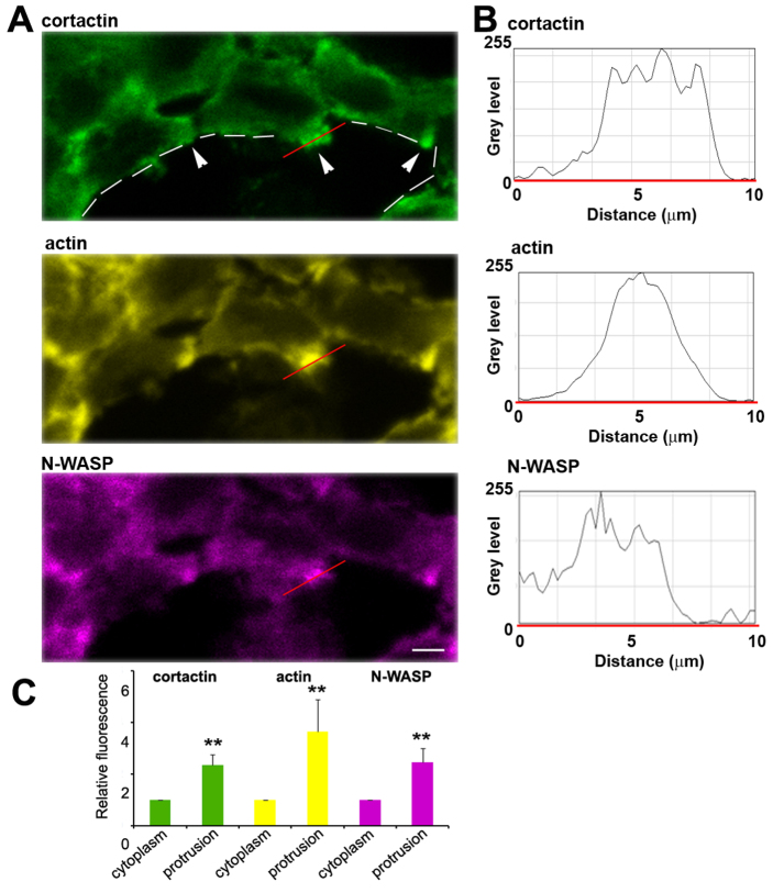
Invadopodia are proteolytic membrane protrusions formed by highly invasive cancer cells, commonly observed on substrate(s) mimicking extracellular matrix. Although invadopodia are proposed to have roles in cancer invasion and metastasis, direct evidence has not been available. We previously reported that neural Wiskott-Aldrich syndrome protein (N-WASP), a member of WASP family proteins that regulate reorganization of the actin cytoskeleton, is an essential component of invadopodia. Here, we report that N-WASP-mediated invadopodium formation is essential in breast cancer invasion, intravasation and lung metastasis. We established stable cell lines based on MTLn3 rat mammary adenocarcinoma cells that either overexpressed a dominant-negative (DN) N-WASP construct or in which N-WASP expression was silenced by a pSuper N-WASP shRNA. Both the N-WASP shRNA and DN N-WASP cells showed a markedly decreased ability to form invadopodia and degrade extracellular matrix. In addition, formation of invadopodia in primary tumors and collagen I degradation were reduced in the areas of invasion (collagen-rich areas in the invasive edge of the tumor) and in the areas of intravasation (blood-vessel-rich areas). Our results suggest that tumor cells in vivo that have a decreased activity of N-WASP also have a reduced ability to form invadopodia, migrate, invade, intravasate and disseminate to lung compared with tumor cells with parental N-WASP levels.


Figures

Fig. 1.
Fig. 1.
Establishment of MTLn3 cell lines expressing DN N-WASP and shRNA constructs and effects on invadopodium formation and activity. (A) Relative expression levels of N-WASP protein measured in lysates of control MTLn3 (pMX and scrambled shRNA) cells or MTLn3 cells stably overexpressing DN N-WASP or N-WASP shRNA. Immunoblotting was done with anti-N-WASP or anti-β-actin antibodies. (B) Representative images of MTLn3 control (pMX and scrambled shRNA) cells and cells expressing DN N-WASP or N-WASP shRNA cultured on a fluorescent gelatin-based matrix. Fixed cells were labeled for anti-cortactin (green) and actin (phalloidin, red). Upper panels show invadopodia (anti-cortactin and phalloidin colocalization – yellow inside cells); lower panels show sites of degraded gelatin substrate (white). Scale bar: 20 μm. The arrowhead points to an invadopodium and its corresponding hole in the matrix. (C) The number of cells with invadopodia (dots where cortactin and actin are colocalized) and the percentage of degraded matrix were calculated in control, DN -N-WASP and N-WASP shRNA cell lines. Error bars indicate the s.e.m. of three separate experiments. In each determination, ≥20 different fields of view with ≥100 cells were analyzed; error bars indicate the s.e.m.; *P<0.05, **P<0.01, Student's t-test.
Fig. 2.
Fig. 2.
Tumor cell invasion in vivo in response to EGF requires functional N-WASP. MTLn3 control cells (pMX or scrambled shRNA) or MTLn3 cells stably expressing DN N-WASP or N-WASP shRNA constructs were injected into the mammary glands of rats and formed orthotopic primary tumors. The ability of these cells to invade from primary tumors into microneedles filled with Matrigel with or without (+/−) 25 nM EGF (see Materials and Methods) was measured by counting the cells recovered from the needle after staining with DAPI. In addition, inhibition of invasion in the absence or presence of 10 μM GM6001, an MMP inhibitor, was measured. **P<0.01, Student's t-test.
Fig. 3.
Fig. 3.
The presence of circulating tumor cells and formation of lung metastasis requires N-WASP in rat and mouse mammary tumors. (A,B) Rat tumor-cell-derived mammary tumors in rat. (A) Number of viable circulating tumor cells per milliliter of blood; (B) Number of lung micrometastases. (C,D) Rat tumor-cell-derived mammary tumors in SCID mice. (C) Number of viable circulating tumor cells per milliliter of blood; (D) number of lung metastases. Error bars indicate the s.e.m.; *P<0.05, **P<0.01, verified by Mann–Whitney test.
Fig. 4.
Fig. 4.
Intravasation of tumor cells in the primary tumor requires N-WASP. (A) At 0 hours, a region of cells was photoconverted (red) in highly vascular regions of the tumors grown from either Dendra2–control-shRNA MTLn3 (top) or Dendra2–N-WASP-shRNA MTLn3 (bottom). At 0 hours and 24 hours z-stacks of 0–100 μm were collected in the green and red channels, and maximum projections are shown. This intravasation assay documents that there is less movement of tumor cells into blood vessels from tumors derived from Dendra2–N-WASP shRNA MTLn3 cells. Scale bar: 70 μm. (B) Number of red tumor cells remaining around blood vessels when normalized to the number at 0 hours. Measurements were based on three or four animals per cell line, 5–11 regions per animal. Error bars indicate the s.e.m. *P<0.05, Student's t-test.
Fig. 5.
Fig. 5.
Cell motility and protrusion formation in vivo requires N-WASP. (A) Motility of control and N-WASP-deficient GFP-labeled cells was monitored over time at different regions of the tumor using time-lapse images taken at 0, 15 and 30 minutes (images at same z-sections are shown). Small colored squares indicate the cell fronts (yellow at 0 minutes, red at 30 minutes) and arrows show the length of the trajectory of the migrating cells. (B) Higher magnifications of regions where cells are sparsely distributed showing that more protrusions are present in control tumors. (CF) Data from both control and N-WASP-deficient tumors showing the number of motile cells per 4D stack (C), velocity of motile cells (D), directionality of motile cells (E) and number of protrusions per 4D stack (F). *P<0.05, **P<0.01, Student's t-test, from three or more different tumors. The 4D stacks were 512×512×100 μm×30 minutes; n=30.
Fig. 6.
Fig. 6.
N-WASP-sensitive protrusions observed in tumors contain invadopodium markers and have degradation activity. (A) Matrix degradation sites detected by immunofluorescence (Collagen 3/4; red) in control shRNA tissue slices (anti-cortactin, green; DAPI, blue). Scale bar: 10 μm. (B) Quantification of the average number of protrusions, and collagen-I-degraded area in control tumors, DN N-WASP and N-WASP-shRNA tumors, as determined with the Collagen 3/4 assay. *P<0.05, Student's t-test. (C,D) Immunohistofluorescence of control shRNA tumors and N-WASP shRNA tumors in (C) the invasive edge and (D) areas next to major blood vessels (perivascular). DAPI is blue, anti-cortactin is green, phalloidin is yellow, Collagen 3/4 is red. The bottom row of images are merged images of all the markers. White arrowheads point to protrusions associated with degradation activity. Scale bars: 10 μm.
Fig. 7.
Fig. 7.
Protrusions observed in tumors are enriched in actin, cortactin and N-WASP. (A) Cerulean–N-WASP–MTLn3 tumor sections (purple) in the area next to a blood vessel (dashed white line), additionally stained by anti-cortactin (green) and phalloidin (yellow). Scale bar: 7 μm. (B) Intensity profiles of cortactin, actin and N-WASP along the red line shown in A illustrating enrichment of these proteins in the protrusion. (C) Levels of cortactin (green), actin (yellow) and N-WASP (purple) in protrusions relative to the average cytoplasmic levels illustrating enrichment of these proteins in protrusions in Cerulean–N-WASP–MTLn3 tumors. Error bars indicate the s.e.m. for >15 protrusions; **P<0.01, Student's t-test.

References

    1. Banzai Y., Miki H., Yamaguchi H., Takenawa T. (2000). Essential role of neural Wiskott-Aldrich syndrome protein in neurite extension in PC12 cells and rat hippocampal primary culture cells. J. Biol. Chem. 275, 11987-11992 - PubMed
    1. Buccione R., Orth J. D., McNiven M. A. (2004). Foot and mouth: podosomes, invadopodia and circular dorsal ruffles. Nat. Rev. Mol. Cell Biol. 5, 647-657 - PubMed
    1. Chudakov D. M., Lukyanov S., Lukyanov K. A. (2007). Tracking intracellular protein movements using photoswitchable fluorescent proteins PS-CFP2 and Dendra2. Nat. Protoc. 2, 2024-2032 - PubMed
    1. Clark E. S., Whigham A. S., Yarbrough W. G., Weaver A. M. (2007). Cortactin is an essential regulator of matrix metalloproteinase secretion and extracellular matrix degradation in invadopodia. Cancer Res. 67, 4227-4235 - PubMed
    1. Condeelis J., Segall J. E. (2003). Intravital imaging of cell movement in tumours. Nat. Rev. Cancer 3, 921-930 - PubMed

Publication types

MeSH terms

Substances