. 2012 Feb 1;125(Pt 3):777-88.
doi: 10.1242/jcs.097956.

Regulation of invadopodia formation and activity by CD147

Affiliations

Regulation of invadopodia formation and activity by CD147

G Daniel Grass et al. J Cell Sci. .

Abstract

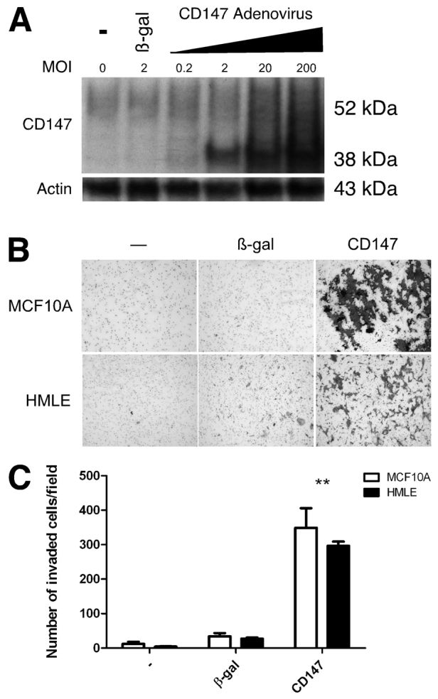
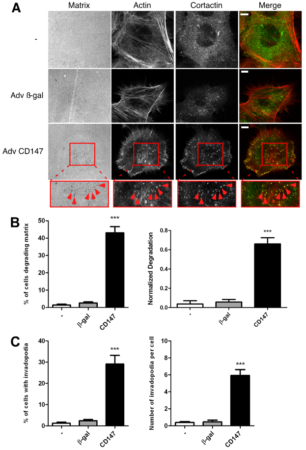
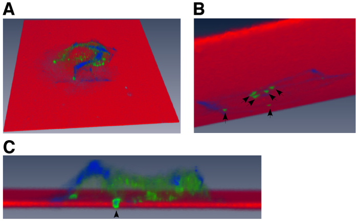
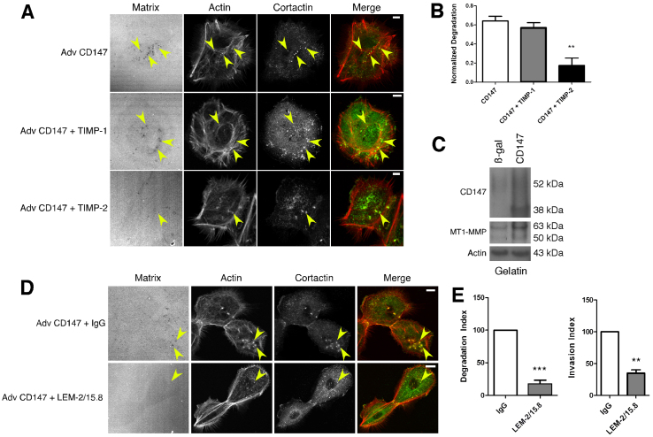
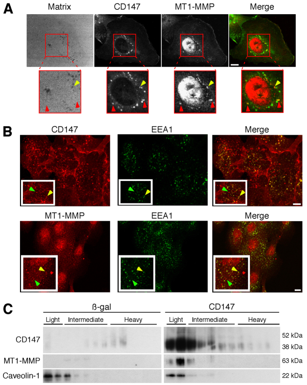
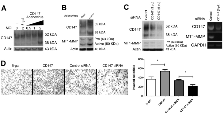
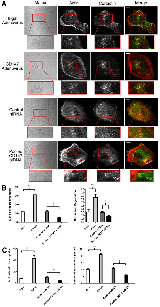
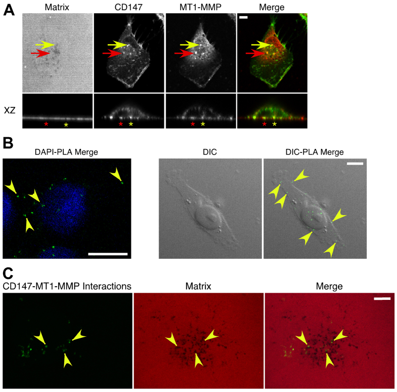
A defining feature of malignant tumor progression is cellular penetration through the basement membrane and interstitial matrices that separate various cellular compartments. Accumulating evidence supports the notion that invasive cells employ specialized structures termed invadopodia to breach these structural barriers. Invadopodia are actin-based, lipid-raft-enriched membrane protrusions containing membrane-type-1 matrix metalloproteinase (MT1-MMP; also known as matrix metalloproteinase 14; MMP14) and several signaling proteins. CD147 (emmprin, basigin), an immunoglobulin superfamily protein that is associated with tumor invasion and metastasis, induces the synthesis of various matrix metalloproteinases in many systems. In this study we show that upregulation of CD147 is sufficient to induce MT1-MMP expression, invasiveness and formation of invadopodia-like structures in non-transformed, non-invasive, breast epithelial cells. We also demonstrate that CD147 and MT1-MMP are in close proximity within these invadopodia-like structures and co-fractionate in membrane compartments with the properties of lipid rafts. Moreover, manipulation of CD147 levels in invasive breast carcinoma cells causes corresponding changes in MT1-MMP expression, invasiveness and invadopodia formation and activity. These findings indicate that CD147 regulates invadopodia formation and activity, probably through assembly of MT1-MMP-containing complexes within lipid-raft domains of the invadopodia.


Figures

Fig. 1.
Fig. 1.
Upregulation of CD147 in non-transformed breast epithelial cells increases invasion. (A) Western blot showing endogenous CD147 protein expression in MCF-10A cells and increased CD147 expression after upregulation with increasing MOI of CD147 adenovirus. An MOI of 2 was used in all subsequent experiments. (B) MCF-10A or HMLE cells that were untreated or infected with β-gal or CD147 adenovirus were plated on Matrigel-coated invasion chambers and analyzed for invasion. (C) Quantification of invasion; the mean number of cells (± s.e.m.) invaded per field in three independent experiments. **P≤0.01.
Fig. 2.
Fig. 2.
CD147 induces the formation of matrix-degrading invadopodia-like structures in non-transformed breast epithelial cells. MCF-10A cells, non-infected or infected with β-gal or CD147 adenovirus, were cultured on a fluorescent gelatin matrix for 15 hours. (A) Representative micrographs of non-infected, β-gal- and CD147-adenovirus-infected cells cultured on fluorescent matrix. After fixation, cells were immunolabeled for cortactin (primary 4F11 antibody followed by secondary Alexa-Fluor-488 antibody) and Alexa-Fluor-647–phalloidin. Actin and the gelatin matrix were pseudo-colored red and blue, respectively allowing easier visualization of colocalization (yellow) of actin (red) and cortactin (green). Invadopodia-mediated matrix degradation appears as dark black foci in the bright fluorescent matrix field. The boxed regions are shown at higher magnification below. These are examples of invadopodia (arrowheads), identified by colocalization of cortactin and actin punctae over areas of degraded matrix. Scale bars: 10 μm. (B,C) Quantification of invadopodia characteristics. (B) Left panel: percentage of cells degrading the matrix. Right panel: normalized degradation, calculated as area of degradation divided by the total cell area defined by the actin channel. (C) Left panel: percentage of cells with active invadopodia, defined as cortactin–actin aggregates over degraded matrix. Right panel: number of invadopodia per cell. Each parameter was calculated by evaluating 10 random fields containing at least 15 cells per field over three independent experiments. Values are means ± s.e.m. ***P≤0.001.
Fig. 3.
Fig. 3.
Three-dimensional volumetric reconstruction of CD147-induced invadopodia-like structures penetrating the underlying matrix. CD147-adenovirus-infected MCF-10A cells cultured on a fluorescent gelatin matrix were analyzed using confocal microscopy. Leica SP5 confocal software was employed to obtain an optimal image stack from single cell images and was further processed with Amira software to reconstruct a 3D volumetric image of a cell producing invadopodia-like protrusions that are penetrating the underlying fluorescent matrix. Cells were stained with Alexa-Fluor-647–phalloidin (blue) to image actin filaments and cortactin (green) to identify invadopodia; the fluorescent matrix is depicted in red. (A) Overhead view of a single cell cultured on fluorescent matrix. (B) View underneath the fluorescent matrix showing approximately six invadopodia (arrows) penetrating the matrix. (C) Magnified cross-section through the cell demonstrating one of the invadopodia-like protrusions (actin and cortactin positive; arrowhead) penetrating the fluorescent matrix. See supplementary material Movies 1 and 2.
Fig. 4.
Fig. 4.
CD147-induced, invadopodia-mediated, matrix degradation is dependent on membrane-type MMPs. (A,B) MCF-10A cells infected with CD147 adenovirus were pre-treated with the protease inhibitors, TIMP-1 (0.5 μg/ml) and TIMP-2 (0.5 μg/ml) for 30 minutes and then plated on fluorescent matrices for 12 hours in the presence of these inhibitors. (A) Representative micrographs of matrix degradation in control and CD147-upregulated cells in the presence of protease inhibitors. Scale bars: 10 μm. (B) Quantification of normalized degradation area, expressed as means ± s.e.m. **P≤0.01. The experiment was repeated three times. (C) Western blot of CD147 and MT1-MMP in aliquots of lysates obtained from MCF-10A cells that were cultured on gelatin and treated with control (β-gal) or CD147 adenovirus. Actin was used as a loading control; n=3. (D,E) MCF-10A cells infected with CD147 adenovirus were pre-treated with function-blocking antibody against MT1-MMP (LEM-2/15.8; 12 μg/ml) or control IgG and then plated on fluorescent matrices for 12 hours in the presence of the blocking antibody or IgG. (D) Representative micrographs of matrix degradation in CD147-upregulated cells in the presence of blocking antibody or IgG. Scale bars: 10 μm. (E) Left panel: quantification of normalized degradation area. Right panel: quantification of invasion through the Matrigel. Values are means ± s.e.m. **P≤0.01; ***P≤0.001; experiments were repeated three times.
Fig. 5.
Fig. 5.
CD147 associates with MT1-MMP in invadopodia-like structures and in membrane sub-fractions with the properties of lipid rafts. (A) Representative micrographs of CD147 and MT1-MMP colocalizing over degraded matrix in CD147-overexpressing MCF-10A cells. Yellow arrows indicate colocalization of CD147 (green) and MT1-MMP (red) over foci of degraded matrix; red arrows indicate colocalization of CD147 and MT1-MMP over areas of matrix that is not degraded. Scale bar: 10 μm. (B) Representative micrographs showing colocalization of CD147 (top panels) or MT1-MMP (bottom panels) with EEA1-positive vesicles. Yellow arrows indicate colocalization of proteins; green arrows indicate lack of colocalization. Scale bars: 10 μm. (C) MCF-10A cells infected with β-gal or CD147 adenovirus were plated on gelatin-coated plates overnight and were subjected to detergent-resistant membrane isolation. Light fractions are from the gradient interface (0–20%) where detergent-resistant membrane domains such as lipid rafts localize; n=2.
Fig. 6.
Fig. 6.
CD147 regulates MT1-MMP expression and Matrigel invasion in invasive breast cancer cells. (A) Western blot showing endogenous CD147 protein expression, CD147 expression following treatment with control adenovirus (β-gal), and CD147 expression following treatment with increasing amounts of recombinant CD147 adenovirus in MDA-MB-231 cells; n=2. An MOI of 2 was chosen for upregulating CD147 in subsequent experiments. β-actin was used as the loading control. (B) Representative western blot of increased MT1-MMP levels following CD147 upregulation in MDA-MB-231 cells; n=3. β-gal treatment was used as the control; β-actin demonstrates equal loading. (C) Left panel: western blot demonstrating that knockdown of CD147 with increasing concentrations (3 and 6 μl) of pooled target-specific siRNA results in decreased MT1-MMP protein expression. Non-specific siRNA was used as a control and β-actin was used to demonstrate equal loading; n=3. Right panel: RT-PCR analysis of CD147 and MT1-MMP mRNA after treatment with control or CD147-targeted siRNAs. (D) Left panel: representative images of Matrigel invasion assay with MDA-MB-231 cells treated with β-gal or CD147 adenovirus or with control or CD147-specific siRNA. Right panel: quantification of invaded cells per field by evaluating four separate fields in three independent experiments. Values are means ± s.e.m. *P≤0.05.
Fig. 7.
Fig. 7.
CD147 regulates invadopodia dynamics in invasive breast cancer cells. (A) Representative micrographs of MDA-MB-231 cells treated with β-gal or CD147 adenovirus or with control or CD147-targeted siRNA. Cells were cultured on Alexa-Fluor-568-conjugated gelatin for 5 hours, fixed and probed for actin (Alexa-Fluor-647–phalloidin) and cortactin (primary 4F11 antibody followed by secondary Alexa-Fluor-488 antibody). Actin and the gelatin matrix were pseudo-colored red and blue, respectively; this allows for easier visualization of colocalization (yellow) of actin (red) and cortactin (green). The left panels show invadopodia-mediated matrix degradation (dark holes). Higher magnifications of the boxed regions show invadopodia in more detail. Scale bars: 10 μm. (B,C) Quantification of invadopodia characteristics for MDA-MB-231 cells. (B) Left panel: percentage of cells degrading the matrix; calculated as cells with at least one area of degradation underneath the cell or near the cell border. Right panel: normalized degradation, calculated as area of degradation divided by the total cell area defined by the actin channel. (C) Left panel: percentage of cells with invadopodia, defined as actin–cortactin aggregates localized over degraded matrix. Right panel: number of invadopodia per cell in cells exhibiting at least one invadopodium. Each parameter was calculated by evaluating 10 random fields containing at least 20 cells per field over three independent experiments. Values are means ± s.e.m. *P≤0.05, **P≤0.01.
Fig. 8.
Fig. 8.
Colocalization of CD147 and MT1-MMP in invasive breast cancer cells. (A) Representative images demonstrating CD147 and MT1-MMP colocalization in MDA-MB-231 cells. Cells were cultured on Alexa-Fluor-568-conjugated gelatin for 5 hours and probed with Alexa-Fluor-488-conjugated CD147 and MT1-MMP followed by an Alexa-Fluor-647 secondary antibody. A polyclonal antibody against MT1-MMP was used for these images; a monoclonal antibody against MT1-MMP gave similar results (supplementary material Fig. S2). MT1-MMP and the gelatin matrix were pseudo-colored red and blue, respectively; this allowed easier visualization of colocalization (yellow) of CD147 (green) and MT1-MMP (red). Yellow arrow indicates colocalization of CD147 and MT1-MMP over foci of degraded matrix; red arrow indicates colocalization of CD147 and MT1-MMP over an area not showing degraded matrix. XZ section: yellow asterisk, CD147–MT1-MMP colocalization over a region of degraded matrix; red asterisk, CD147–MT1-MMP colocalization over an area not showing matrix degradation. n=3. (B,C) A proximity ligation assay (PLA) was employed to detect close protein–protein interactions (≤40 nm) of CD147 and MT1-MMP in MDA-MB-231 cells (see Materials and Methods for details). (B) Left panel: interactions of CD147 and MT1-MMP (small green dots at arrowheads); cells were also stained with DAPI (blue). Right panel: DIC images of a single cell; arrowheads indicate sites of CD147–MT1-MMP interactions, which are restricted to areas over the cell. (C) PLA combined with gelatin degradation assay, showing that CD147–MT1-MMP interactions occurred over areas of degradation (arrowheads), as well as over areas lacking degradation. Scale bars: 10 μm.

References

    1. Annabi B., Lachambre M., Bousquet-Gagnon N., Page M., Gingras D., Beliveau R. (2001). Localization of membrane-type 1 matrix metalloproteinase in caveolae membrane domains. Biochem. J. 353, 547-553 - PMC - PubMed
    1. Artym V. V., Zhang Y., Seillier-Moiseiwitsch F., Yamada K. M., Mueller S. C. (2006). Dynamic interactions of cortactin and membrane type 1 matrix metalloproteinase at invadopodia: defining the stages of invadopodia formation and function. Cancer Res. 66, 3034-3043 - PubMed
    1. Artym V. V., Yamada K. M., Mueller S. C. (2009). ECM degradation assays for analyzing local cell invasion. Methods Mol. Biol. 522, 211-219 - PubMed
    1. Belton R. J., Jr, Chen L., Mesquita F. S., Nowak R. A. (2008). Basigin-2 is a cell surface receptor for soluble basigin ligand. J. Biol. Chem. 283, 17805-17814 - PMC - PubMed
    1. Berditchevski F., Chang S., Bodorova J., Hemler M. E. (1997). Generation of monoclonal antibodies to integrin-associated proteins. Evidence that alpha3beta1 complexes with EMMPRIN/basigin/OX47/M6. J. Biol. Chem. 272, 29174-29180 - PubMed

Publication types

MeSH terms