. 2012 May;11(5):100-7.
doi: 10.1074/mcp.M111.015875. Epub 2012 Mar 4.

Lysine succinylation and lysine malonylation in histones

Affiliations

Lysine succinylation and lysine malonylation in histones

Zhongyu Xie et al. Mol Cell Proteomics. 2012 May.

Abstract

Histone protein post-translational modifications (PTMs) are significant for gene expression and DNA repair. Here we report the identification and validation of a new type of PTM in histones, lysine succinylation. The identified lysine succinylated histone peptides were verified by MS/MS of synthetic peptides, HPLC co-elution, and isotopic labeling. We identified 13, 7, 10, and 7 histone lysine succinylation sites in HeLa, mouse embryonic fibroblast, Drosophila S2, and Saccharomyces cerevisiae cells, respectively. We demonstrated that this histone PTM is present in all eukaryotic cells we examined. Mutagenesis of succinylation sites followed by functional assays implied that histone lysine succinylation can cause unique functional consequences. We also identified one and two histone lysine malonylation sites in HeLa and S. cerevisiae cells, respectively. Our results therefore increase potential combinatorial diversity of histone PTMs and suggest possible new connections between histone biology and metabolism.


Figures

Fig. 1.
Fig. 1.
Detection of lysine succinylation in core histones of different species. A, chemical structure and hypothesized mechanism for lysine succinylation. B, Western blot analysis (top panel) of core histones from S. cerevisiae, D. melanogaster, M. musculus, and H. sapiens cells with or without competition of a peptide library bearing a fixed unmodified lysine (K) or succinylated lysine (KSUCC). The bottom panel shows the loading control.
Fig. 2.
Fig. 2.
Identification of succinyllysine residues in histone proteins. A, illustration of identified histone succinylated sites from S. cerevisiae, D. melanogaster, M. musculus, and H. sapiens. Succinylation sites are shown in bold red type. Three malonyllysine sites identified in yeast and HeLa cells are also indicated. Reported acetylation sites are shown in blue type. B, position of succinylated residues in yeast nucleosome. The crystal structure is from the Protein Data Bank (code 1ID3) and is shown as spheres. DNA is shown as lines. The succinylated residues are labeled in color: yellow, H3K79; orange, H4K31; purple, H4K77; green, H2AK13; red, H2AK21; and blue, H2BK37.
Fig. 3.
Fig. 3.
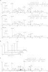
HPLC and mass spectrometric verification of histone succinylation at H4K31 (DNIQGITK+100.0150 PAIR). A, high resolution MS/MS spectra of H4K31 succinylation peptide (DNIQGITK+100.0150 PAIR) from affinity-enriched HeLa histone extract using anti-succinyllysine pan antibody (top), the synthetic DNIQGITKsuccPAIR (middle), and the mixture of them (bottom). The insets show the precursor ions. The label Δ designates b or y ions with water and/or ammonia loss. B, extracted ion chromatograms of the in vivo derived H4K31 peptide (top), its synthetic counterpart (middle), and their mixture (bottom). C, MS and MS/MS spectrum of D4-succinyllysine peptide.
Fig. 4.
Fig. 4.
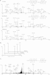
HPLC and mass spectrometric verification of histone succinylation at H4K31 (EIAQDFK+100.0152TDLR). A, high resolution MS/MS spectra of in vivo H3K79 succinylation peptide (EIAQDFK+100.0152TDLR) from affinity-enriched HeLa histone extract (top), the synthetic EIAQDFKsuccTDLR (middle), and the mixture of them (bottom). B, extracted ion chromatograms of the in vivo derived H3K79 peptide (top), its synthetic counterpart (middle), and their mixture (bottom). C, MS and MS/MS spectrum of D4-succinyllysine peptide.
Fig. 5.
Fig. 5.
Phenotypic analysis of mutations on succinylated residues in budding yeast. A, H4K31E mutant has severe growth defect. Circled in red are the spores with K31E mutants. The picture was taken after 4 days of incubation at 30 °C. B, histone H2AK21E mutant is specifically sensitive to methyl methanesulfonate. Two independent colonies containing the corresponding mutations were tested on either YPD or YPD-containing hydroxyurea (HU) or methyl methanesulfonate (MMS). The picture was taken after 3 days of incubation at 30 °C. C, histone H4K77E mutant is temperature-sensitive and loses silencing at both rDNA and telomere.

Similar articles

Cited by

References

    1. Kouzarides T. (2007) Chromatin modifications and their function. Cell 128, 693–705 - PubMed
    1. Dai C., Gu W. (2010) p53 post-translational modification: Deregulated in tumorigenesis. Trends Mol. Med. 16, 528–536 - PMC - PubMed
    1. Garcia B. A., Shabanowitz J., Hunt D. F. (2007) Characterization of histones and their post-translational modifications by mass spectrometry. Curr. Opin. Chem. Biol. 11, 66–73 - PubMed
    1. Sakabe K., Wang Z., Hart G. W. (2010) β-N-acetylglucosamine (O-GlcNAc) is part of the histone code. Proc. Natl. Acad. Sci. U.S.A. 107, 19915–19920 - PMC - PubMed
    1. Kruse J. P., Gu W. (2008) SnapShot: p53 posttranslational modifications. Cell 133, 930-30.e1. - PMC - PubMed

Publication types

MeSH terms