. 2012 May;194(9):2321-33.
doi: 10.1128/JB.00101-12. Epub 2012 Mar 2.

TprC/D (Tp0117/131), a trimeric, pore-forming rare outer membrane protein of Treponema pallidum, has a bipartite domain structure

Affiliations

TprC/D (Tp0117/131), a trimeric, pore-forming rare outer membrane protein of Treponema pallidum, has a bipartite domain structure

Arvind Anand et al. J Bacteriol. 2012 May.

Erratum in

  • J Bacteriol. 2014 Sep;196(18):3360

Abstract

Identification of Treponema pallidum rare outer membrane proteins (OMPs) has been a longstanding objective of syphilis researchers. We recently developed a consensus computational framework that employs a battery of cellular localization and topological prediction tools to generate ranked clusters of candidate rare OMPs (D. L. Cox et al., Infect. Immun. 78:5178-5194, 2010). TP0117/TP0131 (TprC/D), a member of the T. pallidum repeat (Tpr) family, was a highly ranked candidate. Circular dichroism, heat modifiability by SDS-PAGE, Triton X-114 phase partitioning, and liposome incorporation confirmed that full-length, recombinant TprC (TprC(Fl)) forms a β-barrel capable of integrating into lipid bilayers. Moreover, TprC(Fl) increased efflux of terbium-dipicolinic acid complex from large unilamellar vesicles and migrated as a trimer by blue-native PAGE. We found that in T. pallidum, TprC is heat modifiable, trimeric, expressed in low abundance, and, based on proteinase K accessibility and opsonophagocytosis assays, surface exposed. From these collective data, we conclude that TprC is a bona fide rare OMP as well as a functional ortholog of Escherichia coli OmpF. We also discovered that TprC has a bipartite architecture consisting of a soluble N-terminal portion (TprC(N)), presumably periplasmic and bound directly or indirectly to peptidoglycan, and a C-terminal β-barrel (TprC(C)). Syphilitic rabbits generate antibodies exclusively against TprC(C), while secondary syphilis patients fail to mount a detectable antibody response against either domain. The syphilis spirochete appears to have resolved a fundamental dilemma arising from its extracellular lifestyle, namely, how to enhance OM permeability without increasing its vulnerability to the antibody-mediated defenses of its natural human host.


Figures

Fig 1
Fig 1
TprC forms a β-barrel. (A) Tryptophan fluorescence emission spectra of unfolded and folded TprCFl in urea and DDM buffer, respectively. (B) CD spectra of unfolded and folded TprCFl (5 μM) in DDM buffer and OmpG (5 μM) in 50 mM NaCl, 10 mM Tris (pH 7.5), 0.2% n-octyl-β-d-glucopyranoside. (C) Heat modifiability of TprCFl, E. coli OmpG, 326βb, and 326P1-4+. Proteins were stained with GelCode Blue following SDS-PAGE with (+) or without (−) boiling in final sample buffer (SB). Molecular mass standards (kDa) for SDS-PAGE and immunoblot analyses are on the left of each panel. (D) T. pallidum samples (2 × 10⁸ organisms) were dissolved in SB with (+) or without (−) boiling and immunoblotted using rat anti-TprCSp antiserum.
Fig 2
Fig 2
TprC is amphiphilic and trimeric and displays pore-forming activity. (A) Ten micrograms each of TprCFl, 326P1-4+, and folded 326βb was phase partitioned in TX-114 and stained with GelCode Blue following SDS-PAGE. Lanes: aqueous (Aq) and detergent-enriched (Det) phases. (B) Liposomes were reconstituted with 10 μg each of TprCFl, 326P1-4+, or 326βb followed by sucrose density gradient ultracentrifugation. Fractions were subjected to immunoblotting with antisera directed against TprCFl, 326P1-4+, or 326βb following SDS-PAGE. Lanes: top fractions (TF) contain liposome-incorporated material whereas middle and bottom fractions (MF and BF, respectively) contain unincorporated material. (C) BN-PAGE and immunoblot analysis of TprCFl and T. pallidum lysate solubilized in 0.5% DDM with 50 mM Tris (pH 7.0). Molecular mass standards (kDa) are shown on the left. (D) Quenching of LUVs encapsulating Tb(DPA)33− after incubation with 100 nM TprCFl, E. coli OmpF, 326βb, or 326P1-4+ in 50 mM Tris (pH 7.5), 100 mM NaCl supplemented with 5 mM EDTA. Each bar represents the mean ± standard error of the mean for three independent experiments.
Fig 3
Fig 3
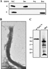
TprC expressed by T. pallidum associates with the peptidoglycan sacculus. (A) TX-114 phase partitioning of T. pallidum lysates (1 × 10⁹ organisms) without (−) or with (+) preincubation with 2% DDM. Lanes: whole cells (WC), TX-114-insoluble material (Ins), aqueous phase (Aq), and detergent-enriched phase (Det). (B) Extensively washed TX-114-insoluble material visualized in negatively stained whole mounts by transmission electron microscopy. (C) Immunoblot analysis of TX-114-insoluble material with anti-TprCSp or immune rabbit serum (IRS). Molecular mass standards (in kilodaltons) are shown at left.
Fig 4
Fig 4
TprC is expressed at low abundance and is accessible to proteinase K in motile treponemes. (A) Quantitative immunoblot analysis of TprC expressed in T. pallidum. T. pallidum lysates in amounts indicated were immunoblotted with anti-TprCSp antiserum followed by densitometric analysis; a standard curve generated from densitometric values obtained for graded amounts of TprCFl was used to determine the copy number of TprC per T. pallidum cell. Molecular mass standards (kDa) are indicated on the left. (B) Immunoblot analysis of TprC, detected by the anti-TprCSp antiserum, in motile treponemes (1.0 × 10⁸ T. pallidum organisms/lane) treated for 1 h with graded concentrations of proteinase K (PK). (C) PK accessibility of TprC and two periplasmic controls in intact and detergent-lysozyme-treated organisms incubated with (+) or without (−) 10 μg/ml of PK. Each lane represents 1.0 × 10⁸ T. pallidum organisms immunoblotted with antisera to TprCSp, TP0453, or TroA. (D) Live imaging of intact treponemes with and without incubation with 100 μg/ml of PK. Asterisks and arrowheads indicate flexing and wave form propagation, respectively. Time scale is 0 to 0.12 s.
Fig 5
Fig 5
TprC antibodies have opsonic activity. (A) Percentages of rabbit peritoneal macrophages containing internalized treponemes. Results shown are means ± standard errors of the means for three independent experiments. (B) Representative micrographs of macrophages incubated with motile treponemes and the indicated sera. Each image is a composite of bright-field images, nuclei stained with DAPI, and FITC-labeled T. pallidum. (C) Percentages of treponemes recovered following incubation of rabbit peritoneal macrophages for 4 h with the indicated sera. Results shown are means ± standard errors of the means for three independent experiments. P values of <0.05 (Student's t test) were considered significant.
Fig 6
Fig 6
TprCC contains the β-barrel of TprC and forms a trimer. (A to C) CD spectroscopy, heat modifiability, and TX-114 phase partitioning of TprCN and TprCC. Aq and Det, aqueous and detergent-enriched phases, respectively. (D) Quenching of Tb(DPA)33− encapsulated within LUVs following incubation with TprCFl, TprCN, TprCC, and E. coli OmpF. (E) SDS-PAGE of unboiled TprCN and TprCC (asterisk indicates TprCC trimer). Numbers at left in panels B and E are molecular masses in kilodaltons.
Fig 7
Fig 7
T. pallidum-infected rabbits, but not humans, mount an antibody response against TprC. (A) Reactivities of normal rabbit serum (NRS), immune rabbit serum (IRS; representative of 3 different animals), normal human serum (NHS), and pooled human secondary syphilitic sera (HSS) against folded, unboiled TprCFl; 326P1-4+; and folded, unboiled 326βb. (B) Immunoblot reactivities of individual patient sera. (C) Immunoblot reactivities of the anti-TprCFl antiserum and IRS used for the opsonophagocytosis assays in Fig. 5 against TprCN and folded, unboiled TprCC. Each immunoblot assay was performed using 100 ng of recombinant protein. Numbers at left are molecular masses in kilodaltons.
Fig 8
Fig 8
Topological models of TprC, TprI, and TprF. (A) Comparison of domain architectures. (B) TprC/D and TprI are proposed to consist of trimers with identical β-barrels and N-terminal periplasmic domains that directly or indirectly tether the barrels to the peptidoglycan (PG) sacculus. TprF is entirely periplasmic. TprCSp is depicted as periplasmic based on its solubility in aqueous buffer. TprI shares with TprF a putative periplasmic fragment not present in TprC.

Similar articles

Cited by

References

    1. Akins DR, et al. 1997. Tromp1, a putative rare outer membrane protein, is anchored by an uncleaved signal sequence to the Treponema pallidum cytoplasmic membrane. J. Bacteriol. 179:5076–5086 - PMC - PubMed
    1. Beher MG, Schnaitman CA, Pugsley AP. 1980. Major heat-modifiable outer membrane protein in gram-negative bacteria: comparison with the ompA protein of Escherichia coli. J. Bacteriol. 143:906–913 - PMC - PubMed
    1. Bennion D, Charlson ES, Coon E, Misra R. 2010. Dissection of β-barrel outer membrane protein assembly pathways through characterizing BamA POTRA 1 mutants of Escherichia coli. Mol. Microbiol. 77:1153–1171 - PMC - PubMed
    1. Bredemeier R, et al. 2007. Functional and phylogenetic properties of the pore-forming beta-barrel transporters of the Omp85 family. J. Biol. Chem. 282:1882–1890 - PubMed
    1. Brusca JS, Radolf JD. 1994. Isolation of integral membrane proteins by phase partitioning with Triton X-114. Methods Enzymol. 228:182–193 - PubMed

Publication types

LinkOut - more resources