. 2012;7(2):e32572.
doi: 10.1371/journal.pone.0032572. Epub 2012 Feb 28.

Cellular traction stresses increase with increasing metastatic potential

Affiliations

Cellular traction stresses increase with increasing metastatic potential

Casey M Kraning-Rush et al. PLoS One. 2012.

Abstract

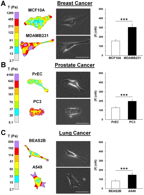
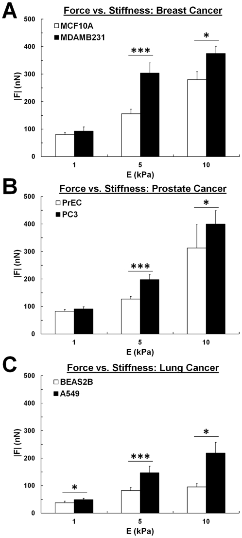
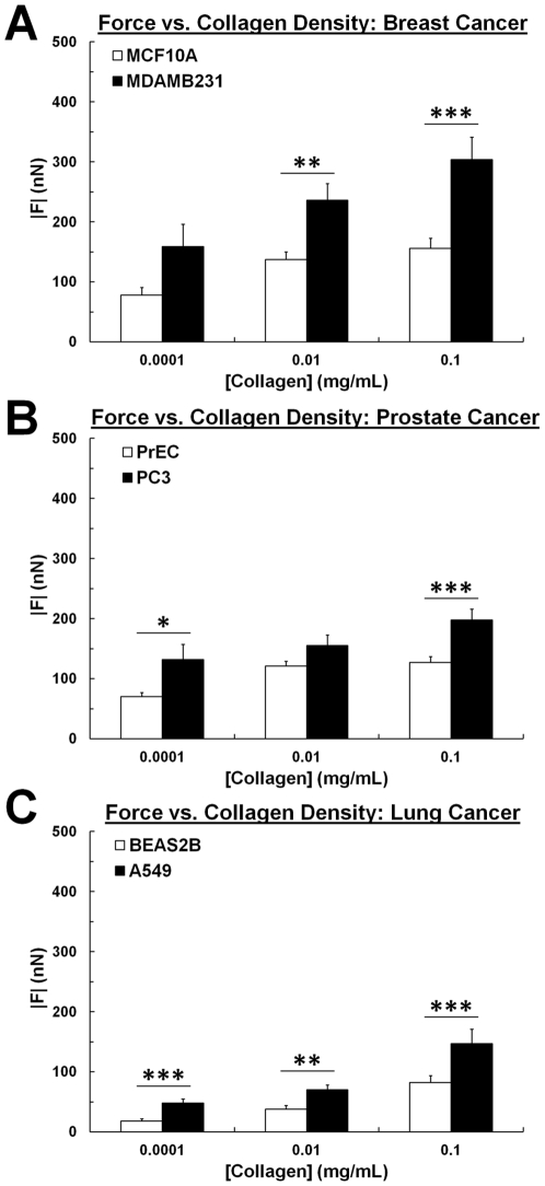
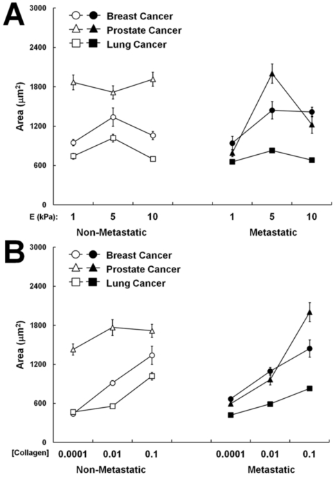
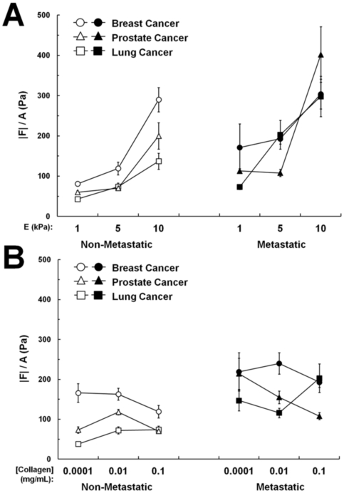
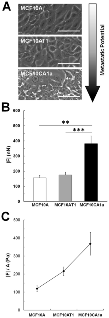
Cancer cells exist in a mechanically and chemically heterogeneous microenvironment which undergoes dynamic changes throughout neoplastic progression. During metastasis, cells from a primary tumor acquire characteristics that enable them to escape from the primary tumor and migrate through the heterogeneous stromal environment to establish secondary tumors. Despite being linked to poor prognosis, there are no direct clinical tests available to diagnose the likelihood of metastasis. Moreover, the physical mechanisms employed by metastatic cancer cells to migrate are poorly understood. Because metastasis of most solid tumors requires cells to exert force to reorganize and navigate through dense stroma, we investigated differences in cellular force generation between metastatic and non-metastatic cells. Using traction force microscopy, we found that in human metastatic breast, prostate and lung cancer cell lines, traction stresses were significantly increased compared to non-metastatic counterparts. This trend was recapitulated in the isogenic MCF10AT series of breast cancer cells. Our data also indicate that increased matrix stiffness and collagen density promote increased traction forces, and that metastatic cells generate higher forces than non-metastatic cells across all matrix properties studied. Additionally, we found that cell spreading for these cell lines has a direct relationship with collagen density, but a biphasic relationship with substrate stiffness, indicating that cell area alone does not dictate the magnitude of traction stress generation. Together, these data suggest that cellular contractile force may play an important role in metastasis, and that the physical properties of the stromal environment may regulate cellular force generation. These findings are critical for understanding the physical mechanisms of metastasis and the role of the extracellular microenvironment in metastatic progression.


Conflict of interest statement

Competing Interests: The authors have declared that no competing interests exist.

Figures

Figure 1
Figure 1. Metastatic cancer cells exert greater forces than non-metastatic cells.
(A) Representative traction maps (left), corresponding phase images (middle), and overall net traction force (|F|, right) of non-metastatic mammary epithelial (MCF10A) and highly metastatic (MDAMB231) cancer cells. (B) Representative traction maps (left), corresponding phase images (middle), and |F| (right) of non-metastatic primary prostate epithelial cells (PrEC) and highly metastatic prostate cancer cells (PC3). (C) Representative traction maps (left), corresponding phase images (middle), and |F| (right) of non-metastatic bronchial epithelial cells (BEAS2B) and metastatic lung adenocarcinoma cells (A549). All cells are on polyacrylamide substrates with Young's Modulus (E) = 5 kPa and type I collagen concentration of 0.1 mg/mL. Scale bar = 50 µm. Mean+SEM; *** indicates p<0.001.
Figure 2
Figure 2. Increased matrix stiffness contributes to increased force generation.
Net traction force (|F|) increases with increasing substrate stiffness (E = 1–10 kPa) for (A) MCF10A non-metastatic mammary epithelial cells and MDAMB231 metastatic cancer cells, (B) PrEC non-metastatic primary prostate epithelial cells and PC3 metastatic prostate cancer cells, and (C) BEAS2B non-metastatic bronchial epithelial cells and A549 metastatic lung cancer cells. Ligand density is maintained at 0.1 mg/mL collagen I. 5 kPa data is from Figure 1. Note also that the metastatic cancer cells (black) exert greater forces than non-metastatic cells (white) at all matrix stiffness levels studied. Mean+SEM; * indicates p<0.05; *** indicates p<0.001.
Figure 3
Figure 3. Increased collagen density contributes to increased force generation.
Net traction force (|F|) increases with collagen density (0.0001–0.1 mg/mL collagen I) for (A) MCF10A non-metastatic mammary epithelial cells and MDAMB231 metastatic cancer cells, (B) PrEC non-metastatic primary prostate epithelial cells and PC3 metastatic prostate cancer cells, and (C) BEAS2B non-metastatic bronchial epithelial cells and A549 metastatic lung cancer cells. Stiffness is maintained at E = 5 kPa. 0.1 mg/mL collagen I data is from Figure 1. Note also that the metastatic cancer cells (black) exert greater forces than non-metastatic cells (white) at all collagen densities studied. Mean+SEM; * indicates p<0.05; ** indicates p<0.01; *** indicates p<0.001.
Figure 4
Figure 4. Cell area is differentially altered by matrix stiffness and collagen density.
Projected cell area shows a biphasic relationship with substrate stiffness (A) and a direct relationship with collagen density (B) in the majority of the metastatic (black) and non-metastatic cells (white) studied. No consistent trend was observed when comparing the projected cell area of complementary metastatic and non-metastatic cells.
Figure 5
Figure 5. Average traction stress increases with stiffness, not collagen density.
|F| of each cell is normalized by its projected area (|F|/A) as a measurement of average traction stress. Average traction stresses increase with increasing substrate stiffness (A) but become relatively uniform with increasing collagen density (B) in both metastatic (black) and non-metastatic (white) cells studied.
Figure 6
Figure 6. Metastatic derivative in a series of isogenic cell lines exerts greater forces.
(A) Phase images of parental untransformed cells (MCF10A), transformed premalignant (MCF10AT1) and highly metastatic (MCF10CA1a) derivatives. (B) Net traction forces increase with increasing metastatic potential, with the highest forces exerted by the metastatic MCF10CA1a cells. (C) Average traction stress (|F|/A) increases with increasing metastatic potential. Mean ± SEM; ** indicates p<0.01; *** indicates p<0.001.

Similar articles

Cited by

References

    1. Steeg PS. Tumor metastasis: mechanistic insights and clinical challenges. Nat Med. 2006;12:895–904. - PubMed
    1. Christofori G. New signals from the invasive front. Nature. 2006;441:444–450. - PubMed
    1. Ravdin PM, Siminoff LA, Davis GJ, Mercer MB, Hewlett J, et al. Computer program to assist in making decisions about adjuvant therapy for women with early breast cancer. J Clin Oncol. 2001;19:980–991. - PubMed
    1. Sidransky D. Emerging molecular markers of cancer. Nat Rev Cancer. 2002;2:210–219. - PubMed
    1. Stacker SA, Caesar C, Baldwin ME, Thornton GE, Williams RA, et al. VEGF-D promotes the metastatic spread of tumor cells via the lymphatics. Nat Med. 2001;7:186–191. - PubMed

Publication types