. 2012 Mar 5:12:10.
doi: 10.1186/1471-213X-12-10.

Genetic and physical interaction of Meis2, Pax3 and Pax7 during dorsal midbrain development

Affiliations

Genetic and physical interaction of Meis2, Pax3 and Pax7 during dorsal midbrain development

Zsuzsa Agoston et al. BMC Dev Biol. .

Abstract

Background: During early stages of brain development, secreted molecules, components of intracellular signaling pathways and transcriptional regulators act in positive and negative feed-back or feed-forward loops at the mid-hindbrain boundary. These genetic interactions are of central importance for the specification and subsequent development of the adjacent mid- and hindbrain. Much less, however, is known about the regulatory relationship and functional interaction of molecules that are expressed in the tectal anlage after tectal fate specification has taken place and tectal development has commenced.

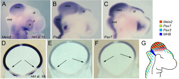
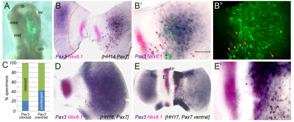
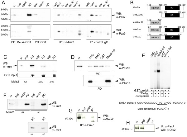
Results: Here, we provide experimental evidence for reciprocal regulation and subsequent cooperation of the paired-type transcription factors Pax3, Pax7 and the TALE-homeodomain protein Meis2 in the tectal anlage. Using in ovo electroporation of the mesencephalic vesicle of chick embryos we show that (i) Pax3 and Pax7 mutually regulate each other's expression in the mesencephalic vesicle, (ii) Meis2 acts downstream of Pax3/7 and requires balanced expression levels of both proteins, and (iii) Meis2 physically interacts with Pax3 and Pax7. These results extend our previous observation that Meis2 cooperates with Otx2 in tectal development to include Pax3 and Pax7 as Meis2 interacting proteins in the tectal anlage.

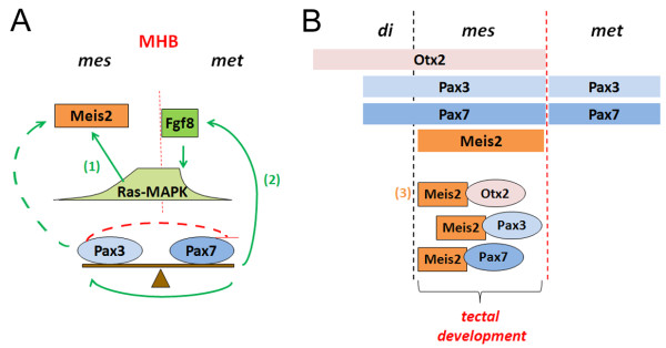
Conclusion: The results described here suggest a model in which interdependent regulatory loops involving Pax3 and Pax7 in the dorsal mesencephalic vesicle modulate Meis2 expression. Physical interaction with Meis2 may then confer tectal specificity to a wide range of otherwise broadly expressed transcriptional regulators, including Otx2, Pax3 and Pax7.


Figures

Figure 1
Figure 1
Meis2, Pax3 and Pax7 are expressed in nested domains in the HH15-19 chick midbrain. (A-G) Expression of Meis2 (A, D), Pax3 (B, E) and Pax7 (C, F) as detected by in-situ hybridization on HH15 whole chick embryos (A-C) or neighboring vibratome cross sections through a HH19 mesencephalic vesicle (D-F). (G) Schematic summary of the expression patterns. di: diencephalic vesicle; le: lens; mes: mesencephalic vesicle; met: metencephalic vesicle; rt: retina. The arrows in (D-F) mark the ventral border of the respective expression domains. Panel (D) was taken from [17].
Figure 2
Figure 2
Pax7 induces Pax3 expression in the mesencephalic vesicle. (A) Representative example of pMES-Pax7 electroporation into the mesencephalic vesicle. GFP expression, indicative of transgene expression, is restricted to the right half of the neural tube 4-6 hours later. (B-B") Flat mount preparation of a HH14 chick neural tube, 16 hours after electroporation of 2 μg/μl pMES-Pax7 into the mesencephalic vesicle; Pax3 expression is in dark blue, Nkx6.1 expression in pink, GFP in green. The dotted lines mark the dorso-ventral boundary, the arrow heads indicate representative examples of ectopic Pax7 positive cells co-expressing Pax3. Ectopic patches of Pax3 expression are restricted to the right, electroporated half of the mesencephalic vesicle. (B') is a higher magnification of (B) with the GFP fluorescent image superimposed onto the preparation. (B") shows the distribution of Pax7-GFP expressing cells in the specimen shown in (B'). (C) Quantification of the results: percent specimens with induced (green bars) and unaltered (blue bars) Pax3 expression following targeted electroporation of pMES-Pax7. (D) Flat mount preparation of a HH18 chick neural tube, 24 hours after electroporation of 2 μg/μl pMES-Pax7. (E) Targeted misexpression of pMES-Pax7 into the ventral mesencephalic vesicle showing cells ectopically expressing Pax3 within the Nkx6.1 domain. (E') is a higher magnification of (E). fb: forebrain; fp: floor plate; mes: mesencephalic vesicle; met: metencephalic vesicle; ov: optic vesicle; otv: otic vesicle. Scale bar (B, B'): 100 μm.
Figure 3
Figure 3
Pax7 represses Meis2 and efnb1 in the mesencephalic vesicle. (A-B") Meis2 expression in HH15 chick embryos electroporated with 2 μg/μl of a control vector carrying only GFP (A) or 2 μg/μl pMES-Pax7 (B, B") into the right wall of the mesencephalic vesicle. The insert in (A) shows a higher magnification and continuous expression of Meis2 in the control embryo. (B') is a higher magnification of the boxed area in (B). Arrowheads point to patches of cells with reduced Meis2 transcripts. In (B") the GFP fluorescence of the specimen shown in (B and B') is superimposed to visualize the extent of Pax7/GFP transfection. (C-D') efnb1 expression in a HH15 chick embryo transfected with a control vector (C) or with pMES-Pax7 (D, D') under identical experimental conditions. (D') is a higher magnification of (D). (E) Quantification of the results: percent specimens with reduced (red bars) and unaltered (blue bars) Meis2 or efnb1 expression following targeted electroporation of pMES-Pax7.
Figure 4
Figure 4
Dose-dependent repression of Meis2, efnb1 and Pax7 by Pax3. (A-C) Meis2 expression in HH15 embryos electroporated with indicated concentrations of pMIWIII-Pax3. Loss of Meis2 transcripts can be seen in random patches of the electroporated right half of the mesencephalic vesicle only after transfection of 2 μg/μl of pMIWIII-Pax3 (C). (D) Loss of efnb1 transcripts following transfection of 2 μg/μl pMIWIII-Pax3. (E, F) Pax7 expression in the mesencephalic vesicle upon transfection of 1 μg/μl (E) and 2 μg/μl (F) pMIWIII-Pax3. The boxed areas in (B, E) are shown at a higher magnification in the upper right corners of the respective panels. (G) Quantification of the results: percent specimens with reduced (red bars) and unchanged (blue bars) expression of Meis2 (left) or Pax7 (right) following electroporation of different concentrations of pMIWIII-Pax3.
Figure 5
Figure 5
Meis2 containing protein complexes in the mesencephalic vesicle. (A) Co-precipitation of Pax3 and Pax7 with Meis2 in GST-pull down (left panels) and immunoprecipitation experiments using a Meis2 specific antibody (right panels). Upper panel: Western Blot with an antibody directed against Pax7; lower panel: Western Blot against Pax3. (B) Schematic representation of the Meis2 deletions N-terminally fused to GST that were used in (A, C, D, E, and F). (C) Pull down experiments using full length Meis2 (Meis2[1-400]), Meis2 lacking the MEINOX-domain (Meis2[199-400]; ΔMD) or Meis2 lacking the homeodomain (Meis2[1-190]; ΔHD) probed for Pax7. (D) GST-pull down with full length Meis2, ΔMD, ΔHD and GST probed for Pbx1b. (E) Specific DNA complex formation on a 26-bp oligonucleodite and purified GST proteins. Complex formation is competed by specific oligomers at a 10×-molar ratio ('competitor'). The sequence of the 23P-labeled oligomer is indicated below. (F) GST-pull down with ΔN and ΔC probed for Pax3 (upper panel) and Pbx1b (lower panel). (G) Immunoprecipitation using a Pax7-specific antibody probed for Meis2. (H) The same blot as in (G) stripped and re-probed for Otx2. Cyto: cytoplasmic extract; In: input; IP: immunoprecipitate; no Ab: precipitation with protein G sepharose beads omitting the antibody; nucl: nuclear extract; PD: pull down; sup: supernatant; wash: final wash step of the precipitates; WB: Western blot: The asterisks in (A) and (C) mark unspecific bands
Figure 6
Figure 6
Model for a possible cooperation of Meis2, Pax3, Pax7 and Otx2during tectal development. See text for details. Red lines indicate negative regulation, green arrows positive regulation. Dashed lines indicate hypothetical direct regulation of the Meis2 promoter/enhancer by different Pax3 concentrations. Solid lines indicate indirect regulation of Meis2 expression via Pax3/7 mediated induction of Fgf8 as previously reported: (1) Regulation of Meis2 expression in response to Ras-MAPK signaling levels reported in [19]; (2) Induction of Fgf8 by Pax3 and Pax7 reported in [11]; (3) Existence of Meis2-Otx2 containing protein complexes in the tectal anlage reported in [17].

References

    1. Acampora D, Mazan S, Lallemand Y, Avantaggiato V, Maury M, Simeone A, Brulet P. Forebrain and midbrain regions are deleted in Otx2-/- mutants due to a defective anterior neuroectoderm specification during gastrulation. Development. 1995;121:3279–3290. - PubMed
    1. Matsuo I, Kuratani S, Kimura C, Takeda N, Aizawa S. Mouse Otx2 functions in the formation and patterning of rostral head. Genes Dev. 1995;9:2646–2658. doi: 10.1101/gad.9.21.2646. - DOI - PubMed
    1. Ang SL, Jin O, Rhinn M, Daigle N, Stevenson L, Rossant J. A targeted mouse Otx2 mutation leads to severe defects in gastrulation and formation of axial mesoderm and to deletion of rostral brain. Development. 1996;122:243–252. - PubMed
    1. Wurst W, Bally-Cuif L. Neural plate patterning: upstream and downstream of the isthmic organizer. Nat Rev Neurosci. 2001;2:99–108. doi: 10.1038/35053516. - DOI - PubMed
    1. Bally-Cuif L, Wassef M. Ectopic induction and reorganization of Wnt-1 expression in quail/chick chimeras. Development. 1994;120:3379–3394. - PubMed

Publication types

MeSH terms

LinkOut - more resources