. 2012 Mar 5:10:17.
doi: 10.1186/1741-7007-10-17.

Cnidocyte discharge is regulated by light and opsin-mediated phototransduction

Affiliations

Cnidocyte discharge is regulated by light and opsin-mediated phototransduction

David C Plachetzki et al. BMC Biol. .

Abstract

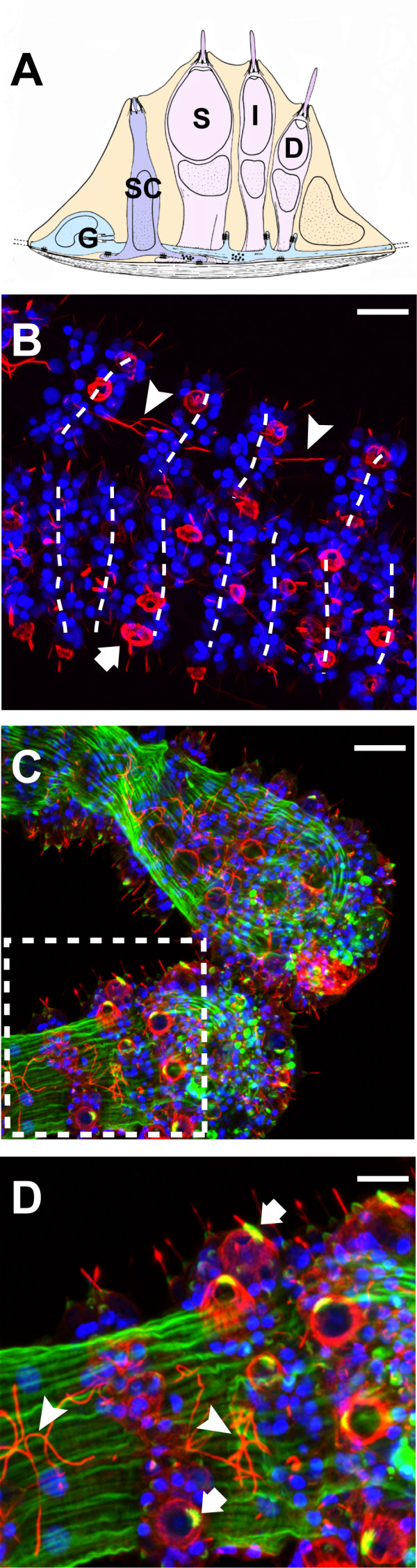
Background: Cnidocytes, the eponymous cell type of the Cnidaria, facilitate both sensory and secretory functions and are among the most complex animal cell types known. In addition to their structural complexity, cnidocytes display complex sensory attributes, integrating both chemical and mechanical cues from the environment into their discharge behavior. Despite more than a century of work aimed at understanding the sensory biology of cnidocytes, the specific sensory receptor genes that regulate their function remain unknown.

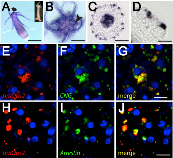
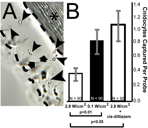
Results: Here we report that light also regulates cnidocyte function. We show that non-cnidocyte neurons located in battery complexes of the freshwater polyp Hydra magnipapillata specifically express opsin, cyclic nucleotide gated (CNG) ion channel and arrestin, which are all known components of bilaterian phototransduction cascades. We infer from behavioral trials that different light intensities elicit significant effects on cnidocyte discharge propensity. Harpoon-like stenotele cnidocytes show a pronounced diminution of discharge behavior under bright light conditions as compared to dim light. Further, we show that suppression of firing by bright light is ablated by cis-diltiazem, a specific inhibitor of CNG ion channels.

Conclusions: Our results implicate an ancient opsin-mediated phototransduction pathway and a previously unknown layer of sensory complexity in the control of cnidocyte discharge. These findings also suggest a molecular mechanism for the regulation of other cnidarian behaviors that involve both photosensitivity and cnidocyte function, including diurnal feeding repertoires and/or substrate-based locomotion. More broadly, our findings highlight one novel, non-visual function for opsin-mediated phototransduction in a cnidarian, the origins of which might have preceded the evolution of cnidarian eyes.


Figures

Figure 1
Figure 1
Cnidocyte morphology and arrangement in H. magnipapillata. (A) A generalized schematic of the hydrozoan battery complex in cross-section. Stenotele cnidocytes (S) are flanked by isorhiza (I) and desmoneme (D) cnidocytes, as well as sensory cells (SC), which form synaptic connections with ganglion cells (G) that link each of the other cnidocyte cell types. (B-D) Confocal z-stacks of H. magnipapillata tentacle midsection (B) and tentacle bulbs (C and D). Neurons, including cnidocytes, are stained red (anti acetylated α-Tubulin) and nuclei are stained blue (DAPI). Musculature is shown in green (phalloidin) in (C and D). Axonal projections (arrow heads) link battery complexes that occur with regular periodicity along the length of the tentacle. Arrows indicate stenotele cnidocytes. (D) is an inset of (C). (A) Redrawn with permission from Westfall [36].
Figure 2
Figure 2
Studies of gene expression suggest a role for opsin-mediated photosensitivity in the regulation of cnidocyte discharge. (A-D) Colorimetric in situ hybridization with HmOps2 probe. Opsin expression is strongly localized to the hypostome (arrow), tentacles, and the ring-like ganglion (arrow head) that surrounds the mouth (A and B). (C and D) Cryosection of tentacle reveals HmOps2 expression in non-cnidocyte cell types. Cnidocytes capsules are clearly visible in these preparations. (E-I) Confocal fluorescence in situ hybridization shows that opsin transcripts co-localize with CNG (E-G) and arrestin (H, I) in battery complexes of the hydra. Blue cells with dark centers are the capsules of stenotele cnidocytes, which stain with DAPI [62]. Signal in (E-I) and (E-G) is located at different focal planes relative to central stenotele cnidocytes. Inset of (A), sense riboprobe negative control. Scale bars in (A) = 1 mm, in (B) = 500 μm, in (C) = 100 μm, in (D) and (E-J) approximately 30 μm.
Figure 3
Figure 3
Behavioral studies of cnidocyte discharge indicate a significant inhibitory effect of light intensity on cnidocyte function. (A) Representative data for cnidocyte discharge assay. In this experiment stenotele cnidocytes were captured using gelatin-coated lengths of monofilament under different experimental conditions. (B) Significantly fewer cnidocysts were recovered from assays conducted in bright blue light (470 nm; 2.8 W/cm2) than in dim trials (470 nm; 0.1 W/cm2). Bright light suppression was ablated by the presence of the CNG inhibitor cis-diltiazem. Area between dashed lines in (A) = gelatin matrix; asterisk = monofilament; arrow heads = stenotele cnidocytes.

Comment in

  • A view to kill.
    Holstein TW. Holstein TW. BMC Biol. 2012 Mar 5;10:18. doi: 10.1186/1741-7007-10-18. BMC Biol. 2012. PMID: 22390773 Free PMC article.
  • Ubiquitin ligases and beyond.
    Dikic I, Robertson M. Dikic I, et al. BMC Biol. 2012 Mar 15;10:22. doi: 10.1186/1741-7007-10-22. BMC Biol. 2012. PMID: 22420755 Free PMC article. No abstract available.

References

    1. Hardie RC, Raghu P. Visual transduction in Drosophila. Nature. 2001;413:186–193. doi: 10.1038/35093002. - DOI - PubMed
    1. Matulef K, Zagotta WN. Cyclic nucleotide-gated ion channels. Annu Rev Cell Dev Biol. 2003;19:23–44. doi: 10.1146/annurev.cellbio.19.110701.154854. - DOI - PubMed
    1. Venkatachalam K, Montell C. TRP channels. Annu Rev Biochem. 2007;76:387–417. doi: 10.1146/annurev.biochem.75.103004.142819. - DOI - PMC - PubMed
    1. Plachetzki DC, Degnan BM, Oakley TH. The origins of novel protein interactions during animal opsin evolution. PLoS One. 2007;2:e1054. doi: 10.1371/journal.pone.0001054. - DOI - PMC - PubMed
    1. Suga H, Schmid V, Gehring WJ. Evolution and functional diversity of jellyfish opsins. Curr Biol. 2008;18:51–55. doi: 10.1016/j.cub.2007.11.059. - DOI - PubMed

Publication types