. 2012 Apr;84(2):203-24.
doi: 10.1111/j.1365-2958.2012.08023.x. Epub 2012 Mar 15.

Escherichia coli low-molecular-weight penicillin-binding proteins help orient septal FtsZ, and their absence leads to asymmetric cell division and branching

Affiliations

Escherichia coli low-molecular-weight penicillin-binding proteins help orient septal FtsZ, and their absence leads to asymmetric cell division and branching

Lakshmi-Prasad Potluri et al. Mol Microbiol. 2012 Apr.

Abstract

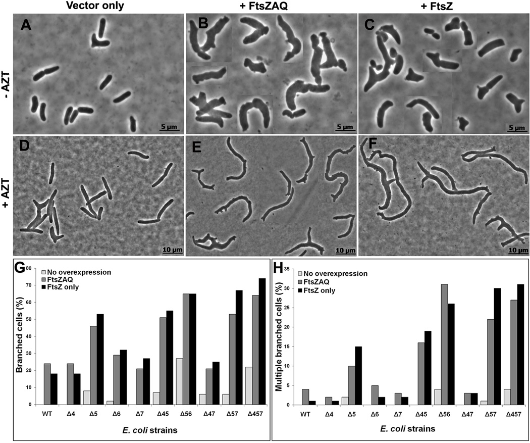
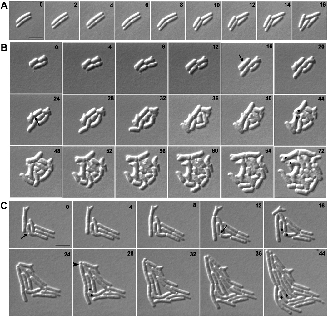
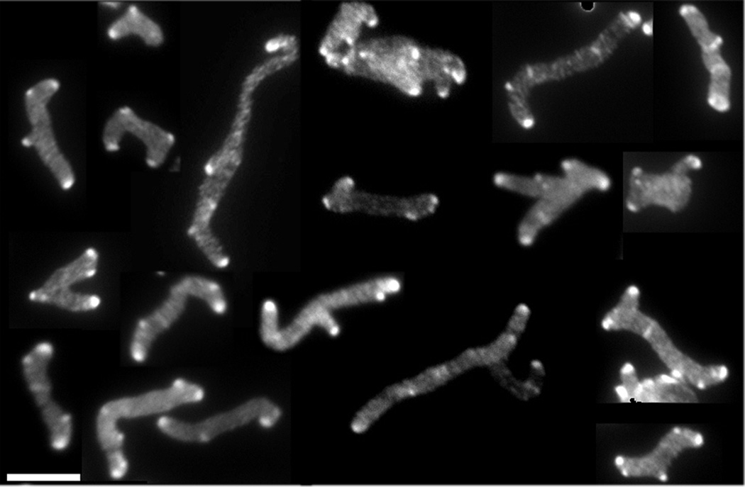
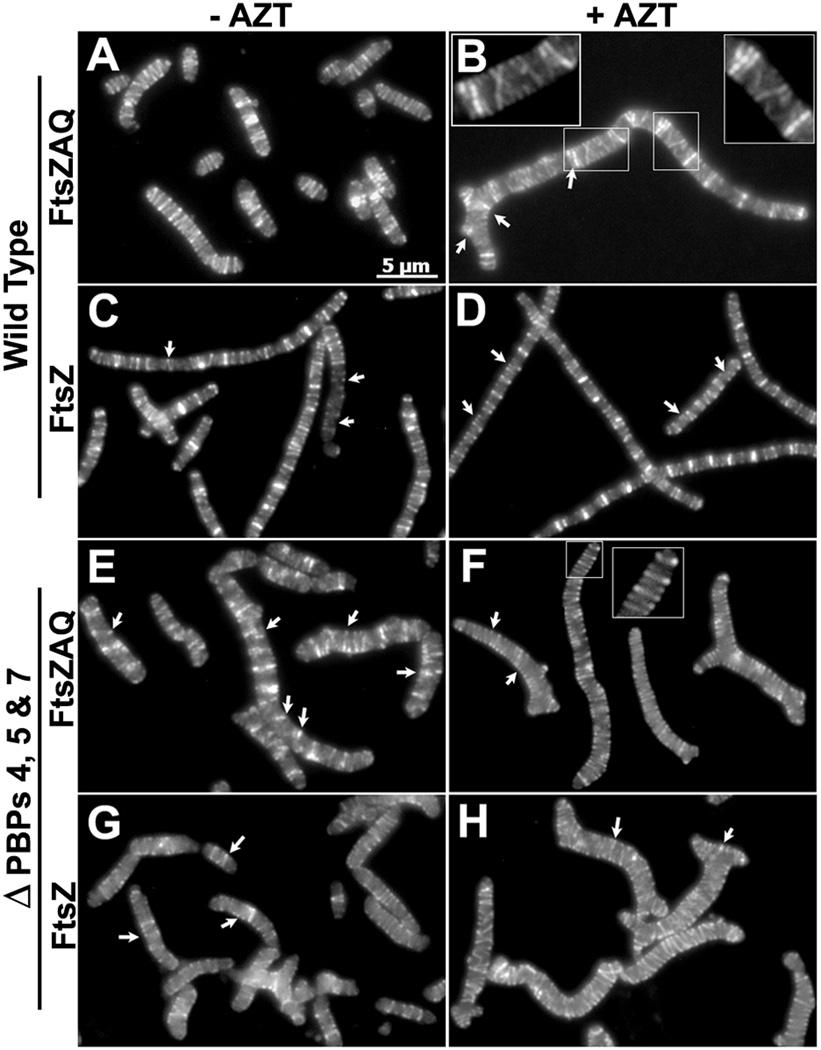
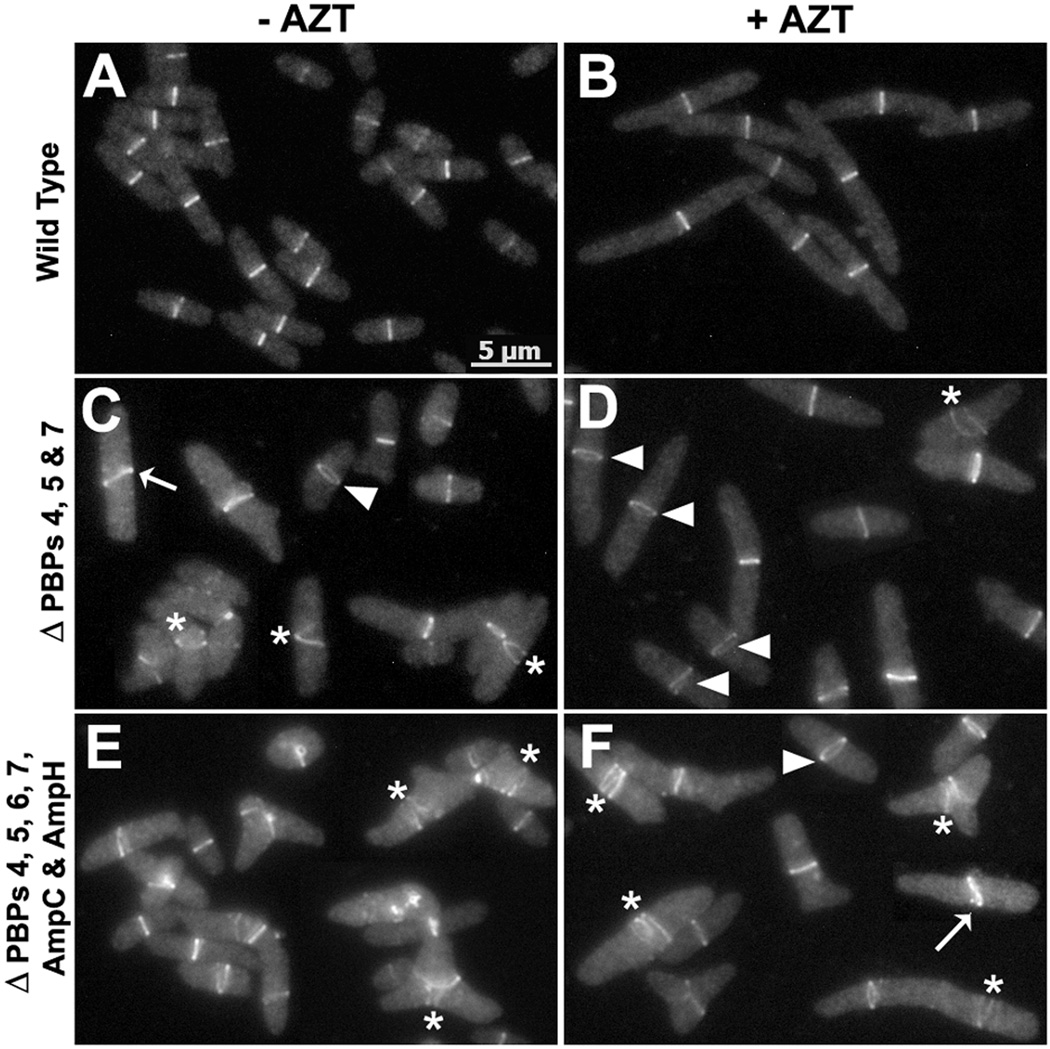
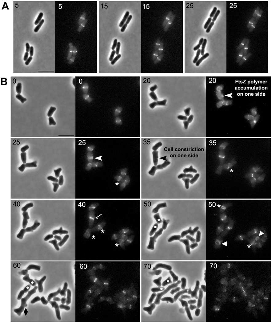
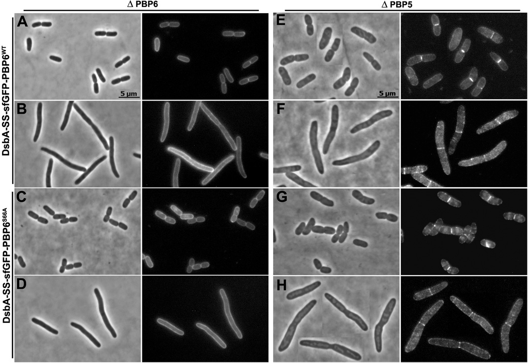
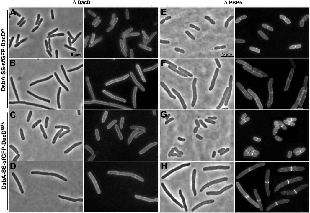
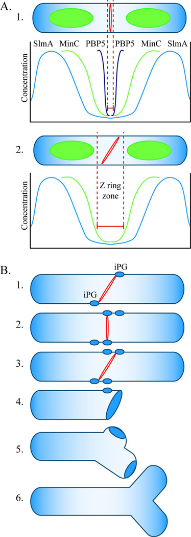
Escherichia coli cells lacking low-molecular-weight penicillin-binding proteins (LMW PBPs) exhibit morphological alterations that also appear when the septal protein FtsZ is mislocalized, suggesting that peptidoglycan modification and division may work together to produce cell shape. We found that in strains lacking PBP5 and other LMW PBPs, higher FtsZ concentrations increased the frequency of branched cells and incorrectly oriented Z rings by 10- to 15-fold. Invagination of these rings produced improperly oriented septa, which in turn gave rise to asymmetric cell poles that eventually elongated into branches. Branches always originated from the remnants of abnormal septation events, cementing the relationship between aberrant cell division and branch formation. In the absence of PBP5, PBP6 and DacD localized to nascent septa, suggesting that these PBPs can partially substitute for the loss of PBP5. We propose that branching begins when mislocalized FtsZ triggers the insertion of inert peptidoglycan at unusual positions during cell division. Only later, after normal cell wall elongation separates the patches, do branches become visible. Thus, a relationship between the LMW PBPs and cytoplasmic FtsZ ultimately affects cell division and overall shape.


Figures

Fig. 1
Fig. 1. FtsZ overexpression exacerbates branching in LMW PBP mutants
E. coli CS315-1, which lacks PBPs 4, 5 and 7, harboring plasmids pBR322 (A, D) pZAQ (B, E) and pLPZ (C, F), were grown in LB-Tet medium until they reached an OD600 of 0.2. At this OD, cells were either harvested for microscopy (A, B, C) or were further grown for one mass doubling (MD) in the presence of 1 µg/ml of aztreonam to filament the cells (D, E, F). Panels A–C and D–F have different magnifications. The scale bars in panels A–C denote 5 µm and in panels D–F denote 10 µm. Panel G shows the percentage of cells with branches, including single and multiple protrusions. Panel H shows the percentage of cells with multiple branches (more than 3 poles per cell). In panels G and H, the percentage of branched cells in different strains harboring pBR322, pZAQ and pLPZ plasmids are represented by light, grey and black bars, respectively. At least 500 cells were counted for each strain. Panels G and H represent results averaged from two independent experiments.
Fig. 2
Fig. 2. Branches arise from abnormal septation
E. coli strains A) CS109 (wild type), B) CS612-1 (Δ PBPs 4, 5, 6, 7, AmpC and AmpH), and C) CS109 harboring pLPZ, were grown and imaged as described in Experimental Procedures. Arrows in panels B and C represent abnormal cell constrictions. In panels B and C, dots were used to follow abnormal cell poles produced by abnormal cell constrictions and which gave rise to branches. The arrow head in panel C marks a mini-cell. The numbers on each image indicates the time in minutes. The scale bar equals 5 µm.
Fig. 3
Fig. 3. All branches have inert PG at their poles
E. coli CS703-1, a mutant lacking PBPs 1a, 4, 5, 6, 7, AmpC and AmpH, was grown in LB containing 100 µg/ml of D-cys for three generations, after which cells were chased in the absence of D-cys by growing them in LB plus 1 µg/ml of aztreonam (to inhibit cell division) for 2 h (~3 mass doublings). After the chase, sacculi were isolated and immunolabelled to detect D-cys residues, as described (de Pedro et al., 1997). This figure is a mosaic of images taken from different fields of view. The scale bar equals 5 µM.
Fig. 4
Fig. 4. FtsZ overexpression produces spiral and helical FtsZ rings and bands
E. coli LP18-1 (wild type) harboring pZAQ (A, B) or pLPZ (C, D), and E. coli LP1 (ΔPBPs 4, 5 and 7) harboring pZAQ (E, F) or pLPZ (G, H), were grown and imaged as described in Experimental Procedures. Cells in panels B, D, F and H were grown for 1 mass doubling in the presence of 1 µg/ml of aztreonam. The inset in panel B is an enlarged portion of the figure to show the FtsZ spirals more clearly. White arrows represent incomplete Z-rings and FtsZ arcs. All images have same magnification; the scale bar in panel A equals 5 µm.
Fig. 5
Fig. 5. LMW PBP mutants have aberrant Z rings
E. coli strains LP18-1 (wild type) (A, B), LP1 (ΔPBPs 4, 5 and 7) (C, D) and LP17-1 (ΔPBPs 4, 5, 6, 7, ampC and ampH) (E, F) were grown and imaged as described in Experimental Procedures. Cells in panels B, D and F were incubated for ~1 mass doubling in the presence of 1 µg/ml of aztreonam. FtsZ polymers that appear to be spirals or helices, incomplete Z rings, arcs and FtsZ polymers that did not form compact rings were considered to be spirals, and are illustrated by the Z ring marked by asterisks (*). Rings that are broader (take up more space than normal along the long axis of the cell) are represented by a triangle (▲); these Z-rings may be slanted but appear as rings instead of bands, probably because they are oriented differently. Slanted rings that are not perpendicular to the long axis of the cell are illustrated by those marked with white arrows. All images have the same magnification; the scale bar in panel A equals 5 µm.
Fig. 6
Fig. 6. Abnormal FtsZ polymers are responsible for abnormal cell constriction
E. coli strains LP18-1 (wild type) (A) and LP1 (Δ PBPs 4, 5 and 7) (B) were grown and imaged as described in Experimental Procedures. White arrow heads in panel B (at time 20 and 25 min) represent abnormal FtsZ polymers that formed on one side of the cell which eventually resulted in abnormal cell constriction (black arrowhead at 35 min). These events gave rise to abnormal poles, leading to the formation of a branch (white dots are inserted to mark and follow the development of selected abnormal cell poles). Abnormal FtsZ polymers are denoted by symbols described in Fig. 5. The numbers in each panel indicate time in minutes. Only selected time points were included in this figure for clarity (see Fig. S6 for a compilation of all time points). The scale bar in panels A and B equals 5 µm.
Fig. 7
Fig. 7. sfGFP-PBP6 localizes to the septum in cells lacking PBP5
E. coli CS17-1 (ΔPBP6) expressing DsbA-SS-sfGFP-PBP6 from pLP602 (A and B), or DsbA-SS-sfGFP-PBP6S66A from pLP607 (C and D). E. coli CS12-7 (Δ PBP5) expressing DsbA-SS-sfGFP-PBP6 from pLP602 (E and F), or DsbA-SS-sfGFP-PBP6S66A from pLP607 (G and H). Cells were grown and imaged as described in Experimental Procedures. Cells in panels B, D, F and H were filamented by treating with 1 µg/ml of aztreonam for 1 mass doubling. Each panel has a phase contrast image on the left and the corresponding fluorescence image on the right. All images have same magnification; the scale bars in panels A and E equal 5 µm.
Fig. 8
Fig. 8. sfGFP-DacD localizes to the septum in cells lacking PBP5
E. coli CS18-1 (ΔDacD) expressing DsbA-SS-sfGFP-DacD from pLP652 (A and B), or DsbA-SS-sfGFP-DacDS63A from pLP656 (C and D). E. coli CS12-7 (ΔPBP5) expressing DsbA-SS-sfGFP-DacD from pLP652 (E and F), or DsbA-SS-sfGFP-DacDS63A from pLP656 (G and H). Cells in panels B, D, F and H were filamented by treating with 1 µg/ml of aztreonam for 1 MD. Each panel has a phase contrast image on the left and the corresponding fluorescence image on the right. The fluorescence images in panels E, F, G and H were processed similarly. All images have same magnification, and the scale bars in panels A and E equal 5 µm.
Fig. 9
Fig. 9. Model for FtsZ ring localization and cell branching in E. coli
A. FtsZ ring localization. A1. In wild type E. coli, the FtsZ ring (red ring) is restricted to the center of an elongating cell (“Z ring zone” between dotted red lines) by three mechanisms. The SlmA protein is associated with nucleoids (green ovoids), and its concentration is lowest near the cell’s center and near the poles (blue line). This FtsZ inhibitor restricts Z ring formation to a relatively broad area at these three sites. The MinC protein prevents polymerization of FtsZ at the poles and restricts Z ring formation to a small region near the cell’s midpoint, where the concentration of this inhibitor is lowest (green line). PBP5 (black line) fine tunes the location of Z rings to an even narrower zone at the cell’s center. The action of PBP5 is probably aided by other LMW PBPs. A2. In the absence of PBP5, FtsZ can polymerize in a larger midcell zone to form a slanted Z ring (shown) or multiple Z rings (not shown). Before invagination begins, the orientation of midcell Z rings can vary within the permissive zone. B. Mechanism of cell branching. B1. In the absence of PBP5, slanted FtsZ rings can form at the center of the cell and initiate synthesis of inert peptidoglycan (iPG) (blue ovals). B2. Before invagination begins, the Z ring can reorient and initiate the synthesis of iPG at sites well separated from the first. B3. A non-perpendicular Z ring triggers invagination. B4. The daughter cells have one pole consisting of iPG, plus at least one other patch of iPG some distance away. B5. During subsequent cell growth, insertion of peptidoglycan into the sidewall separates the true pole from the patch of iPG. B6. Cell wall elongation around the iPG patch creates a branch and an ectopic pole.

Comment in

References

    1. Aarsman ME, Piette A, Fraipont C, Vinkenvleugel TM, Nguyen-Disteche M, den Blaauwen T. Maturation of the Escherichia coli divisome occurs in two steps. Mol. Microbiol. 2005;55:1631–1645. - PubMed
    1. Åkerlund T, Nordström K, Bernander R. Branched Escherichia coli cells. Mol. Microbiol. 1993;10:849–858. - PubMed
    1. Begg K, Nikolaichik Y, Crossland N, Donachie WD. Roles of FtsA and FtsZ in activation of division sites. J Bacteriol. 1998;180:881–884. - PMC - PubMed
    1. Bernhardt TG, de Boer PA. SlmA, a nucleoid-associated, FtsZ binding protein required for blocking septal ring assembly over chromosomes in E. coli. Molec. Cell. 2005;18:555–564. - PMC - PubMed
    1. Bi E, Lutkenhaus J. Isolation and characterization of ftsZ alleles that affect septal morphology. J. Bacteriol. 1992;174:5414–5423. - PMC - PubMed

Publication types

MeSH terms