Review
. 2012 Mar;76(1):66-112.
doi: 10.1128/MMBR.05011-11.

Phylogenetic framework and molecular signatures for the main clades of the phylum Actinobacteria

Affiliations
Review

Phylogenetic framework and molecular signatures for the main clades of the phylum Actinobacteria

Beile Gao et al. Microbiol Mol Biol Rev. 2012 Mar.

Abstract

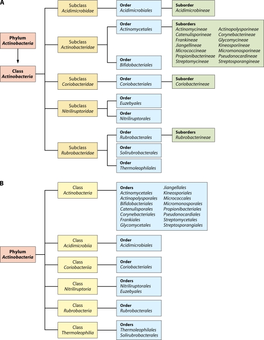
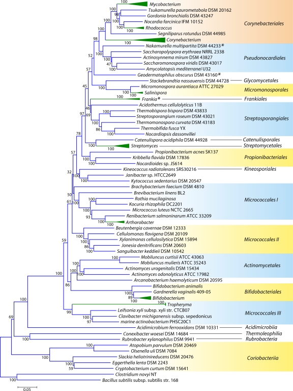
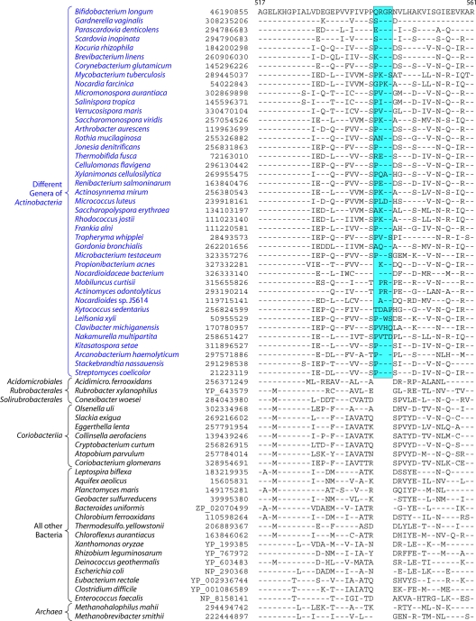
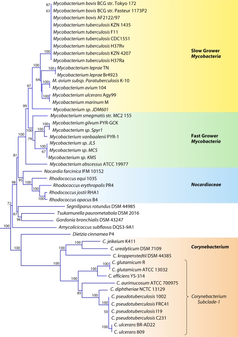
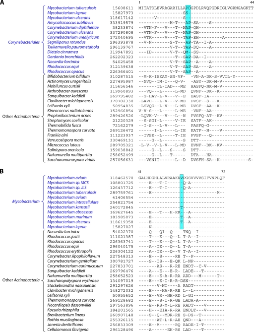
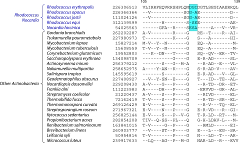
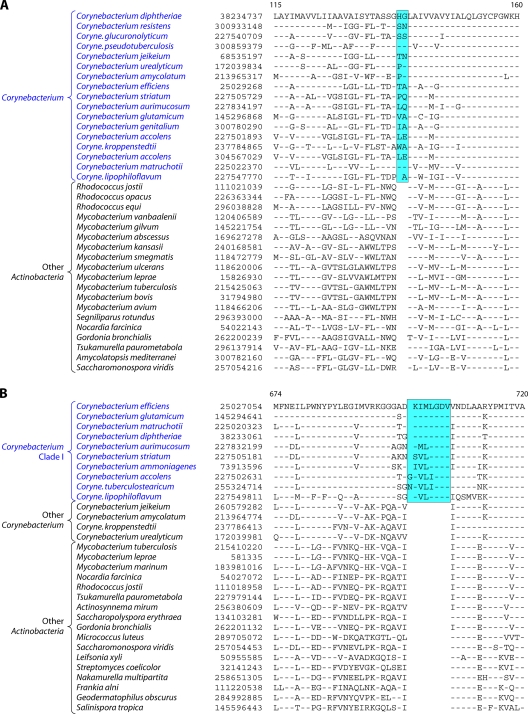
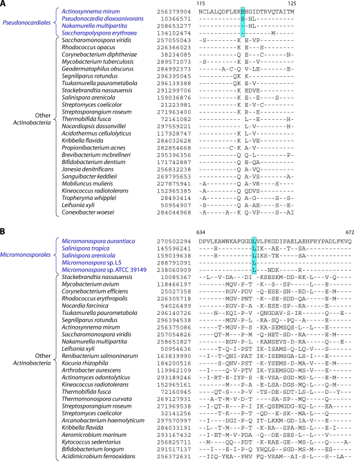
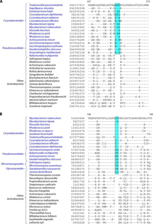
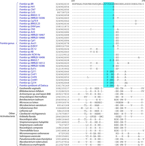
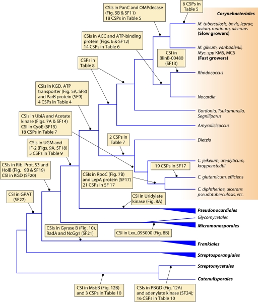
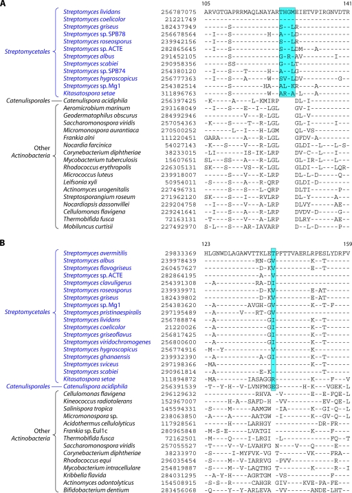
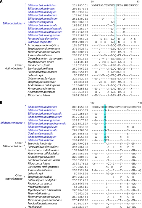
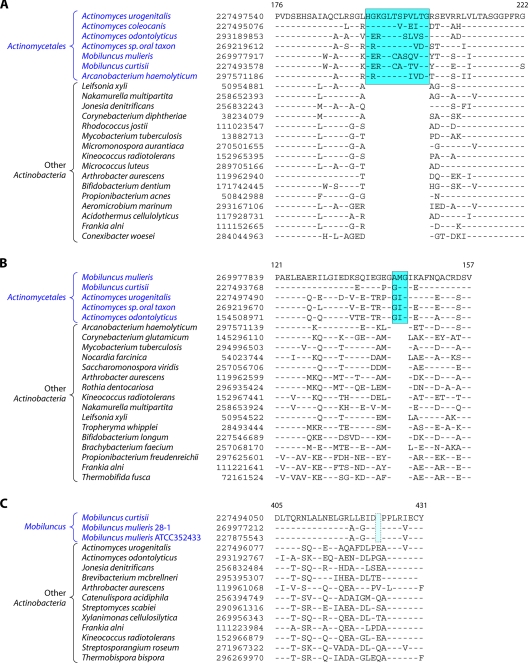
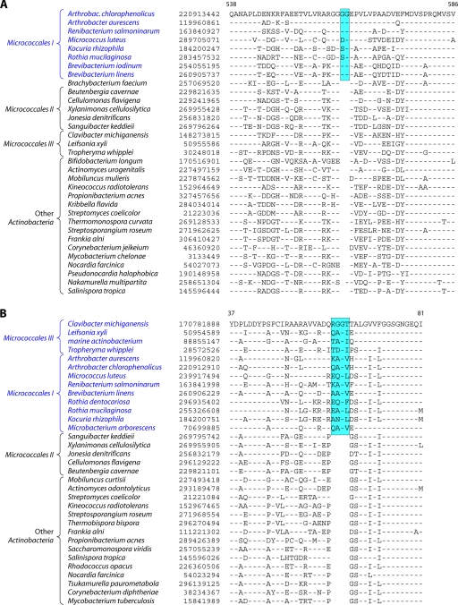
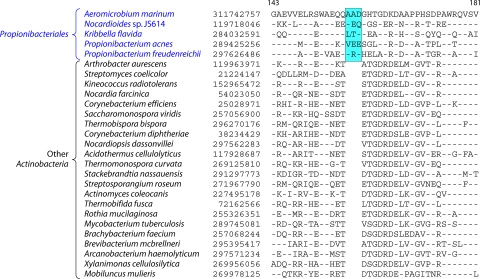
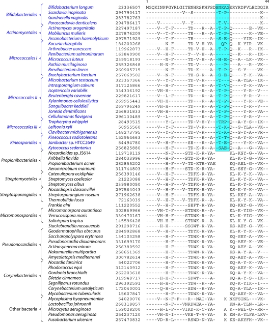
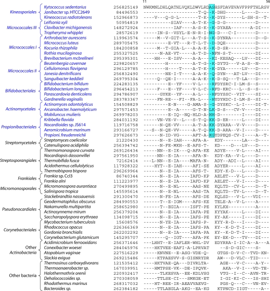
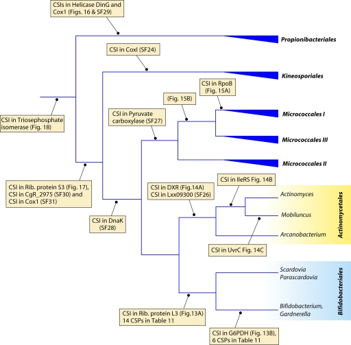
The phylum Actinobacteria harbors many important human pathogens and also provides one of the richest sources of natural products, including numerous antibiotics and other compounds of biotechnological interest. Thus, a reliable phylogeny of this large phylum and the means to accurately identify its different constituent groups are of much interest. Detailed phylogenetic and comparative analyses of >150 actinobacterial genomes reported here form the basis for achieving these objectives. In phylogenetic trees based upon 35 conserved proteins, most of the main groups of Actinobacteria as well as a number of their superageneric clades are resolved. We also describe large numbers of molecular markers consisting of conserved signature indels in protein sequences and whole proteins that are specific for either all Actinobacteria or their different clades (viz., orders, families, genera, and subgenera) at various taxonomic levels. These signatures independently support the existence of different phylogenetic clades, and based upon them, it is now possible to delimit the phylum Actinobacteria (excluding Coriobacteriia) and most of its major groups in clear molecular terms. The species distribution patterns of these markers also provide important information regarding the interrelationships among different main orders of Actinobacteria. The identified molecular markers, in addition to enabling the development of a stable and reliable phylogenetic framework for this phylum, also provide novel and powerful means for the identification of different groups of Actinobacteria in diverse environments. Genetic and biochemical studies on these Actinobacteria-specific markers should lead to the discovery of novel biochemical and/or other properties that are unique to different groups of Actinobacteria.


Figures

Fig 1
Fig 1
Current taxonomic outline for the phylum Actinobacteria based upon the List of Prokaryotic Names with Standing in Nomenclature (http://www.bacterio.cict.fr/classifphyla.html#Actinobacteria) (A) and proposed taxonomy for Actinobacteria in the forthcoming Bergey's Manual of Systematic Bacteriology (191) (B).
Fig 2
Fig 2
Phylogenetic tree for 98 actinobacterial species whose genomes have been sequenced, based upon concatenated sequences for 35 conserved proteins. Many genera for which sequence information is available from multiple species are represented by triangles in this tree. The sizes of the triangles reflect the number of species that have been sequenced, and more detailed trees for some of these groups are presented in other figures. The tree shown is based on neighbor-joining (NJ) analysis, and the numbers at the nodes represent the bootstrap scores of the nodes. Similar branching patterns for most of these groups can also be observed in a maximum likelihood tree. The asterisks mark the Frankiales species that branch in different positions in this tree.
Fig 3
Fig 3
Partial sequence alignment of the protein glucosamine-fructose-6-phosphate aminotransferase (GFT) showing a 4-aa insert that is uniquely present in different genera belonging to the class Actinobacteria but is not found in Coriobacteriia, Rubrobacter, Acidimicrobiia, and Thermoleophilia or any other prokaryotic organism. Sequence information for several other CSIs that are specifically found in most Actinobacteria is presented in Files S2 to S6 in the supplemental material and Table 2. The dashes in this as well as all other sequence alignments indicate identity with the amino acid on the top line. The numbers on the top lines indicate the sequence region where this CSI is found in the species shown at the top. The second column shows the GenBank accession number or GenBank identification (gi) number for the sequences. Sequence information for a limited number of Actinobacteria is shown in this alignment. However, detailed information regarding the presence or absence of this CSI in various sequenced genera of Actinobacteria is provided in the Table 2.
Fig 4
Fig 4
Bootstrapped neighbor-joining tree for Corynebacteriales species based upon concatenated sequences for the RpoB, RpoC, and gyrase B proteins. The distinctness of a number of clades seen in this tree is independently supported by many identified CSIs and CSPs.
Fig 5
Fig 5
(A) Partial sequence alignment of a macrolide ABC transporter ATP-binding protein showing a 2-aa conserved indel that is uniquely present in various Corynebacteriales species. Information for two other CSIs that are specific for Corynebacteriales is provided in Files S8 and S9 in the supplemental material. Sequence information for most of the CSIs is shown for a limited number of species; however, unless otherwise indicated, they are specific for the indicated groups. (B) Excerpt from the sequence alignment of the pantoate beta-alanine ligase (PanC) protein showing a 1-aa conserved insert that is specific for Mycobacterium species but not found in any other Actinobacteria. Sequence information for another Mycobacterium-specific CSI in the protein OMP-decarboxylase is presented in File S11 in the supplemental material.
Fig 6
Fig 6
Partial sequence alignments of an ATP-binding protein showing a 3-aa CSI that is uniquely found in Rhodococcus-Nocardia species. Another CSI that is specific for Rhodococcus-Nocardia is shown in File S12 in the supplemental material.
Fig 7
Fig 7
(A) Partial sequence alignments of the protein phosphoribose diphosphate:decaprenyl-phosphate phosphoribosyltransferase acyl-CoA carboxylase acetate kinase showing a 2-aa conserved insert that is uniquely found in various Corynebacterium species but not in any other bacteria. The acetate kinase and CyoE proteins also contain CSIs that are specific for the genus Corynebacterium (see Files S14 and S15 in the supplemental material). (B) Partial sequence alignments of the RNA polymerase β′-subunit (RpoC) showing a 7- to 8-aa conserved insert that is specifically found in clade I Corynebacterium species (Fig. 4). Another CSI that is specific for clade I Corynebacterium species is present in the GTP-binding protein LepA (see File S16 in the supplemental material).
Fig 8
Fig 8
Excerpts from sequence alignments of the uridylate kinase protein (A) and the hypothetical protein Lxx093000 (B) showing two conserved inserts that are specific for species of the orders Pseudonocardiales and Micromonosporales, respectively.
Fig 9
Fig 9
(A) Partial sequence alignments of the protein UDP-galactopyranose mutase showing a 3-aa CSI that is uniquely shared by various species of the orders Corynebacteriales and Pseudonocardiales but that is not found in other Actinobacteria. (B) Partial sequence alignments of DNA polymerase HolB showing a CSI that is uniquely shared by various species of the orders Corynebacteriales, Pseudonocardiales, Micromonosporales, and Glycomycetales, indicating that species from these groups shared a common ancestor exclusive of other Actinobacteria. Sequence information for other CSIs that are specific for these actinobacterial orders is presented in Files S18 to S20 and S22 in the supplemental material.
Fig 10
Fig 10
Excerpts from the sequence alignment of the gyrase B protein showing a 7-aa insert in a conserved region that is uniquely present in various Frankia species but is not found in other Actinobacteria. Two other CSIs that are also largely specific for the genus Frankia are shown in File S21 in the supplemental material.
Fig 11
Fig 11
Summary diagram showing the evolutionary relationships among the orders Corynebacteriales, Pseudonocardiales, Micromonosporales, Glycomycetales, Frankiales, Streptosporangiales, Streptomycetales, and Catenulisporales based upon phylogenetic trees (Fig. 2 and 4, and see File S10 in the supplemental material) and various identified CSIs and CSPs. OMPdecase, OMP-decarboxylase; GPAT, glutamine phosphoribosyl amidotransferase; SF11, File S11 in the supplemental material.
Fig 12
Fig 12
Excerpts from sequence alignments of two proteins showing CSIs that are specific for the order Streptomycetales or shared with Catenulisporales. (A) A 4-aa insert in the porphobilinogen deaminase (PBGD) protein that is specific for various Streptomycetales species, including Kitasatospora setae. The adenylate kinase protein also contains a CSI that is specific for the Streptomycetales (see File S24 in the supplemental material). (B) A 1-aa insert in the lipid A biosynthesis lauroyl acyltransferase (MsbB) protein that is uniquely shared by various Streptomycetales species and Catenulispora acidiphila.
Fig 13
Fig 13
Partial sequence alignments of ribosomal protein L3 (A) and glucose-6-phosphate dehydrogenase (G6PDH) (B) showing two CSIs consisting of a 1-aa deletion and a 1-aa insert, respectively, that are specific for Bifidobacteriales species. The CSI in the ribosomal protein is present in all sequenced Bifidobacteriales species, whereas that in G6PDH is found only in Bifidobacterium and Gardnerella species.
Fig 14
Fig 14
(A and B) Partial sequence alignments of the deoxy-d-xylulose 5-phosphate reductoisomerase (DXR) (A) and isoleucine tRNA synthetase (IleRS) (B) proteins depicting 12-aa and 3-aa inserts, respectively, in highly conserved regions that are uniquely present in various sequenced Actinomycetales species. (C) Sequence alignment of the excision endonuclease UvrC showing a 1-aa deletion that is specific for the genus Mobiluncus. Information for another CSI that is specific for Actinomycetales is provided in File S26 in the supplemental material.
Fig 15
Fig 15
Partial sequence alignments of the RNA polymerase β-subunit showing a 2-aa insert in a conserved region that is specific for cluster I Micrococcales species (A) and a 4-aa insert in ribose-5-phosphate isomerase that is uniquely shared by both cluster I and cluster II Micrococcales species (B). Another CSI that is specific for Micrococcales can be found in File S27 in the supplemental material.
Fig 16
Fig 16
Excerpts from sequence alignments for the helicase DinG showing a CSI consisting of a 3-aa insert that is uniquely present in various sequenced Propionibacteriales species. Sequence information for another CSI that is specific for Propionibacteriales can be found in File S29 in the supplemental material.
Fig 17
Fig 17
Partial sequence alignment of ribosomal protein S3 showing a 5-aa conserved insert that is commonly shared by various sequenced species of the orders Bifidobacteriales, Actinomycetales, Micrococcales, and Kineosporiales but which is not found in any other Actinobacteria or in other phyla of bacteria. Information for two other CSIs showing similar specificities is provided in Files S30 and S31 in the supplemental material.
Fig 18
Fig 18
Partial sequence alignment of the triosephosphate isomerase protein showing a 2-aa conserved insert that is commonly shared by various sequenced species of the orders Bifidobacteriales, Actinomycetales, Micrococcales, Kineosporiales, and Propionibacteriales but which is not found in any other Actinobacteria.
Fig 19
Fig 19
Summary diagram showing evolutionary relationships among the orders Bifidobacteriales, Actinomycetales, Micrococcales, Kineosporiales, and Propionibacteriales based upon phylogenetic trees (Fig. 2, and see File S25 in the supplemental material) and various identified CSIs and CSPs. Rib., ribosomal.
Fig 20
Fig 20
Structures of the S-adenosyl-l-homocysteine hydrolase (PDB accession number 3CE6) (240) (A and B) and serine hydroxymethyltransferase (PDB accession number 3H7F) (C and D) proteins from M. tuberculosis showing the locations in protein structures of the 9-aa and 5-aa actinobacterium-specific inserts that are found in these proteins (see Files S5 and S6 in the supplemental material). While panels A and C show ribbon representations, panels B and D depict the surface representations of these protein structures. The inserts in these proteins are shown in magenta.
None
None

References

    1. Abdallah AM, et al. 2006. A specific secretion system mediates PPE41 transport in pathogenic mycobacteria. Mol. Microbiol. 62:667–679 - PubMed
    1. Abt B, et al. 2010. Complete genome sequence of Cellulomonas flavigena type strain (134). Stand. Genomic Sci. 3:15–25 - PMC - PubMed
    1. Adekambi T, et al. 2011. Core gene set as the basis of multilocus sequence analysis of the subclass Actinobacteridae. PLoS One 6:e14792. - PMC - PubMed
    1. Akiva E, Itzhaki Z, Margalit H. 2008. Built-in loops allow versatility in domain-domain interactions: lessons from self-interacting domains. Proc. Natl. Acad. Sci. U. S. A. 105:13292–13297 - PMC - PubMed
    1. Alam MT, Merlo ME, Takano E, Breitling R. 2010. Genome-based phylogenetic analysis of Streptomyces and its relatives. Mol. Phylogenet. Evol. 54:763–772 - PubMed

Publication types

MeSH terms

LinkOut - more resources