Review
. 2012 Mar 1;26(5):417-32.
doi: 10.1101/gad.183509.111.

On PAR with PARP: cellular stress signaling through poly(ADP-ribose) and PARP-1

Affiliations
Review

On PAR with PARP: cellular stress signaling through poly(ADP-ribose) and PARP-1

Xin Luo et al. Genes Dev. .

Abstract

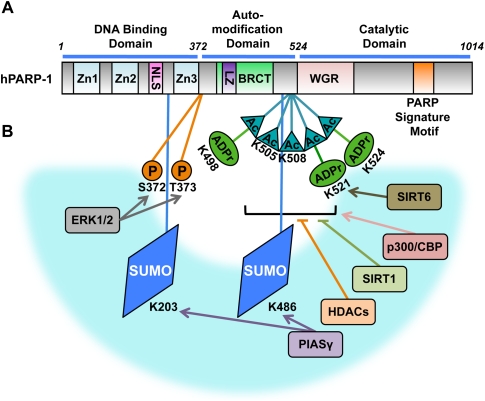
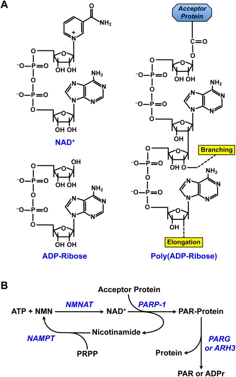
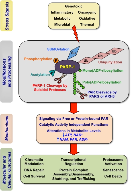
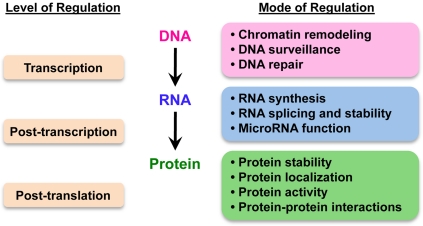
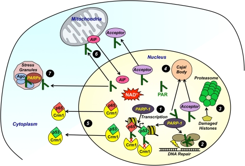
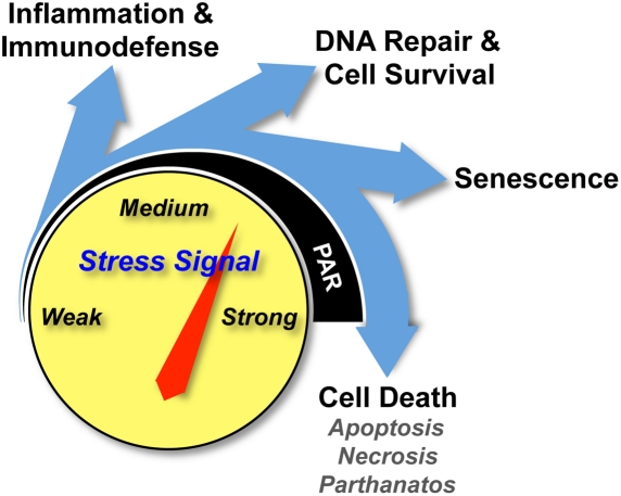
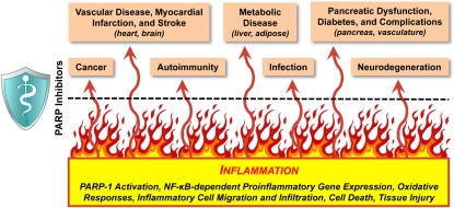
Cellular stress responses are mediated through a series of regulatory processes that occur at the genomic, transcriptional, post-transcriptional, translational, and post-translational levels. These responses require a complex network of sensors and effectors from multiple signaling pathways, including the abundant and ubiquitous nuclear enzyme poly(ADP-ribose) (PAR) polymerase-1 (PARP-1). PARP-1 functions at the center of cellular stress responses, where it processes diverse signals and, in response, directs cells to specific fates (e.g., DNA repair vs. cell death) based on the type and strength of the stress stimulus. Many of PARP-1's functions in stress response pathways are mediated by its regulated synthesis of PAR, a negatively charged polymer, using NAD(+) as a donor of ADP-ribose units. Thus, PARP-1's functions are intimately tied to nuclear NAD(+) metabolism and the broader metabolic profile of the cell. Recent studies in cell and animal models have highlighted the roles of PARP-1 and PAR in the response to a wide variety of extrinsic and intrinsic stress signals, including those initiated by oxidative, nitrosative, genotoxic, oncogenic, thermal, inflammatory, and metabolic stresses. These responses underlie pathological conditions, including cancer, inflammation-related diseases, and metabolic dysregulation. The development of PARP inhibitors is being pursued as a therapeutic approach to these conditions. In this review, we highlight the newest findings about PARP-1's role in stress responses in the context of the historical data.


Figures

Figure 1.
Figure 1.
Structural and functional organization of PARP-1, and site-specific post-translational modifications. (A) Human PARP-1 is a 116-kDa protein comprising (1) an N-terminal DNA-binding domain, which contains three zinc-binding domains (Zn1, Zn2, and Zn3) and a NLS; (2) a central automodification domain, which contains a leucine zipper (LZ) motif and a BRCT motif; and (3) a C-terminal catalytic domain, which contains a WGR domain and the “PARP signature” motif required for NAD+ binding and the catalysis of PAR synthesis. (B) Key post-translational modifications of PARP-1 are illustrated on the PARP-1 functional organization schematic from A. Four types of post-translational modifications are shown: phosphorylation (P), SUMOylation (SUMO), acetylation (Ac), and mono(ADP-ribosyl)ation or poly(ADP-ribosyl)ation (ADPr). The Ser (S), Lys (K), or Thr (T) residues that are the sites of covalent modification are indicated. Enzymes that add (arrows) or remove (blunt lines) the specific post-translational modifications, as described in the text, are shown in the blue arc.
Figure 2.
Figure 2.
NAD+ metabolism and the synthesis of PAR. (A, counterclockwise from top left) Chemical structures of NAD+, ADP-ribose (ADRr), and PAR. The positions on the ribose moieties where the PAR polymer is elongated and branched from are indicated. In the example shown, PAR is covalently attached to an aspartic or glutamic acid residue on the acceptor proteins. PAR may also be attached to lysine residues. (B) The PARP-1-dependent NAD+ metabolic cycle. NMN is synthesized from nicotinamide (NAM) and 5-phosphoribosyl-1-pyrophosphate (PRPP) by nicotinamide phosphoribosyltransferase (NAMPT). NAD+ is synthesized from ATP and NMN by NMNAT (NMNAT-1 is the nuclear member of the NMNAT group of enzymes). PARP-1 catalyzes the covalent attachment of PAR on acceptor proteins using NAD+ as a donor of ADP-ribose units, with the concomitant release of nicotinamide. Covalently attached PAR can be hydrolyzed to free PAR or mono(ADP-ribose) (ADPr) by PARG, which possesses both endoglycosidic and exoglycosidic activities, and ARH3, which also has PARG activity.
Figure 3.
Figure 3.
PARP-1 at the crossroads of cellular stress responses. Cells are regularly exposed to a wide variety of extrinsic and intrinsic stress signals, including those initiated by oxidative, nitrosative, genotoxic, oncogenic, thermal, inflammatory, and metabolic stresses. PARP-1 senses these stresses and propagates different stress signals to execute diverse downstream molecular and cellular actions. PARP-1 and related PARP family members function at the intersection of converging stress signaling pathways. (Ac) Acetylation; (P) phosphorylation; (Su) SUMOylation; (Ub) ubiquitylation; (NAM) nicotinamide.
Figure 4.
Figure 4.
PARP-1 modulates the molecular biology and biochemistry of stress responses at multiple levels. PARP-1 modulates cellular stress responses through a series of regulatory processes that occur at the genomic, transcriptional, post-transcriptional, translational, and post-translational levels.
Figure 5.
Figure 5.
Molecular and cellular actions of PARP-1 and related PARPs. Key molecular and cellular actions of PARP-1 and related PARPs are illustrated: (1) modulating chromatin structure and regulating gene transcription, (2) facilitating the assembly and function of the DNA repair machinery, (3) activating proteasomes to remove damaged histones, (4) facilitating protein transport into Cajal bodies, (5) preventing the nuclear export of p65 and p53 through a Crm1-dependent mechanism, (6) promoting the release of AIF from mitochondria and transport to nucleus, and (7) regulating the assembly and function of stress granules (PARP family members). Details are provided in the text.
Figure 6.
Figure 6.
PARP-1 functions as a cellular rheostat. PARP-1 promotes different cellular responses to different types and levels of stress signals. As the strength of stress stimulus increases, the levels of PARP-1 activity and PAR synthesis increase, leading to different cellular outcomes.
Figure 7.
Figure 7.
Anti-inflammatory therapeutic effects of PARP inhibitors. Inflammation is a common thread underlying many diseases and conditions. The therapeutic effects of PARP inhibitors in various diseases may share a common mechanism; namely, the inhibition of NF-κB-dependent inflammatory pathways.

References

    1. Ahel I, Ahel D, Matsusaka T, Clark AJ, Pines J, Boulton SJ, West SC 2008. Poly(ADP-ribose)-binding zinc finger motifs in DNA repair/checkpoint proteins. Nature 451: 81–85 - PubMed
    1. Altmeyer M, Messner S, Hassa PO, Fey M, Hottiger MO 2009. Molecular mechanism of poly(ADP-ribosyl)ation by PARP1 and identification of lysine residues as ADP-ribose acceptor sites. Nucleic Acids Res 37: 3723–3738 - PMC - PubMed
    1. Ame JC, Spenlehauer C, de Murcia G 2004. The PARP superfamily. Bioessays 26: 882–893 - PubMed
    1. Andrabi SA, Kang HC, Haince JF, Lee YI, Zhang J, Chi Z, West AB, Koehler RC, Poirier GG, Dawson TM, et al. 2011. Iduna protects the brain from glutamate excitotoxicity and stroke by interfering with poly(ADP-ribose) polymer-induced cell death. Nat Med 17: 692–699 - PMC - PubMed
    1. Asher G, Reinke H, Altmeyer M, Gutierrez-Arcelus M, Hottiger MO, Schibler U 2010. Poly(ADP-ribose) polymerase 1 participates in the phase entrainment of circadian clocks to feeding. Cell 142: 943–953 - PubMed

Publication types

Substances

LinkOut - more resources