

The MRI findings of a de Garengeot hernia
- PMID: 22391502
- PMCID: PMC3473985
- DOI: 10.1259/bjr/27759683
The MRI findings of a de Garengeot hernia
Abstract
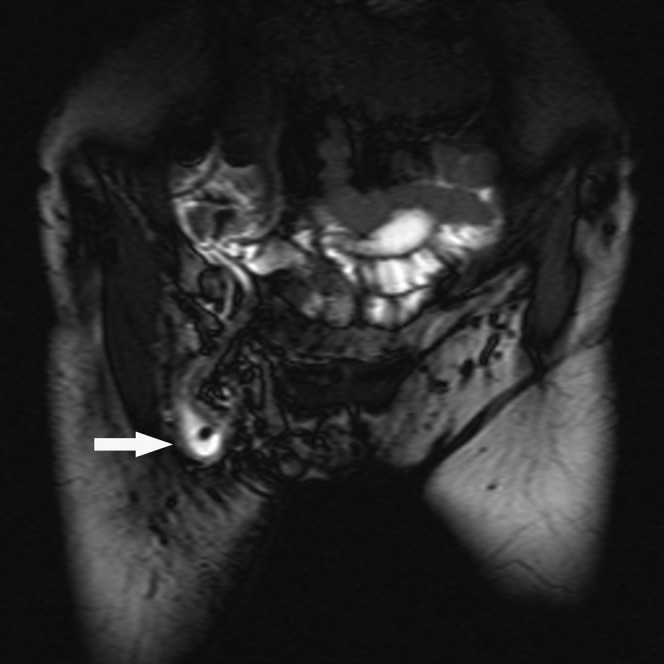
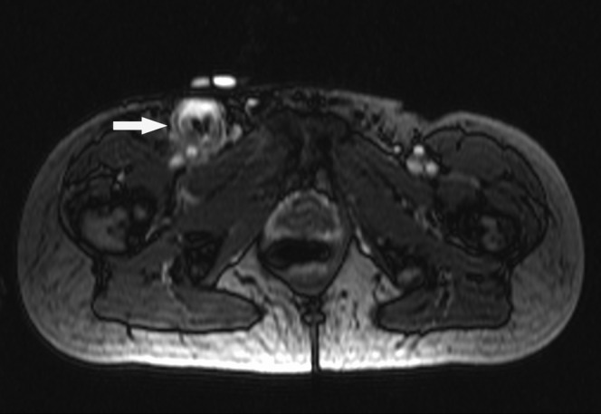
The presence of the appendix within a femoral hernia is rare. It was first described by the French surgeon Jacques Croissant de Garengeot in 1731. This phenomenon accounts for 0.8-1% of all femoral hernias. Acute appendicitis occurring within a femoral hernia is even rarer and is difficult to diagnose pre-operatively. This type of hernia is termed a de Garengeot hernia. The ultrasonographic and CT imaging features of de Garengeot hernias have been described previously. We report a case of a 57-year-old female who presented with a painful right-sided groin mass. She underwent MRI of the inguinal region, which successfully diagnosed this rare hernia pre-operatively. To our knowledge, this is the first description of a de Garengeot hernia diagnosed using MRI.
Figures
References
-
- Sharma H, Jha PK, Shekhawat NS, Memon B, Memon MA. De Garengeot hernia: an analysis of our experience. Hernia 2007;11:235–8 - PubMed
-
- Shadbolt CL, Heinze SB, Dietrich RB. Imaging of groin masses: inguinal anatomy and pathological conditions revisited. Radiographics 2001;21:S261–71 - PubMed
-
- Filatov J, Ilibitzki A, Davidovitch S, Soudack M. Appendicitis within a femoral hernia: sonographic appearance. J Ultrasound Med 2006;25:1233–5 - PubMed
