

Case Reports
doi: 10.1259/bjr/95600238.
Contrast-enhanced ultrasound in testicular trauma: role in directing exploration, debridement and organ salvage
Affiliations
- PMID: 22391504
- PMCID: PMC3473993
- DOI: 10.1259/bjr/95600238
Item in Clipboard
Case Reports
Contrast-enhanced ultrasound in testicular trauma: role in directing exploration, debridement and organ salvage
Br J Radiol.
2012 Mar.
Abstract
We describe the use of contrast-enhanced ultrasound as an additional imaging technique during an ultrasound examination of a traumatised testis, allowing for confident testicular preserving surgery to be performed.
Figures

Longitudinal image of the right testis, with an enlarged upper pole (star) and several areas of low reflectivity at the site of trauma (arrows).
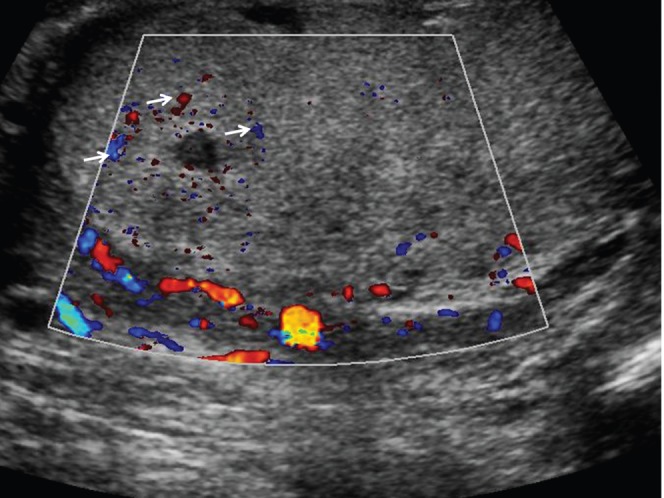
Longitudinal image of the right testis, with colour Doppler mode, demonstrating a paucity of colour Doppler flow to the entire right testis, with pockets of colour signal (arrows); likely to be artefact.
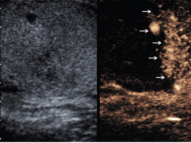
Longitudinal image of the right testis with microbubble contrast imaging, 24 s after administration of SonoVue™ (Bracco SpA, Milan, Italy), demonstrating a clear demarcation between vascularised and non-vascularised tissue (arrows).
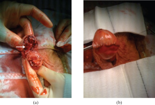
(a) At surgical exploration, the tissue of the upper third of the testis (arrow) is “dusky” without demonstrable blood flow. This ischaemic area is excised with preservation of the remainder of the testis. (b) Following non-viable tissue excision, the testis is reconstructed and secured as shown.
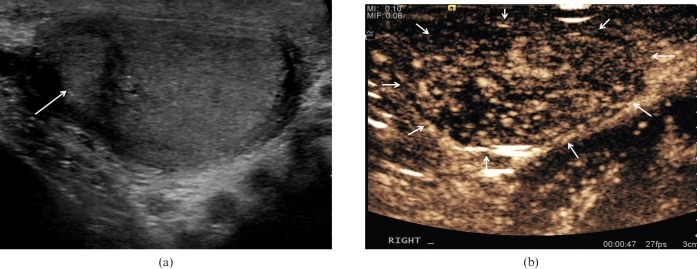
(a) Longitudinal image of the right testis 3 months post-surgery demonstrating an area of altered reflectivity at the upper aspect of the testis (arrow). (b) Contrast-enhanced ultrasound of the right testis at 47 s after administration of SonoVue™ (Bracco SpA, Milan, Italy), demonstrating perfusion of the entire remaining right testis.
References
-
- Buckley JC, McAninch JW. Use of ultrasound for the diagnosis of testicular injuries in blunt scrotal trauma. J Urol 2006;175:175–8 - PubMed
-
- Herbener TE. Ultrasound in the assessment of the acute scrotum. J Clin Ultrasound 1996;24:405–21 - PubMed
-
- Allen TD, Elder JS. Shortcomings of color Doppler sonography in the diagnosis of testicular torsion. J Urol 1995;154:1508–10 - PubMed
-
- Wilson SR, Greenbaum LD, Goldberg BB. Contrast-enhanced ultrasound: what is the evidence and what are the obstacles? Am J Roentgenol 2009;193:55–60 - PubMed
-
- Bhandary P, Abbit PL, Watson L. Ultrasound diagnosis of traumatic testicular rupture. J Clin Ultrasound 1992;20:346–8 - PubMed
Publication types
MeSH terms
LinkOut - more resources
Full Text Sources
Medical
