. 2013 Jan 31;32(5):599-609.
doi: 10.1038/onc.2012.81. Epub 2012 Mar 5.

Histone deacetylase inhibitors suppress mutant p53 transcription via histone deacetylase 8

Affiliations

Histone deacetylase inhibitors suppress mutant p53 transcription via histone deacetylase 8

W Yan et al. Oncogene. .

Abstract

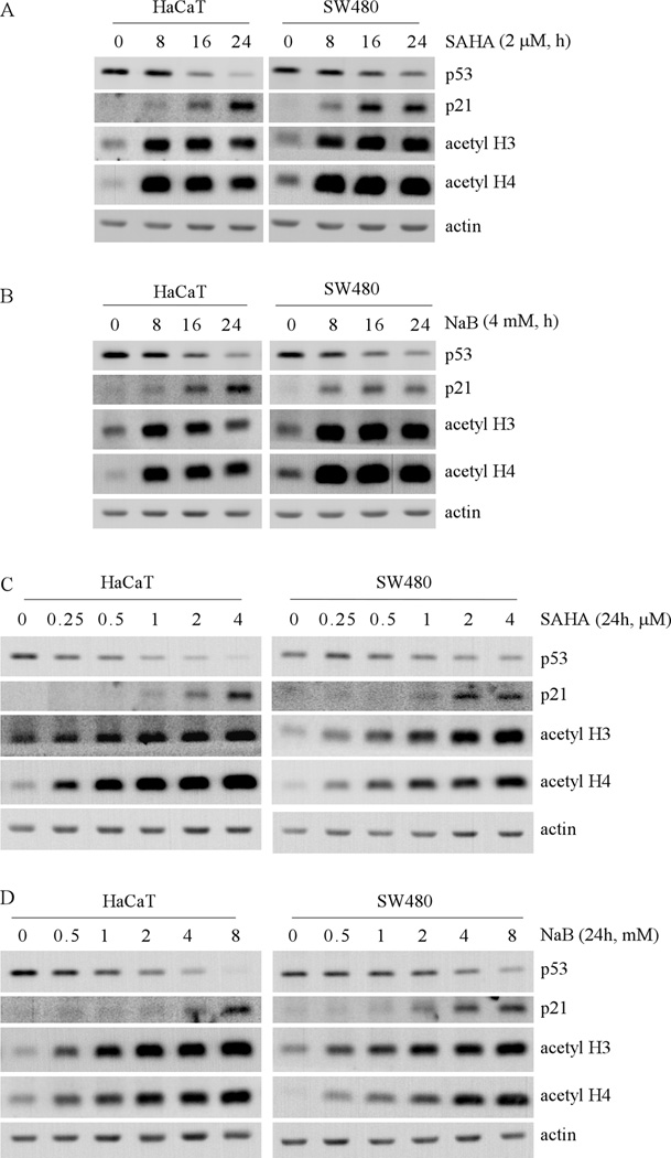
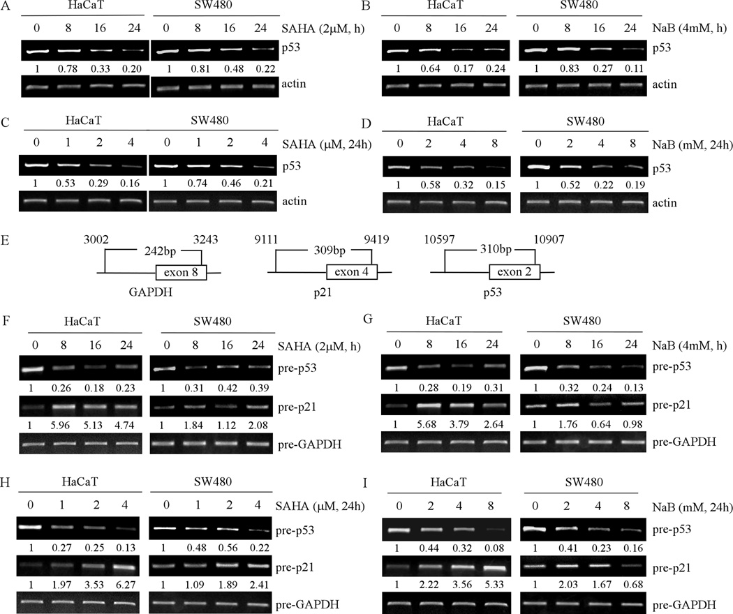
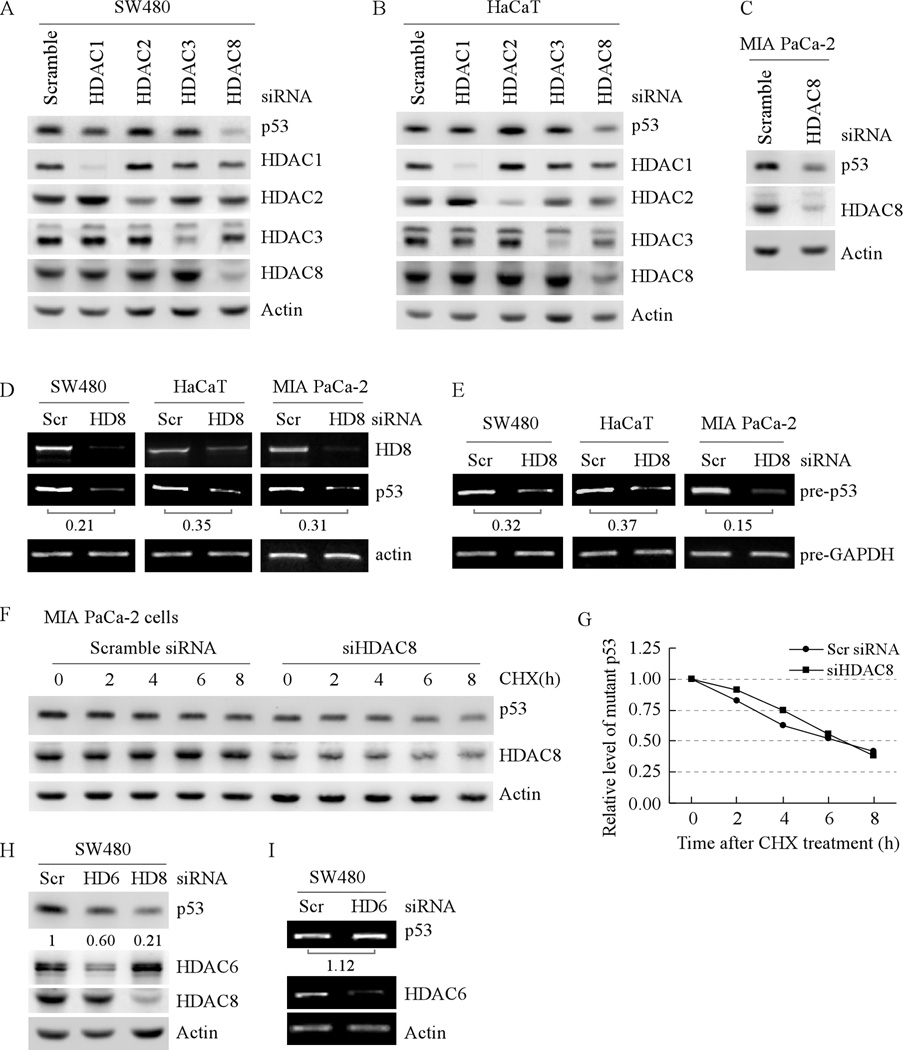
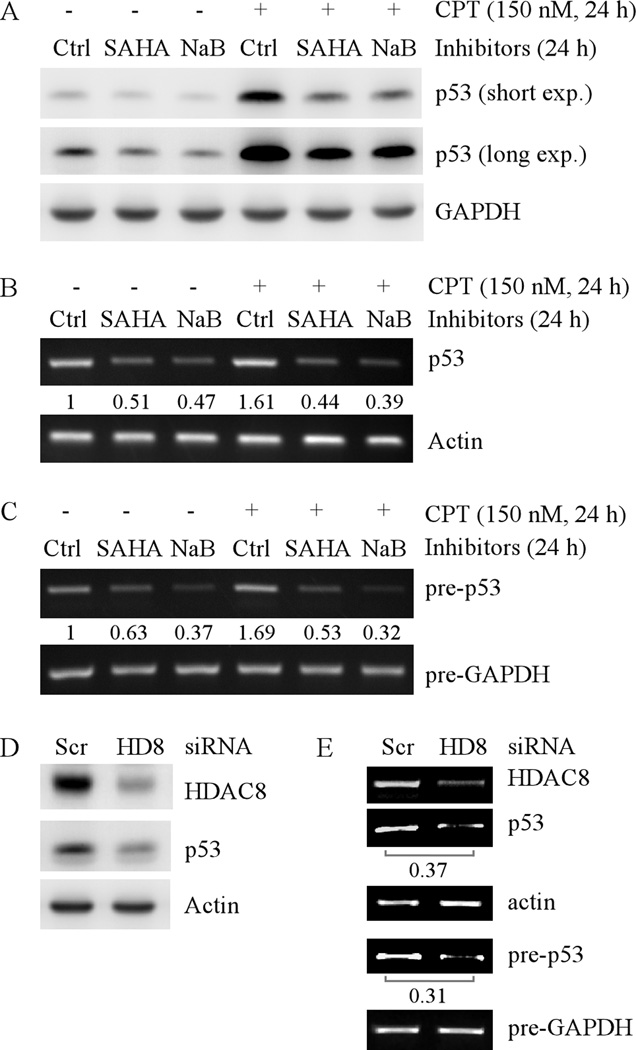
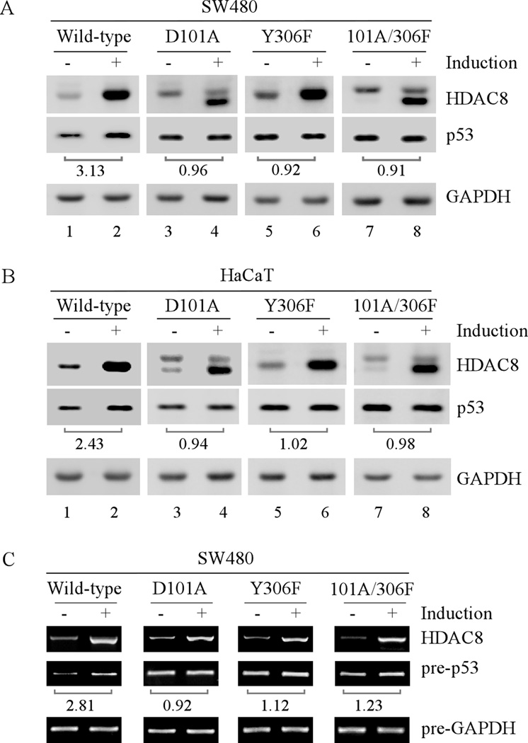
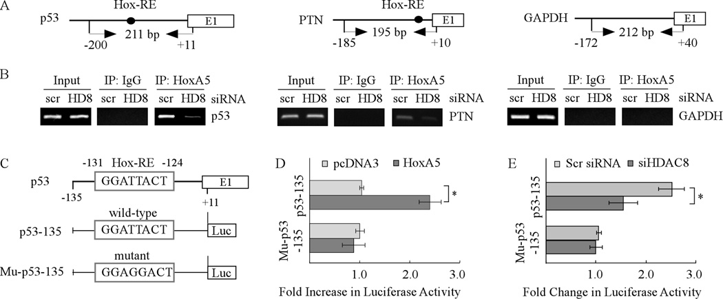
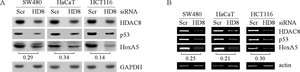
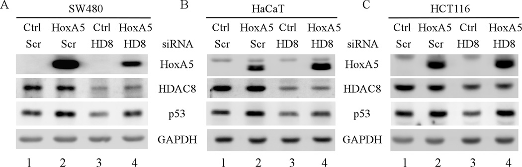
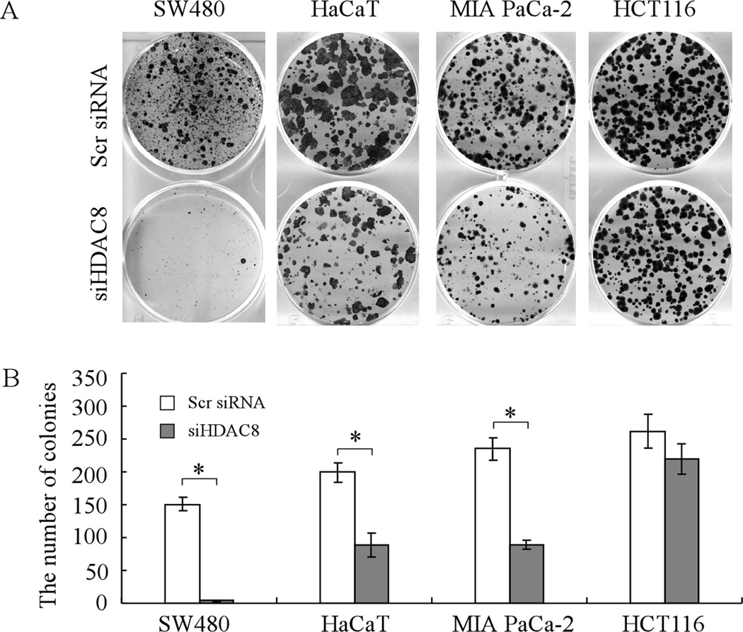
Mutation of the p53 gene is the most common genetic alteration in human cancer and contributes to malignant process by enhancing transformed properties of cells and resistance to anticancer therapy. Mutant p53 is often highly expressed in tumor cells at least, in part, due to its increased half-life. However, whether mutant p53 expression is regulated by other mechanisms in tumors is unclear. Here we found that histone deacetylase (HDAC) inhibitors suppress both wild-type and mutant p53 transcription in time- and dose-dependent manners. Consistent with this, the levels of wild-type and mutant p53 proteins are decreased upon treatment with HDAC inhibitors. Importantly, we found that upon knockdown of each class I HDAC, only HDAC8 knockdown leads to decreased expression of wild-type and mutant p53 proteins and transcripts. Conversely, we found that ectopic expression of wild-type, but not mutant HDAC8, leads to increased transcription of p53. Furthermore, we found that knockdown of HDAC8 results in reduced expression of HoxA5 and consequently, attenuated ability of HoxA5 to activate p53 transcription, which can be rescued by ectopic expression of HoxA5. Because of the fact that HDAC8 is required for expression of both wild-type and mutant p53, we found that targeted disruption of HDAC8 expression remarkably triggers proliferative defect in cells with a mutant, but not wild-type, p53. Together, our data uncover a regulatory mechanism of mutant p53 transcription via HDAC8 and suggest that HDAC inhibitors and especially HDAC8-targeting agents might be explored as an adjuvant for tumors carrying a mutant p53.


Conflict of interest statement

Conflict of interest

The authors declare no conflict of interest.

Figures

Fig. 1
Fig. 1. HDAC inhibitors decrease the level of mutant p53 protein in time- and dose-dependent manners
(A) Western blots were prepared with extracts from HaCat (left panel) and SW480 (right panel) cells untreated or treated with 2 µM SAHA for 8 to 24 h, and then probed with antibodies against p53, p21, acetyl-H3, acetyl-H4 and actin, respectively. (B) The experiments were performed as in (A) except that cells were treated with 4 mM NaB. (C) Western blots were prepared with extracts from HaCaT (left panel) and SW480 (right panel) cells untreated or treated with 0.25 to 4 µM SAHA for 24 h, and then probed with antibodies as in (A). (D) The experiments were performed as in (C) except that the cells were treated with 0.5 to 8 mM NaB for 24 h.
Fig. 2
Fig. 2. HDAC inhibitors decrease the level of mature and precursor mRNAs of mutant p53 in time- and dose-dependent manners
(A) RT-PCR was performed with total RNAs isolated from HaCaT (left panel) and SW480 (right panel) cells untreated or treated with 2 µM SAHA for 8 to 24 h. Actin mRNA was amplified for a loading control. (B) The experiments were performed as in (A) except that the cells were treated with 4 mM NaB. (C) RT-PCR was performed with total RNAs isolated from HaCaT (left panel) and SW480 (right panel) cells untreated or treated with 0.25 to 4 µM SAHA for 24 h. (D) The experiments were performed as in (C) except that cells were treated with 0.5 to 8 mM NaB for 24 h. (E) Schematic presentation of primers used in (F–I) to amplify precursor mRNAs of p53, p21 and GAPDH. (F–I) RT-PCR was performed with total RNAs isolated from HaCaT (left panel) and SW480 (right panel) cells, which were untreated or treated as in (A–D). Prior to reverse transcription, total RNAs were treated with DNase I to remove genomic DNA.
Fig. 3
Fig. 3. Knockdown of HDAC8 decreases the level of mutant p53 protein and transcript
(A) The level of mutant p53 protein in SW480 cells is decreased by knockdown of HDAC8 but not by HDAC1-3. Western blots were prepared with extracts from SW480 cells transfected with scrambled siRNA, siRNA against HDAC1-3, or siRNA against HDAC8 for 3 d. The blots were then probed with antibodies against p53, HDAC1-3, HDAC8 and actin, respectively. (B) The experiment was performed as in (A) except that HaCaT cells were used. (C) The experiment was performed as in (A) except that MIA PaCa-2 cells were used. (D) Knockdown of HDAC8 decreases the level of mature mutant p53 transcript in SW480 (left panel), HaCaT (middle panel) and MIA PaCa-2 (right panel) cells. RT-PCR was performed with total RNAs isolated from SW480, HaCaT, and MIA PaCa-2 cells, which were transfected with scrambled siRNA or siRNA against HDAC8 for 3 d. Actin mRNA was amplified for a loading control. (E) RT-PCR was performed as in (D) except that total RNAs were treated with DNaseI to remove genomic DNA prior to reverse transcription and that the primers for precursor mRNAs were used. The precursor mRNA of GAPDH was amplified as a loading control. (F) The half-life of mutant p53 protein is not changed by HDAC8 knockdown. Western blots were prepared with extracts from MIA PaCa-2 cells that were transfected with scrambled siRNA, or siRNA against HDAC8 for 3 d, and then treated with cycloheximide (50 µg/ml) for 0–8 h. The blots were then probed with antibodies against p53, HDAC8, and actin, respectively. (G) The relative levels of mutant p53 protein measured in (F) were normalized by levels of actin protein and then plotted versus time. (H) The level of mutant p53 protein in SW480 cells is decreased by knockdown of HDAC6 and HDAC8. Western blots were prepared with extracts from SW480 cells transfected with scrambled siRNA, or siRNA against HDAC6 or HDAC8 for 3 d. The blots were then probed with antibodies against p53, HDAC6, HDAC8 and actin, respectively. (I) Knockdown of HDAC6 has little if any effect on the level of mutant p53 transcript in SW480 cells. RT-PCR was performed as in (D).
Fig. 4
Fig. 4. HDAC inhibitors or knockdown of HDAC8 decreases the levels of wild-type p53 protein and transcript
(A) Western blots were prepared with extracts from HCT116 cells untreated or treated with 2 µM SAHA or 4 mM NaB for 24 h in the absence or presence of CPT. The blots were then probed with antibodies against p53 and actin, respectively. (B) RT-PCR was performed with total RNAs isolated from HCT116 cells, which were treated as in (A). Actin mRNA was amplified for a loading control. (C) Experiments were performed as in (B), except that the total RNAs were treated with DNaseI to remove genomic DNA prior to reverse transcription and primers for precursor mRNA were used for PCR. (D) Western blots were prepared with extracts from HCT116 cells, which were transfected with scrambled siRNA or siRNA against HDAC8 for 3 d. The blots were then probed with antibodies against HDAC8, p53 and actin, respectively. (E) RT-PCR for mature and precursor mRNAs was performed with total RNAs isolated from HCT116 cells treated as in (D).
Fig. 5
Fig. 5. Ectopic expression of HDAC8 increases mutant p53 expression
(A) Western blots were prepared with extracts from SW480 cells uninduced or induced to express wild-type or a mutant HDAC8 for 48 h. The blots were then probed with antibodies against HDAC8, p53 and GAPDH, respectively. The relative fold increase of mutant p53 protein by ectopic expression of HDAC8 over the control shown below the corresponding bands was calculated after normalized by levels of actin protein. (B) The experiment was performed as in (A) except that HaCaT cells were used. (C) RT-PCR was performed with total RNAs isolated from SW480 cells treated as in (A). Mature mRNA of HDAC8 and precursor mRNAs of p53 and GAPDH were amplified. The fold increase of precursor p53 transcript by ectopic expression of HDAC8 over the control shown below the corresponding bands was calculated similarly as that in (A).
Fig. 6
Fig. 6. HDAC8 knockdown inhibits the ability of HoxA5 to activate the p53 promoter
(A) Schematic presentation of the HoxA5 (left), PTN (middle), and GAPDH (right) promoters and the location of PCR primers used for ChIP assay. (B) HDAC8 knockdown inhibits the binding of HoxA5 to the p53 promoter. SW480 cells were transfected with scrambled siRNA or siRNA against HDAC8 for 3 d, and then HoxA5-DNA complexes were captured with anti-HoxA5 along with rabbit IgG as a control. The binding of HoxA5 protein to PTN or GAPDH promoter was measured as a positive or negative control. (C) Schematic presentation of luciferase reporter constructs including the location of a HoxA5 binding site in the p53 promoter. The reporter construct mu-p53-135 carries two nucleotide substitutions in which TT were substituted with GG. (D) Wild-type but not mutant HoxA5-binding site in the p53 promoter is responsive to HoxA5. The dual luciferase assay was performed as described in the Materials and Methods. (E) HDAC8 knockdown inhibits the luciferase activity under the control of the p53 promoter with wild-type but not mutant HoxA5-binding site. The experiment was performed as in (D) except that siRNA against HDAC8 or a scrambled siRNA was co-transfected with a luciferase reporter.
Fig. 7
Fig. 7. Knockdown of HDAC8 decreases the levels of HoxA5 protein and transcript
(A) Western blots were prepared with extracts from SW480, HaCaT, and HCT116 cells, which were transfected with scrambled siRNA or siRNA against HDAC8 for 3 d. The blots were then probed with antibodies against HDAC8, p53, HoxA5, and actin, respectively. (B) RT-PCR was performed with total RNAs isolated from SW480, HaCaT, and HCT116 cells, which were treated as in (A). Actin mRNA was amplified for a loading control.
Fig. 8
Fig. 8. Ectopic expression of HoxA5 restores p53 expression in HDAC8-knockdown cells
(A) Western blots were prepared with extracts from SW480 cells which were transfected with scrambled siRNA or siRNA against HDAC8 along with or without overexpression of HoxA5 for 3 d. The blots were probed with antibodies against HoxA5, HDAC8, p53 and GAPDH, respectively. (B–C) The experiments were performed as in (A), except that HaCaT (B) and HCT116 (C) cells were used.
Fig. 9
Fig. 9. Knockdown of HDAC8 inhibits cell proliferation of tumor cells harboring a mutant p53
(A) Mutant p53 cell lines SW480, HaCaT, and MIA PaCa-2 and wild-type p53 cell line HCT116 were transfected with scrambled siRNA or siRNA against HDAC8 for 1 d, and then split and cultured in fresh medium for the next 15–20 days. The colonies were fixed with methanol/glacial acetic acid (7:1) and stained with 0.1% of crystal violet. (B) Quantification of the number of colonies with a diameter of >0.5 mm from three separate experiments. * represents p<0.05.

References

    1. Minucci S, Pelicci PG. Histone deacetylase inhibitors and the promise of epigenetic (and more) treatments for cancer. Nat Rev Cancer. 2006;6:38–51. - PubMed
    1. Halkidou K, Gaughan L, Cook S, Leung HY, Neal DE, Robson CN. Upregulation and nuclear recruitment of HDAC1 in hormone refractory prostate cancer. Prostate. 2004;59:177–189. - PubMed
    1. Song J, Noh JH, Lee JH, Eun JW, Ahn YM, Kim SY, et al. Increased expression of histone deacetylase 2 is found in human gastric cancer. APMIS. 2005;113:264–268. - PubMed
    1. Huang BH, Laban M, Leung CH, Lee L, Lee CK, Salto-Tellez M, et al. Inhibition of histone deacetylase 2 increases apoptosis and p21Cip1/WAF1 expression, independent of histone deacetylase 1. Cell Death Differ. 2005;12:395–404. - PubMed
    1. Hrzenjak A, Moinfar F, Kremser ML, Strohmeier B, Staber PB, Zatloukal K, et al. Valproate inhibition of histone deacetylase 2 affects differentiation and decreases proliferation of endometrial stromal sarcoma cells. Mol Cancer Ther. 2006;5:2203–2210. - PubMed

Publication types

MeSH terms