. 2013 Jan 24;32(4):453-61.
doi: 10.1038/onc.2012.70. Epub 2012 Mar 5.

S6K1 determines the metabolic requirements for BCR-ABL survival

Affiliations

S6K1 determines the metabolic requirements for BCR-ABL survival

J F Barger et al. Oncogene. .

Abstract

In chronic myelogenous leukemia, the constitutive activation of the BCR-ABL kinase transforms cells to an addicted state that requires glucose metabolism for survival. We investigated S6K1, a protein kinase that drives glycolysis in leukemia cells, as a target for counteracting glucose-dependent survival induced by BCR-ABL. BCR-ABL potently activated S6K1-dependent signaling and glycolysis. Although S6K1 knockdown or rapamycin treatment suppressed glycolysis in BCR-ABL-transformed cells, these treatments did not induce cell death. Instead, loss of S6K1 triggered compensatory activation of fatty-acid oxidation, a metabolic program that can support glucose-independent cell survival. Fatty-acid oxidation in response to S6K1 inactivation required the expression of the fatty-acid transporter carnitine palmitoyl transferase 1c, which was recently linked to rapamycin resistance in cancer. Finally, addition of an inhibitor of fatty-acid oxidation significantly enhanced cytotoxicity in response to S6K1 inactivation. These data indicate that S6K1 dictates the metabolic requirements mediating BCR-ABL survival and provide a rationale for combining targeted inhibitors of signal transduction, with strategies to interrupt oncogene-induced metabolism.


Figures

Figure 1
Figure 1
BCR-ABL activates growth factor-independent glycolysis through S6K1. A. IL-3-dependent FL5.12 cells were transduced with vector control or BCR-ABL-expressing retrovirus, then cultured in the presence or absence of IL-3 for 3 hours. Glycolytic release of 3H2O from 5-3H-glucose in the absence of growth factor was increased by BCR-ABL. The mean ±standard deviation is plotted. B. BCR-ABL strongly activated S6K1. Vector control and BCR-ABL cells were cultured for 3 hours in the absence of growth factor, then restimulated for 30 minutes where indicated for analysis of S6K1 phosphorylation at T389 and the phosphorylation of ribosomal protein S6 at serines 235/236. C. S6K1 knockdown (inset) impaired glycolysis in BCR-ABL+ FL5.12 cells cultured in the absence of IL-3. siRNA pools targeting S6K1 or Non-targeting siRNA were transfected where indicated. D - F. Rapamycin (Rapa., 20 nM) and imatinib (1μM) suppressed glycolysis in BCR-ABL+ FL5.12 cells (D and E), human BCR-ABL+ KBM7 cells (F) and human BCR-ABL+ K562 cells (G). Inset, Both imatinib and rapamycin reduced S6K1 activity as indicated by reduced phosphorylation of small ribosomal protein S6 (pS6).
Figure 2
Figure 2
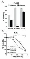
BCR-ABL-activated survival is glucose-dependent. A. Viability of BCR-ABL+ FL5.12 cells cultured in cytokine-free medium containing 0.02mM glucose (-Glucose) for 48 hours was significantly reduced compared to cells cultured in 10mM glucose (+Glucose). Survival of cells expressing an activated mutant of Akt (myrAkt), which mediates a glucose-dependent form of survival is shown for comparison. For reference, the glucose-independent survival of cells expressing Bcl-xL is also shown. The mean of triplicate viability measurements ±standard deviation were determined by propidium iodide exclusion in a flow cytometer. B. BCR-ABL+ K562 cells require glucose for long-term survival. Cell death in response to low glucose and imatinib were comparable.
Figure 3
Figure 3
Glucose independent survival upon loss of S6K1. A. S6K1 is not required for cytokine-independent survival in BCR-ABL+ FL5.12 cells. Decreased viability in myrAkt+ FL5.12 cells upon loss of S6K1 is shown for comparison. The siRNA pool targeting S6K1 is indicated by (+); Non-targeting siRNA is indicated by (-). B. Efficacy of S6K1 knockdown in treatments shown in A. C. S6K1 knockdown BCR-ABL+ cells exhibited a survival advantage when cultured under low glucose conditions (0.02 mM Glucose, indicated as -Glucose). D. Increased ATP in S6K1-knockdown cells cultured under low glucose conditions for 3 hours (left). Cells were not yet committed to programmed cell death at the time point measured, as illustrated by the absence of cleaved Caspase 3 at 3 hours (right). E-G. Rapamycin inactivation of S6K1 triggered a survival advantage in low glucose conditions in BCR-ABL+ FL5.12 (E), BCR-ABL+ primary murine bone marrow cells (F), and K562 cells (G).
Figure 4
Figure 4
S6K1 deficiency does not impair BCR-ABL leukemogenesis. A. Bone marrow cells from S6K1+/+ or S6K1−/− mice were transduced with BCR-ABL then transplanted into lethally irradiated recipients. Symptom-free survival is plotted. A trend towards more aggressive disease was observed in mice transplanted with BCR-ABL+ S6K1−/− cells, although the trend is not statistically significant (log-rank test). B. Little change in disease characteristics in mice that received S6K1−/− vs. S6K1+/+ BCR-ABL transformed cells. Shown are spleen wet weight (left), frequency of myeloid cells in the spleen (center), and frequency of BCR-ABL+ (GFP+) cells in the bone marrow (right) at the time of organ harvest.
Figure 5
Figure 5
FAO is activated in BCR-ABL+ cells upon S6K1 inactivation. A. BCR-ABL+ FL5.12 cells were cultured in the absence of cytokine and under low glucose conditions in the presence of 3H palmitate. S6K1 knockdown increased 3H2O release from 3H-palmitate in BCR-ABL+ FL5.12 cells. The CPT1 inhibitor etomoxir, which blocks mitochondrial FAO, counteracted this effect. The mean ±standard deviation of triplicate measurements is shown. B,C. Rapamycin increased FAO release of 3H from 3H-palmitate, which was inhibited by etomoxir in human BCR-ABL+ cell lines K562 (B) and KBM7 (C).
Figure 6
Figure 6
Requirement for FAO to mediate glucose-independent survival upon S6K1 inactivation. A. S6K1 knockdown improved survival under low glucose conditions and the FAO inhibitor etomoxir prevented this survival advantage in BCR-ABL+ FL5.12 cells. B. The AMPK agonist AICAR induced FAO-dependent survival in low glucose conditions, which was comparable to the survival observed in response to S6K1 knockdown. The FAO inhibitor etomoxir prevented survival in response to either AICAR or S6K1 knockdown. C. Cpt1c knockdown specifically reduced glucose-independent survival in S6K1-knockdown cells. siRNA targeting Cpt1a and Cpt1c (indicated by 1a and 1c, respectively) was transfected, followed by measurement of cell viability after 48 hours of culture in low glucose cytokine-free medium.
Figure 7
Figure 7
Coordinated metabolic inactivation enhances cytotoxicity in response to rapamycin. A. In the presence of full concentrations of glucose, neither S6K1 knockdown nor etomoxir induced significant cytotoxicity. Combination of S6K1 knockdown with 200 μM etomoxir was markedly cytotoxic for BCR-ABL+ FL5.12 cells measured at 48 hours. B. siS6K1 enhanced the sensitivity to a range of doses of etomoxir in BCR-ABL+ FL5.12 cells cultured in full glucose, suggesting a synergistic effect. Viability was measured at 48 hours.

References

    1. Hsu PP, Sabatini DM. Cancer cell metabolism: Warburg and beyond. Cell. 2008;134:703–7. - PubMed
    1. Plas DR, Talapatra S, Edinger AL, Rathmell JC, Thompson CB. Akt and Bcl-xL Promote Growth Factor-independent Survival through Distinct Effects on Mitochondrial Physiology. J Biol Chem. 2001;276:12041–8. - PubMed
    1. Rathmell JC, Fox CJ, Plas DR, Hammerman PS, Cinalli RM, Thompson CB. Akt-directed glucose metabolism can prevent Bax conformation change and promote growth factor-independent survival. Mol Cell Biol. 2003;23:7315–28. - PMC - PubMed
    1. Buzzai M, Bauer DE, Jones RG, Deberardinis RJ, Hatzivassiliou G, Elstrom RL, et al. The glucose dependence of Akt-transformed cells can be reversed by pharmacologic activation of fatty acid beta-oxidation. Oncogene. 2005;24:4165–73. - PubMed
    1. Schafer ZT, Grassian AR, Song L, Jiang Z, Gerhart-Hines Z, Irie HY, et al. Antioxidant and oncogene rescue of metabolic defects caused by loss of matrix attachment. Nature. 2009;461:109–13. - PMC - PubMed

Publication types

MeSH terms