. 2012 Jun;7(2):477-87.
doi: 10.1007/s11481-012-9343-6. Epub 2012 Mar 6.

S-nitrosoglutathione prevents experimental cerebral malaria

Affiliations

S-nitrosoglutathione prevents experimental cerebral malaria

Graziela M Zanini et al. J Neuroimmune Pharmacol. 2012 Jun.

Abstract

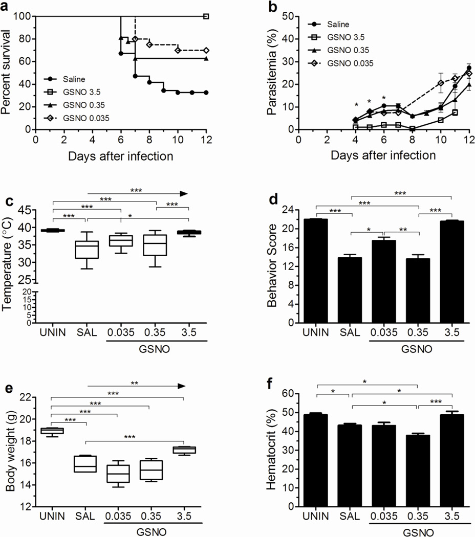
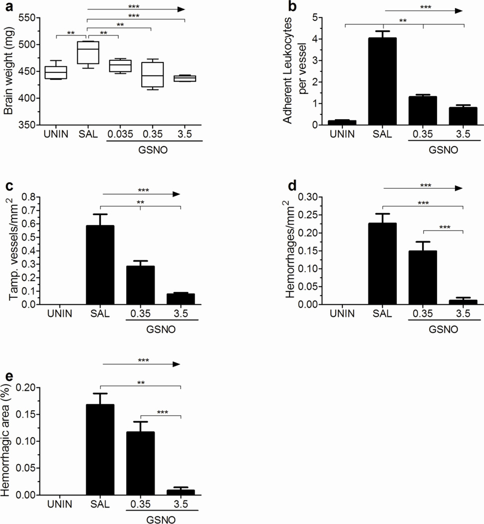
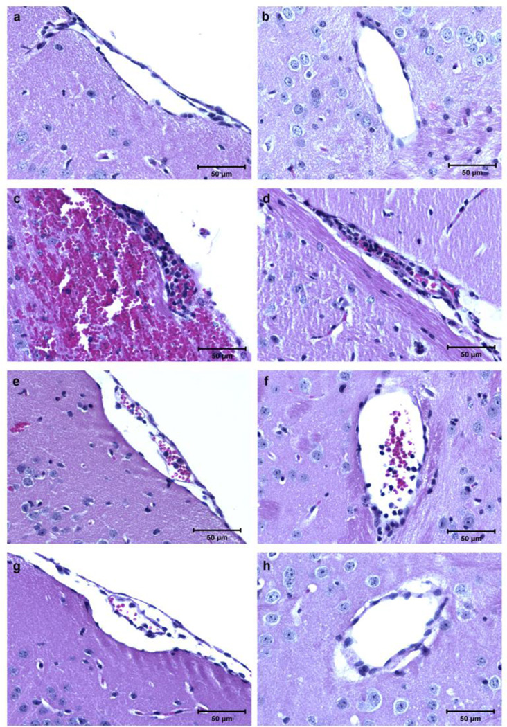
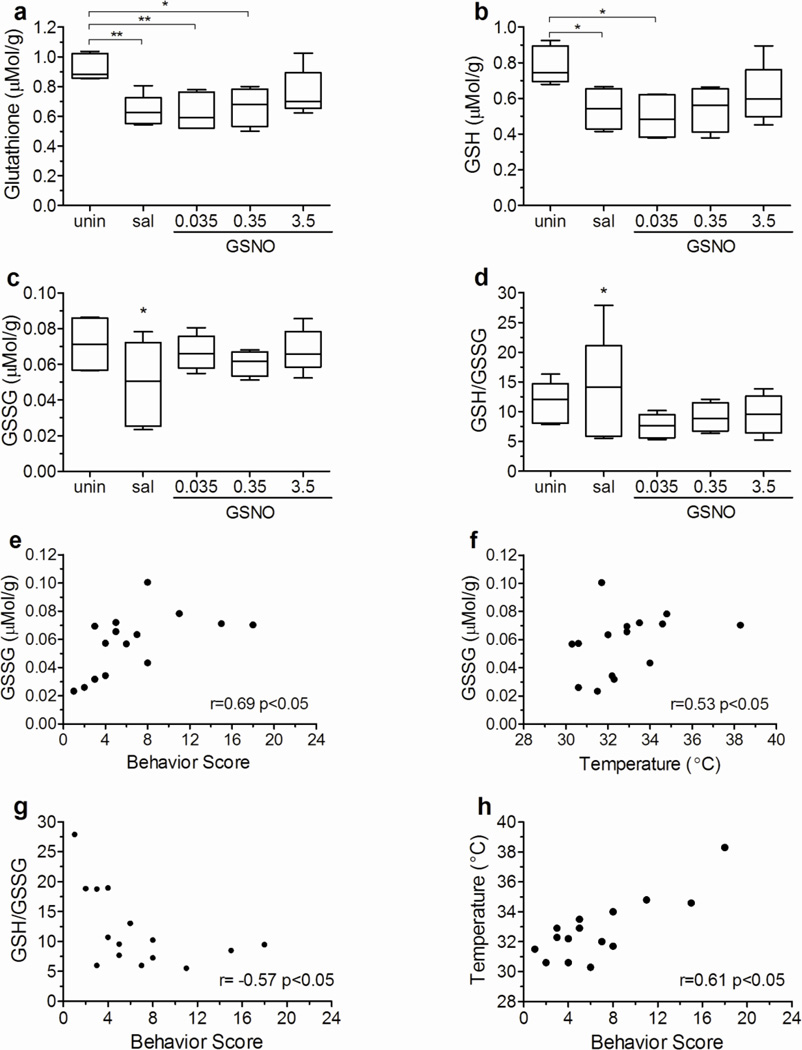
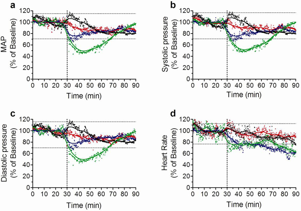
Administration of the exogenous nitric oxide (NO) donor dipropylenetriamine-NONOate (DPTA-NO) to mice during Plasmodium berghei ANKA (PbA) infection largely prevents development of experimental cerebral malaria (ECM). However, a high dose (1 mg/mouse twice a day) is necessary and causes potent side effects such as marked hypotension. In the present study we evaluated whether an alternative, physiologically relevant NO donor, S-nitrosoglutathione (GSNO), was able to prevent ECM at lower doses with minimal side effects. Prophylactic treatment with high (3.5 mg), intermediate (0.35 mg) or low (0.035 mg) doses of GSNO decreased incidence of ECM in PbA-infected mice, decreasing also edema, leukocyte accumulation and hemorrhage incidence in the brain. The high dose inhibited parasite growth and also induced transient hypotension. Low and intermediate doses had no or only mild effects on parasitemia, blood pressure, and heart rate compared to saline-treated mice. PbA infection decreased brain total and reduced (GSH) glutathione levels. Brain levels of oxidized (GSSG) glutathione and the GSH/GSSG ratio were positively correlated with temperature and motor behavior. Low and intermediate doses of GSNO failed to restore the depleted brain total glutathione and GSH levels, suggesting that ECM prevention by GSNO was probably related to other effects such as inhibition of inflammation and vascular protection. These results indicate that ECM is associated with depletion of the brain glutathione pool and that GSNO is able to prevent ECM development in a wide range of doses, decreasing brain inflammation and inducing milder cardiovascular side effects.


Figures

Figure 1
Figure 1. GSNO prevents ECM development
Cumulative survival (A), course of parasitemia (B), rectal temperature (C), and motor behavior score (D) of PbA-infected mice treated with saline (n=55) or GSNO at 0.035 (n=18), 0.35 (n=28), and 3.5 (n=20) mg/mouse. Body weight (E, n=4–6 per group) and hematocrit (F, n=cumulative survival) were also evaluated. Rectal temperature, motor behavior score, body weight and hematocrit were measured on day 6 of infection. *p<0.05, **p<0.01, ***p<0.001, arrows indicate the presence of a linear trend.
Figure 2
Figure 2. GSNO increases NO bioavailability in PbA infected mice
Exhaled NO (A) of uninfected and PbA-infected mice treated with saline or GSNO at 0.035, 0.35, and 3.5 mg/mouse was measured 1 hour after the morning treatment on day 5 of infection (n≥5 per group). Plasma nitrite (F) of uninfected and PbA-infected mice treated with saline or GSNO at 0.035, 0.35, and 3.5 mg/mouse was measured on samples collected prior to the morning dosing on day 6 of infection (n≥7 per group). *p<0.05, **p<0.01, ***p<0.001, arrows indicate the presence of a linear trend.
Figure 3
Figure 3. GSNO decreases brain edema and inflammation in PbA infected mice
Mice receiving GSNO at 0.35 and 3.5 mg/mouse did not show an increase in total brain weight (A) upon infection. Treated mice also presented a smaller number of adherent leukocytes per meningeal vessel (B), a lesser number of tamponated vessel in brain parenchyma (C), and a small hemorrhagic burden as reflected by a decreased number of hemorrhages/mm2 (D) and hemorrhagic area (E, defined as the area occupied by hemorrhages in relation to the total area analyzed) of brain parenchyma in a dose response manner on day 6 of infection. n= 4–5 per group, *p<0.05, **p<0.01, ***p<0.001, arrows indicate the presence of a linear trend.
Figure 4
Figure 4. Inhibition of inflammation in the brain of PbA-infected mice treated with GSNO
(A and B) Pial (A) and parenchymal (B) vessels of uninfected control mice, showing no accumulation or adhesion of leukocytes; (C and D) pial (C) and parenchymal (D) vessels of mice receiving saline with ECM, plugged with leukocytes (black arrows); an extensive subarachnoid hemorrhage is seen in panel C (white arrows); (E and F) pial (C) and parenchymal (F) vessels of mice receiving GSNO at 0.35 mg/mouse showing a less intense leukocyte adhesion (black arrows); (G and H) pial (G) and parenchymal (H) vessels of mice receiving GSNO at 3.5 mg/mouse, a small number of adherent leukocytes is shown in panel G (black arrows), and a clean parenchymal vessel is shown in panel H. Magnitude of leukocyte accumulation and adherence was heterogeneous among vessels in mice receiving saline and GSNO at 0.35 and 3.5 mg/mouse. All sections were stained with H&E (magnification, ×400).
Figure 5
Figure 5. PbA infection decreases brain total glutathione and GSH levels, but GSSG levels and GSH/GSSG ratio are correlated with ECM development
Brain levels of total glutathione (A), GSH (B), and GSSG (C) and GSH/GSSG ratio of uninfected and PbA infected mice receiving saline or GSNO at 0.035, 0.35, and 3.5 mg/mouse. Brain GSSG levels were positively correlated with motor behavior score (E) and temperature (F), and negatively correlated with the GSH/GSSG ratio (G) in PbA infected mice. Temperature was also positively correlated with motor behavior score (H). All measurements were made on day 6 of infection. *p<0.05, **p<0.01, r= spearman correlation coefficient.
Figure 6
Figure 6. GSNO effects on cardiovascular parameters
Changes in mean arterial pressure (MAP, A), systolic pressure (B), diastolic pressure (C), and heart rate (D) following one IP injection of saline (black dots and lines), and GSNO at 0.035 (red dots and lines), 0.35 (blue dots and lines), and 3.5 (green dots and lines). Vertical doted lines represent the time when the IP injection was given and separate the baseline period from the experimental period. Results are expressed as the percentage change in relation to the mean of the baseline period for each group. Horizontal doted lines represent the range of values falling within the mean plus and minus two standard deviations (SD) of the baseline value calculated for saline treated group. N = 4 per group.

References

    1. Aoyama K, Watabe M, Nakaki T. Regulation of neuronal glutathione synthesis. J Pharmacol Sci. 2008;108(3):227–238. - PubMed
    1. Astrand A, Bohlooly YM, Larsdotter S, Mahlapuu M, Andersen H, Tornell J, Ohlsson C, Snaith M, Morgan DG. Mice lacking melanin-concentrating hormone receptor 1 demonstrate increased heart rate associated with altered autonomic activity. Am J Physiol Regul Integr Comp Physiol. 2004;287(4):R749–R758. - PubMed
    1. Bauer PR, Van Der Heyde HC, Sun G, Specian RD, Granger DN. Regulation of endothelial cell adhesion molecule expression in an experimental model of cerebral malaria. Microcirculation. 2002;9(6):463–470. - PubMed
    1. Beare NA, Harding SP, Taylor TE, Lewallen S, Molyneux ME. Perfusion abnormalities in children with cerebral malaria and malarial retinopathy. J Infect Dis. 2009;199(2):263–271. - PMC - PubMed
    1. Cabrales P, Zanini GM, Meays D, Frangos JA, Carvalho LJ. Murine cerebral malaria is associated with a vasospasm-like microcirculatory dysfunction, and survival upon rescue treatment is markedly increased by nimodipine. Am J Pathol. 2010;176(3):1306–1315. - PMC - PubMed

Publication types

LinkOut - more resources